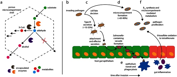
Upon arriving in the host gut, enteric pathogens face a daunting challenge: to proliferate in an environment already rich in commensal microbes and poor in available nutrients. Recent evidence suggests that Salmonella and other enteric bacteria conduct a coordinated assault employing two complimentary systems: bacterial microcompartments and the type III secretion system. While a portion of invading bacteria construct subcellular metabolic organelles designed to utilize unique nutrients, the remaining invading cells induce intestinal inflammation, remodeling the chemical environment of the gut to render it more favorable to Salmonella proliferation (Fig 1).
Fig 1. Bacterial microcompartment function and the coordinated invasion of the host gut by Salmonella enterica.
(a): A substrate molecule enters the microcompartment and is converted to an aldehyde species, which is trapped in the microcompartment shell before being converted either to an alcohol or to a Coenzyme A-conjugated species [32]. (b) The invading pathogen population enters the gut. (c) Each pathogen cell undergoes a fate decision between type III secretion (~10%–35% of cells) and microcompartment formation (~65%–90% of cells). (d) Type III secretion-competent cells invade the host epithelium while microcompartment-competent cells form microcompartments and synthesize vitamin B12. (e) Type III secretion-competent cells traverse the epithelium and undergo phagocytosis in the lamina propria. (f) Gut inflammation causes thiosulfate oxidation to tetrathionate, allowing microcompartment-mediated metabolism and pathogen proliferation.
What Are Bacterial Microcompartments and What Are They For?
Despite the received wisdom that eukarya possess intracellular organelles and bacteria do not, bacteria do use organelles called bacterial microcompartments to spatially segregate metabolism. Rather than a phospholipid membrane, however, these organelles are bound by a porous protein monolayer made up of trimeric, pentameric, and hexameric shell proteins. A suite of metabolic enzymes, including those required for cofactor regeneration, are encapsulated in the microcompartment lumen. The mechanism of microcompartment assembly remains elusive, but it is known that some enzymes are localized to the microcompartment through interactions with the inner face of the microcompartment shell [1,2]. Many pathogens possess microcompartments, including Salmonella enterica, Escherichia coli, Listeria monocytogenes, Yersinia enterocolitica, and Shigella flexneri, and microcompartment genes have been found in as many as 20% of sequenced bacterial genomes [3,4]. Bacteria are known to use a variety of microcompartment systems to metabolize compounds such as 1,2-propanediol [5], ethanolamine [6], and L-fucose and L-rhamnose [7]. All of these metabolic pathways proceed through toxic aldehyde intermediates, and it is proposed that the microcompartment shell functions to protect the rest of the bacterial cell contents from these toxic compounds as well as to sequester a private pool of the requisite cofactor molecules [8]. Compartmentalized metabolic processes may impart a competitive advantage to invading pathogens over the existing gut microbiota, which typically lack microcompartment operons and thus are unable to utilize the substrates metabolized in the microcompartments [9,10]. For example, S. enterica subsp. enterica, serovar Typhimurium dedicates approximately 2% of its genome to 1,2-propanediol and ethanolamine metabolism and the synthesis of associated cofactors, suggesting that these processes confer a significant competitive advantage at some point in the pathogen’s life cycle [11,12].
Microcompartment systems, such as the ethanolamine utilization microcompartment, enhance E. coli and S. enterica proliferation in diverse settings, including in food products, in a Caenorhabditis elegans model of infection, during growth on bovine intestinal content, and in the gut of a mouse model of Salmonella infection [9,13,14]. This indicates that there are many circumstances in which microcompartments may provide a competitive advantage to pathogens. Evidence suggests that the gut microbiota plays a central role in preventing host colonization by pathogens, possibly by sequestering critical nutrients [15]. In a model of S. Typhimurium infection of the mouse gut, for example, antibiotic treatment to reduce the abundance of native gut microbes renders the host more susceptible to Salmonella infection [16], and mice with a compromised microbiota clear S. Typhymurium from the gut much less effectively following nonfatal infection [17]. Gaining a unique metabolic capacity may help pathogens sidestep microbiotic defense mechanisms by creating a new nutritional niche in the host gut, but microcompartment-mediated metabolism also requires a unique micronutrient: the cofactor vitamin B12.
The B12 Synthesis Paradox: Is 1,2-Propanediol Utilization an Aerobic or Anaerobic Process?
1,2-propanediol and ethanolamine utilization both require vitamin B12, and the B12 biosynthetic genes in S. enterica were found to be transcriptionally co-regulated with the 1,2-propanediol utilization operon [18]. This raised an apparent paradox: 1,2-propanediol and ethanolamine metabolism were once thought to occur only in aerobic conditions, whereas B12 synthesis is a strictly anaerobic process [11,19]. This puzzle was solved by the discovery that 1,2-propanediol and ethanolamine metabolism can proceed using tetrathionate as an electron acceptor in place of molecular oxygen [12]. Tetrathionate, in turn, is a product of the oxidation of thiosulfate, an abundant molecule in the gut produced by the inactivation of H2S. Salmonella or other pathogenic bacteria in the gut might thus synthesize vitamin B12 anaerobically while simultaneously respiring to tetrathionate instead of oxygen. Under normal gut conditions, oxidation of thiosulfate to tetrathionate is minimal; however, this reaction is accelerated by inflammation when the gut is rendered a more oxidizing environment [20]. The oxidizing environment of the inflamed gut may favor microcompartment-mediated metabolism by Salmonella and other pathogens, allowing their proliferation at the expense of the gut microbiota [14]. Indeed, many pathogenic bacteria possess a mechanism to induce just such an oxidative dysbiosis—the type III secretion system [21].
Type III Secretion and Microcompartment-Mediated Metabolism: Does Salmonella Conduct a Coordinated Assault on the Gut Microbiota?
Upon encountering environmental cues that are indicative of the host intestinal tract (e.g., high osmolarity, low pH, and low oxygen concentration), many enteric pathogens, including Salmonella and Shigella spp., express type III secretion systems, which function to mechanically penetrate the intestinal epithelium and translocate various effector proteins into host cells [22]. These effectors not only mediate internalization of the Salmonella cell into a Salmonella-containing vacuole but also induce an inflammatory response throughout the host gut. This inflammatory response increases the rate of thiosulfate oxidation and hence the concentration of tetrathionate in the gut [23,20]. Not all the invading Salmonella cells, however, express the type III secretion system. Experiments examining the transcriptional regulation of type III secretion system promoters indicate that only a fraction of a given population expresses the type III secretion system even in appropriate inducing conditions [24]. What, then, is the role of the non-induced cells? This population is believed to remain in the gut in order to exploit the ensuing inflammation and gain a foothold in the metabolic competition between invaders and commensals [25,26].
Interestingly, 1,2-propanediol represses expression of the type III secretion system master regulator hilA, suggesting that cells may undergo a fate decision between type III secretion system-mediated epithelium invasion (leading to bacterial cell death) and 1,2-propanediol or ethanolamine metabolism (leading to proliferation) [23,27]. Furthermore, propionate, a downstream product of 1,2-propanediol metabolism, down-regulates another type III secretion system master regulator, HilD, at the post-translational level; it is proposed that endogenous propionate in the gut is primarily responsible for this phenomenon [28]. We additionally propose that post-translational modification of HilD as a result of intracellular propionate production may be a means of down-regulating type III secretion in response to 1,2-propanediol utilization microcompartment expression.
Does the Paradigm of Nutritional Competition Extend beyond Micronutrients?
Nutritional immunity, the modulation of micronutrient concentrations by the host to prevent colonization by pathogens, is well characterized for transition metal micronutrients [29,30]. It seems likely that this paradigm, in which host and commensal processes are under selective pressure to sequester critical nutrients from pathogens, extends beyond micronutrients such as iron and copper ions to other small molecules as well. Bacterial microcompartments may therefore represent another step in the nutritional arms race between pathogens and commensal species. The coordinated induction of the type III secretion system and bacterial microcompartments in separate bacterial populations allows the pathogen population to induce and exploit an inflamed state in the host gut, allowing colonization followed by diarrhea favorable for subsequent transmission to other hosts [31]. The proposed interplay between type III secretion and bacterial microcompartments suggests that pathogens “dumpster diving” in the gut can develop specialized metabolic mechanisms to utilize compounds otherwise considered to be waste by the gut microbiota. These strategies may involve the coordinated action of multiple cellular processes across the invading bacterial population.
Acknowledgments
We are grateful to current and former members of the Tullman-Ercek laboratory for stimulating discussions about this topic, which lies at the intersection of our research interests. We also wish to thank Kevin Metcalf, Leah Sibener, Lisa Burdette, Nicholas Jakobson, and Stanley Herrmann for critical reading of the manuscript.
We apologize to those whose work we were unable to discuss due to the space limitations of this format.
Funding Statement
Supported by: the National Science Foundation (award MCB1150567 to DTE), the Army Research Office (grant W911NF-15-1-0144 to DTE), a gift from ExxonMobil Corporation (to DTE), the Hellman Family Faculty Fund, and a UC Berkeley Fellowship (CMJ). The funders had no role in study design, data collection and analysis, decision to publish, or preparation of the manuscript.
References
- 1. Fan C, Cheng S, Liu Y, Escobar CM, Crowley CS, Jefferson RE, et al. Short N-terminal sequences package proteins into bacterial microcompartments. Proc Natl Acad Sci. 2010;107: 7509–7514. 10.1073/pnas.0913199107 [DOI] [PMC free article] [PubMed] [Google Scholar]
- 2. Fan C, Cheng S, Sinha S, Bobik TA. Interactions between the termini of lumen enzymes and shell proteins mediate enzyme encapsulation into bacterial microcompartments. Proc Natl Acad Sci. 2012;109: 14995–15000. 10.1073/pnas.1207516109 [DOI] [PMC free article] [PubMed] [Google Scholar]
- 3. Kerfeld CA, Heinhorst S, Cannon GC. Bacterial Microcompartments. Annu Rev Microbiol. 2010;64: 391–408. 10.1146/annurev.micro.112408.134211 [DOI] [PubMed] [Google Scholar]
- 4. Axen SD, Erbilgin O, Kerfeld CA. A Taxonomy of Bacterial Microcompartment Loci Constructed by a Novel Scoring Method. Tanaka MM, editor. PLoS Comput Biol. 2014;10: e1003898 10.1371/journal.pcbi.1003898 [DOI] [PMC free article] [PubMed] [Google Scholar]
- 5. Bobik TA, Havemann GD, Busch RJ, Williams DS, Aldrich HC. The Propanediol Utilization (pdu) Operon of Salmonella enterica Serovar Typhimurium LT2 Includes Genes Necessary for Formation of Polyhedral Organelles Involved in Coenzyme B12-Dependent 1, 2-Propanediol Degradation. J Bacteriol. 1999;181: 5967–5975. [DOI] [PMC free article] [PubMed] [Google Scholar]
- 6. Kofoid E, Rappleye C, Stojiljkovic I, Roth J. The 17-Gene Ethanolamine (eut) Operon of Salmonella typhimurium Encodes Five Homologues of Carboxysome Shell Proteins. J Bacteriol. 1999;181: 5317–5329. [DOI] [PMC free article] [PubMed] [Google Scholar]
- 7. Erbilgin O, McDonald KL, Kerfeld CA. Characterization of a Planctomycetal Organelle: a Novel Bacterial Microcompartment for the Aerobic Degradation of Plant Saccharides. Appl Environ Microbiol. 2014;80: 2193–2205. 10.1128/AEM.03887-13 [DOI] [PMC free article] [PubMed] [Google Scholar]
- 8. Sampson EM, Bobik TA. Microcompartments for B12-Dependent 1,2-Propanediol Degradation Provide Protection from DNA and Cellular Damage by a Reactive Metabolic Intermediate. J Bacteriol. 2008;190: 2966–2971. 10.1128/JB.01925-07 [DOI] [PMC free article] [PubMed] [Google Scholar]
- 9. Bertin Y, Girardeau JP, Chaucheyras-Durand F, Lyan B, Pujos-Guillot E, Harel J, et al. Enterohaemorrhagic Escherichia coli gains a competitive advantage by using ethanolamine as a nitrogen source in the bovine intestinal content. Environ Microbiol. 2011;13: 365–377. 10.1111/j.1462-2920.2010.02334.x [DOI] [PubMed] [Google Scholar]
- 10. Rivera-Chávez F, Bäumler AJ. The Pyromaniac Inside You: Salmonella Metabolism in the Host Gut. Annu Rev Microbiol. 2015;69: 31–48. 10.1146/annurev-micro-091014-104108 [DOI] [PubMed] [Google Scholar]
- 11. Roth JR, Lawrence JG, Bobik TA. Cobalamin (coenzyme B12): synthesis and biological significance. Annu Rev Microbiol. 1996;50: 137–181. 10.1146/annurev.micro.50.1.137 [DOI] [PubMed] [Google Scholar]
- 12. Price-Carter M, Tingey J, Bobik TA, Roth JR. The Alternative Electron Acceptor Tetrathionate Supports B12-Dependent Anaerobic Growth of Salmonella enterica Serovar Typhimurium on Ethanolamine or 1,2-Propanediol. J Bacteriol. 2001;183: 2463–2475. 10.1128/JB.183.8.2463-2475.2001 [DOI] [PMC free article] [PubMed] [Google Scholar]
- 13. Srikumar S, Fuchs TM. Ethanolamine Utilization Contributes to Proliferation of Salmonella enterica Serovar Typhimurium in Food and in Nematodes. Appl Environ Microbiol. 2011;77: 281–290. 10.1128/AEM.01403-10 [DOI] [PMC free article] [PubMed] [Google Scholar]
- 14. Thiennimitr P, Winter SE, Winter MG, Xavier MN, Tolstikov V, Huseby DL, et al. Intestinal inflammation allows Salmonella to use ethanolamine to compete with the microbiota. Proc Natl Acad Sci. 2011;108: 17480–17485. 10.1073/pnas.1107857108 [DOI] [PMC free article] [PubMed] [Google Scholar]
- 15. Stecher B, Hardt W-D. The role of microbiota in infectious disease. Trends Microbiol. 2008;16: 107–114. [DOI] [PubMed] [Google Scholar]
- 16. Keeney KM, Yurist-Doutsch S, Arrieta M-C, Finlay BB. Effects of Antibiotics on Human Microbiota and Subsequent Disease. Annu Rev Microbiol. 2014;68: 217–235. 10.1146/annurev-micro-091313-103456 [DOI] [PubMed] [Google Scholar]
- 17. Endt K, Stecher B, Chaffron S, Slack E, Tchitchek N, Benecke A, et al. The Microbiota Mediates Pathogen Clearance from the Gut Lumen after Non-Typhoidal Salmonella Diarrhea. PLoS Pathog. 2010;6: e1001097 10.1371/journal.ppat.1001097 [DOI] [PMC free article] [PubMed] [Google Scholar]
- 18. Bobik TA, Ailion M, Roth JR. A single regulatory gene integrates control of vitamin B12 synthesis and propanediol degradation. J Bacteriol. 1992;174: 2253–2266. [DOI] [PMC free article] [PubMed] [Google Scholar]
- 19. Jeter RM, Olivera BM, Roth JR. Salmonella typhimurium synthesizes cobalamin (vitamin B12) de novo under anaerobic growth conditions. J Bacteriol. 1984;159: 206–213. [DOI] [PMC free article] [PubMed] [Google Scholar]
- 20. Winter SE, Thiennimitr P, Winter MG, Butler BP, Huseby DL, Crawford RW, et al. Gut inflammation provides a respiratory electron acceptor for Salmonella . Nature. 2010;467: 426–429. 10.1038/nature09415 [DOI] [PMC free article] [PubMed] [Google Scholar]
- 21. Galán JE, Collmer A. Type III Secretion Machines: Bacterial Devices for Protein Delivery into Host Cells. Science. 1999;284: 1322–1328. 10.1126/science.284.5418.1322 [DOI] [PubMed] [Google Scholar]
- 22. Galán JE. Salmonella Interactions with Host Cells: Type III Secretion at Work. Annu Rev Cell Dev Biol. 2001;17: 53–86. 10.1146/annurev.cellbio.17.1.53 [DOI] [PubMed] [Google Scholar]
- 23. Ackermann M, Stecher B, Freed NE, Songhet P, Hardt W-D, Doebeli M. Self-destructive cooperation mediated by phenotypic noise. Nature. 2008;454: 987–990. 10.1038/nature07067 [DOI] [PubMed] [Google Scholar]
- 24. Sturm A, Heinemann M, Arnoldini M, Benecke A, Ackermann M, Benz M, et al. The Cost of Virulence: Retarded Growth of Salmonella Typhimurium Cells Expressing Type III Secretion System 1. Ausubel FM, editor. PLoS Pathog. 2011;7: e1002143 10.1371/journal.ppat.1002143 [DOI] [PMC free article] [PubMed] [Google Scholar]
- 25. Stecher B, Robbiani R, Walker AW, Westendorf AM, Barthel M, Kremer M, et al. Salmonella enterica Serovar Typhimurium Exploits Inflammation to Compete with the Intestinal Microbiota. PLoS Biol. 2007;5: e244 10.1371/journal.pbio.0050244 [DOI] [PMC free article] [PubMed] [Google Scholar]
- 26. Lupp C, Robertson ML, Wickham ME, Sekirov I, Champion OL, Gaynor EC, et al. Host-Mediated Inflammation Disrupts the Intestinal Microbiota and Promotes the Overgrowth of Enterobacteriaceae. Cell Host Microbe. 2007;2: 119–129. [DOI] [PubMed] [Google Scholar]
- 27. Nakayama S, Watanabe H. Mechanism of hilA Repression by 1,2-Propanediol Consists of Two Distinct Pathways, One Dependent on and the Other Independent of Catabolic Production of Propionate, in Salmonella enterica Serovar Typhimurium. J Bacteriol. 2006;188: 3121–3125. 10.1128/JB.188.8.3121-3125.2006 [DOI] [PMC free article] [PubMed] [Google Scholar]
- 28. Hung C-C, Garner CD, Slauch JM, Dwyer ZW, Lawhon SD, Frye JG, et al. The Intestinal Fatty Acid Propionate Inhibits Salmonella Invasion through the Post-translational Control of HilD. Mol Microbiol. 2013;87: 1045–1060. 10.1111/mmi.12149 [DOI] [PMC free article] [PubMed] [Google Scholar]
- 29. Skaar EP. The Battle for Iron between Bacterial Pathogens and Their Vertebrate Hosts. PLoS Pathog. 2010;6: e1000949 10.1371/journal.ppat.1000949 [DOI] [PMC free article] [PubMed] [Google Scholar]
- 30. Hood MI, Skaar EP. Nutritional immunity: transition metals at the pathogen-host interface. Nat Rev Microbiol. 2012;10: 525–537. 10.1038/nrmicro2836 [DOI] [PMC free article] [PubMed] [Google Scholar]
- 31. Gopinath S, Carden S, Monack D. Shedding light on Salmonella carriers. Trends Microbiol. 2012;20: 320–327. [DOI] [PubMed] [Google Scholar]
- 32. Kerfeld CA, Erbilgin O. Bacterial microcompartments and the modular construction of microbial metabolism. Trends Microbiol. 2014; [DOI] [PubMed] [Google Scholar]


