Figure 3.
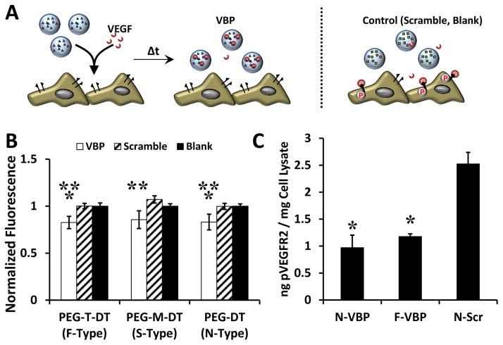
VBP microspheres reduced VEGF-dependent metabolic activity and VEGFR activation in HUVEC culture A: Schematic of the VEGF sequestering by VBP microspheres (or Scramble or Blank microspheres) in HUVEC culture containing VEGF. B: Relative HUVEC metabolic activity (given as normalized fluorescence intensity of each condition relative to the Blank microsphere condition of each crosslinker type) upon addition of VEGF-containing medium to Blank microspheres (containing no peptide), VBP, or Scramble and varying crosslinker identity. Two-way analysis of variance was performed (microsphere peptide identity p-value< 0.0001, microsphere crosslink type p-value> 0.05, interaction p-value> 0.05) with post-hoc Student’s t-test. Statistical significance is denoted compared to Scramble (**) and Blank (*) microspheres at each respective crosslinker type or between conditions in brackets for p-value < 0.05 using Student’s t-test. Error bars represent the standard deviation about the mean for six replicates per condition. C: Amount of phosphorylated VEGFR2 (in ng) measured via ELISA normalized to the total protein content of the cell lysate (in mg) after treatment of HUVECs with microsphere supernatants from microspheres (F-VBP, N-VBP, or N-Scramble) that were pre-incubated in 10 ng/mL VEGF for 2 days in culture medium containing 2 vol.% FBS in M199. Data is presented as mean + /− standard deviation for three replicates per condition, and statistical significance is denoted relative to N-Scramble control at p-value < 0.05 (*) using one-way ANOVA and Tukey’s post-hoc test.

