

Abstract
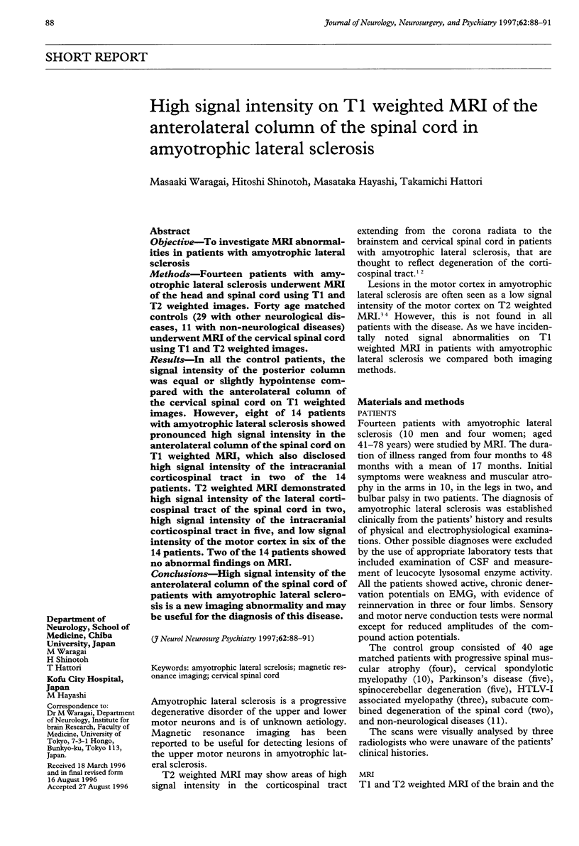
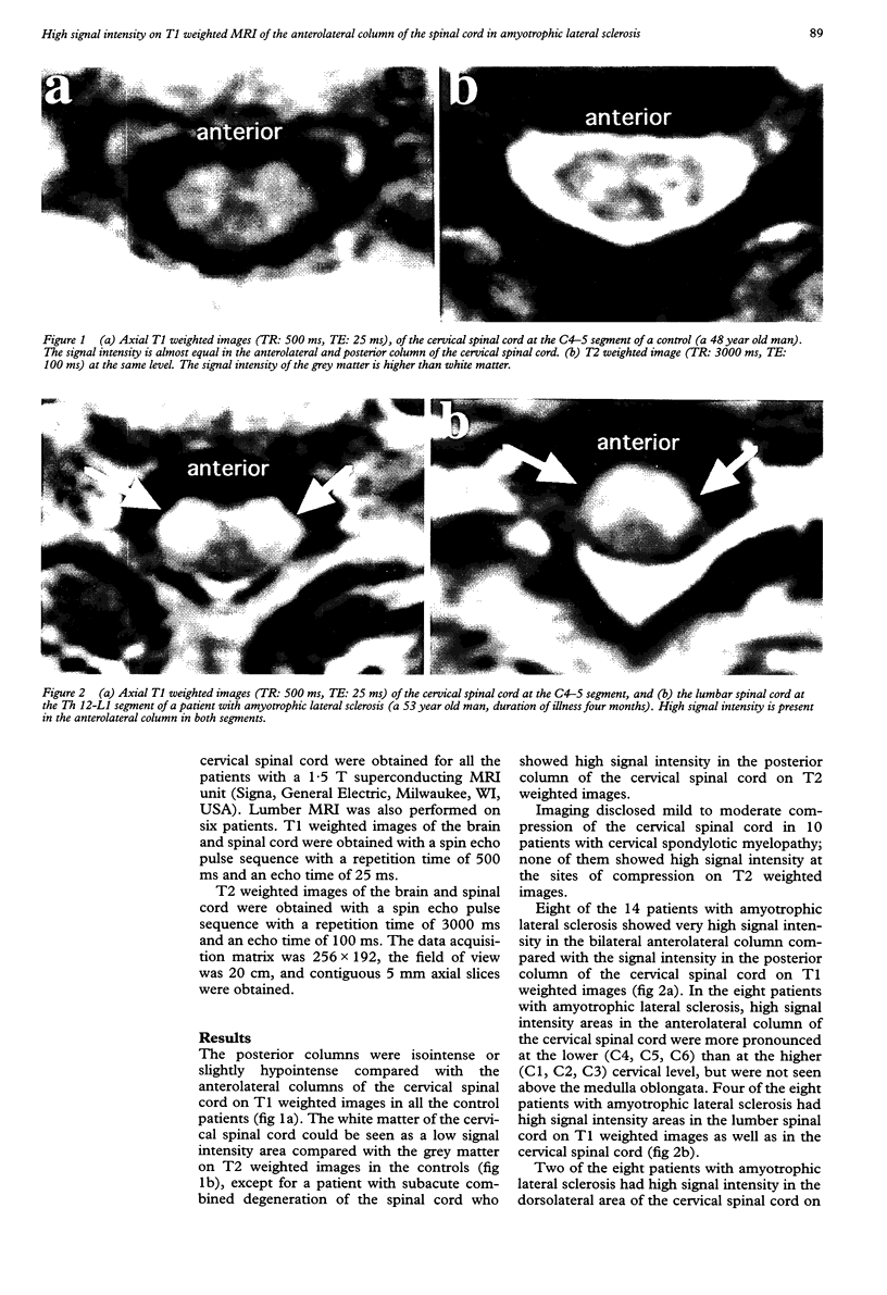
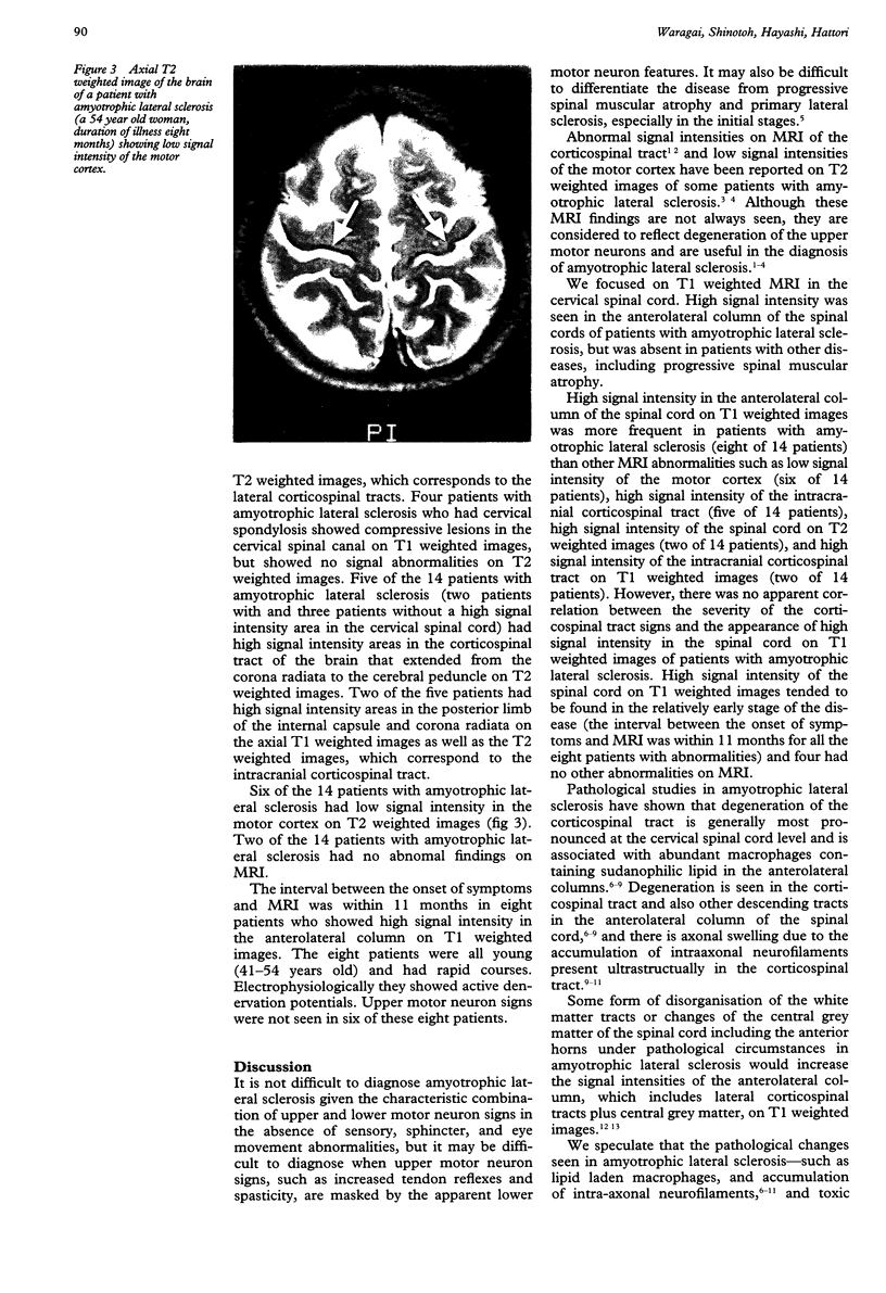
OBJECTIVE: To investigate MRI abnormalities in patients with amyotrophic lateral sclerosis. METHODS: Fourteen patients with amyotrophic lateral sclerosis underwent MRI of the head and spinal cord using T1 and T2 weighted images. Forty age matched controls (29 with other neurological diseases, 11 with non-neurological diseases) underwent MRI of the cervical spinal cord using T1 and T2 weighted images. RESULTS: In all the control patients, the signal intensity of the posterior column was equal or slightly hypointense compared with the anterolateral column of the cervical spinal cord on T1 weighted images. However, eight of 14 patients with amyotrophic lateral sclerosis showed pronounced high signal intensity in the anterolateral column of the spinal cord on T1 weighted MRI, which also disclosed high signal intensity of the intracranial corticospinal tract in two of the 14 patients. T2 weighted MRI demonstrated high signal intensity of the lateral corticospinal tract of the spinal cord in two, high signal intensity of the intracranial corticospinal tract in five, and low signal intensity of the motor cortex in six of the 14 patients. Two of the 14 patients showed no abnormal findings on MRI. CONCLUSIONS: High signal intensity of the anterolateral column of the spinal cord of patients with amyotrophic lateral sclerosis is a new imaging abnormality and may be useful for the diagnosis of this disease.
Full text
PDF
Images in this article
Selected References
These references are in PubMed. This may not be the complete list of references from this article.
- Breuer A. C., Lynn M. P., Atkinson M. B., Chou S. M., Wilbourn A. J., Marks K. E., Culver J. E., Fleegler E. J. Fast axonal transport in amyotrophic lateral sclerosis: an intra-axonal organelle traffic analysis. Neurology. 1987 May;37(5):738–748. doi: 10.1212/wnl.37.5.738. [DOI] [PubMed] [Google Scholar]
- Brownell B., Oppenheimer D. R., Hughes J. T. The central nervous system in motor neurone disease. J Neurol Neurosurg Psychiatry. 1970 Jun;33(3):338–357. doi: 10.1136/jnnp.33.3.338. [DOI] [PMC free article] [PubMed] [Google Scholar]
- Czervionke L. F., Daniels D. L., Ho P. S., Yu S. W., Pech P., Strandt J. A., Williams A. L., Haughton V. M. The MR appearance of gray and white matter in the cervical spinal cord. AJNR Am J Neuroradiol. 1988 May-Jun;9(3):557–562. [PMC free article] [PubMed] [Google Scholar]
- Eisen A., Kim S., Pant B. Amyotrophic lateral sclerosis (ALS): a phylogenetic disease of the corticomotoneuron? Muscle Nerve. 1992 Feb;15(2):219–224. doi: 10.1002/mus.880150215. [DOI] [PubMed] [Google Scholar]
- Friedman D. P., Tartaglino L. M. Amyotrophic lateral sclerosis: hyperintensity of the corticospinal tracts on MR images of the spinal cord. AJR Am J Roentgenol. 1993 Mar;160(3):604–606. doi: 10.2214/ajr.160.3.8430564. [DOI] [PubMed] [Google Scholar]
- Ho P. S., Yu S. W., Czervionke L. F., Sether L. A., Wagner M., Pech P., Haughton V. M. MR appearance of gray and white matter at the cervicomedullary region. AJNR Am J Neuroradiol. 1989 Sep-Oct;10(5):1051–1055. [PMC free article] [PubMed] [Google Scholar]
- Ishikawa K., Nagura H., Yokota T., Yamanouchi H. Signal loss in the motor cortex on magnetic resonance images in amyotrophic lateral sclerosis. Ann Neurol. 1993 Feb;33(2):218–222. doi: 10.1002/ana.410330214. [DOI] [PubMed] [Google Scholar]
- Mirowitz S. A., Westrich T. J., Hirsch J. D. Hyperintense basal ganglia on T1-weighted MR images in patients receiving parenteral nutrition. Radiology. 1991 Oct;181(1):117–120. doi: 10.1148/radiology.181.1.1909445. [DOI] [PubMed] [Google Scholar]
- Oba H., Araki T., Ohtomo K., Monzawa S., Uchiyama G., Koizumi K., Nogata Y., Kachi K., Shiozawa Z., Kobayashi M. Amyotrophic lateral sclerosis: T2 shortening in motor cortex at MR imaging. Radiology. 1993 Dec;189(3):843–846. doi: 10.1148/radiology.189.3.8234713. [DOI] [PubMed] [Google Scholar]
- Okamoto K., Hirai S., Shoji M., Senoh Y., Yamazaki T. Axonal swellings in the corticospinal tracts in amyotrophic lateral sclerosis. Acta Neuropathol. 1990;80(2):222–226. doi: 10.1007/BF00308929. [DOI] [PubMed] [Google Scholar]
- Perl D. P., Gajdusek D. C., Garruto R. M., Yanagihara R. T., Gibbs C. J. Intraneuronal aluminum accumulation in amyotrophic lateral sclerosis and Parkinsonism-dementia of Guam. Science. 1982 Sep 10;217(4564):1053–1055. doi: 10.1126/science.7112111. [DOI] [PubMed] [Google Scholar]
- Shinotoh H., Snow B. J., Hewitt K. A., Pate B. D., Doudet D., Nugent R., Perl D. P., Olanow W., Calne D. B. MRI and PET studies of manganese-intoxicated monkeys. Neurology. 1995 Jun;45(6):1199–1204. doi: 10.1212/wnl.45.6.1199. [DOI] [PubMed] [Google Scholar]
- Terao S., Sobue G., Yasuda T., Kachi T., Shimada N., Oguri C., Mitsuma T. Magnetic resonance imaging of spinal pyramidal tract degeneration in amyotrophic lateral sclerosis. J Neurol. 1995 Feb;242(3):178–180. doi: 10.1007/BF00936893. [DOI] [PubMed] [Google Scholar]
- Yase Y. The pathogenesis of amyotrophic lateral sclerosis. Lancet. 1972 Aug 12;2(7772):292–296. doi: 10.1016/s0140-6736(72)92903-0. [DOI] [PubMed] [Google Scholar]