Abstract
A good LED phosphor must possess strong enough absorption, high quantum yields, colour purity, and quenching temperatures. Our synthesized Y2Mo4O15:Eu3+ phosphors possess all of these properties. Excitation of these materials with near-UV or blue radiation yields bright red emission and the colour coordinates are relatively stable upon temperature increase. Furthermore, samples doped with 50% Eu3+ showed quantum yields up to 85%, what is suitable for commercial application. Temperature dependent emission spectra revealed that heavily Eu3+ doped phosphors possess stable emission up to 400 K and lose half of the efficiency only at 515 K. In addition, ceramic disks of Y2Mo4O15:75%Eu3+ phosphor with thickness of 0.71 and 0.98 mm were prepared and it turned out that they efficiently convert radiation of 375 and 400 nm LEDs to the red light, whereas combination with 455 nm LED yields purple colour.
Solid state light sources based on the blue emitting InGaN semiconductor chips became a revolution in lighting industry after the discovery of efficient blue emitting diode by S. Nakamura in 19911. However, the blend of the blue light emitted by diode and yellow emitted by a Y3Al5O12:Ce3+ (YAG:Ce) phosphor usually yields a cold white light due to deficiency of the red component in the spectrum. To overcome this issue some red phosphors are added to the light source. Frequently used nitride based red phosphors are very expensive and require complicated synthesis techniques. Another way to produce solid state white light sources is to employ near UV emitting LED chip and coat it with red, green and blue phosphor. The advantage of such approach is much broader phosphor selection than for blue LED excitation. Inorganic materials doped with rare earth ions are mostly used as activators in the mentioned phosphors. Since there are some efficient blue (BaMgAl10O17:Eu2+) and green (SrSi2O2N2:Eu2+, Ba2SiO4:Eu2+)2,3 phosphors, the main problems arise with finding a suitable and relatively inexpensive red-emitting phosphor. Moreover, the requirements for LED phosphors are also high, for instance, strong absorption of LED radiation, high thermal quenching temperature, high quantum yield, excellent chemical and thermal stability and absence of emission saturation at high fluxes4. Unfortunately, it is very hard to find materials that meet all the aforementioned criteria and, therefore, the reports of efficient Eu3+ doped phosphors with high thermal stability is scarce.
Trivalent europium doped materials are usually considered as good red-emitting phosphor candidates for LEDs. On the other hand, Eu3+ ions typically possess rather low absorption strength due to the spin and parity forbidden nature of their intraconfigurational [Xe]4f6 → [Xe]4f6 transitions5. However, in molybdates, tungstates, niobates and vanadates these transitions, especially at shorter wavelengths (<400 nm), become rather strong due to admixing with low lying charge transfer (CT) band6,7,8. The position of the CT band depends on the host material as do the emission spectra of Eu3+ ions. Thus by selecting the appropriate host material one should be able to obtain a desired absorption strength and emission profile.
Furthermore, the rare earth doped molybdates and tungstates also attract much attention for application in areas such as lasers9, scintillators10, upconverters11, and bio-imaging12 to mention just a few. This shows the versatility of molybdate and tungstate based compounds. Thus the research of these compounds is very intensive and new structures are reported every so often11,13.
In this work, Y2Mo4O15:Eu3+ compounds were investigated as a potential phosphors for blue and near-UV LEDs. These materials showed good colour saturation, high luminous efficacies, and high external quantum yields. Phosphors also possessed high thermal quenching temperature what is favourable in application with high power LEDs.
Experimental
Y2Mo4O15:Eu3+ (where Eu3+ concentration is 0%, 1%, 5%, 10%, 25%, 50%, and 75%) powders were synthesized by high temperature solid state reaction. The stoichiometric amounts of Y2O3 (99.99% Tailorlux), Eu2O3 (99.99% Tailorlux), and MoO3 (99+% Acros Organics) were thoroughly mixed in an agate mortar employing some acetone as the grinding medium. The blends of starting materials were dried, transferred to the porcelain crucibles and sintered at 700 °C for 12 h in air. An attempt to synthesize isostructural Eu2Mo4O15 under the same conditions failed.
The Y2Mo4O15:75%Eu3+ ceramic disks with a thickness of 0.71 and 0.98 mm were prepared by applying 30 kN force (ø 8 mm disk) on the phosphor powder for 3 min. Subsequently, the obtained pellets were placed on alumina plate and sintered at 700 °C for 4 h in air.
Powder XRD data for phase identification were collected in the range 10° ≤ 2θ ≤ 80° (step width 0.02° and scanning speed 5°/min.) using Ni-filtered Cu Kα radiation on a Rigaku MiniFlexII diffractometer.
Powder XRD data for Rietveld refinement were collected in the range 10° ≤ 2θ ≤ 100° (step width 0.01° and integration time 3 s) using Ni-filtered Cu Kα radiation on a Bruker D8 Advance diffractometer with Bragg-Brentano focusing geometry and position sensitive LynxEYE detector. The Rietveld refinement was carried out on FullProf Suite Program (3.00) software.
FTIR spectra were taken with PerkinElmer Frontier ATR-FTIR Spectrometer equipped with a liquid nitrogen cooled MCT detector. Step width was 2 cm−1 and each measurement consisted of 25 scans.
The SEM pictures of phosphor powders and ceramic disks were taken with FE-SEM Hitachi SU-70. The accelerating voltage was 2 kV.
Reflection spectra were recorded on an Edinburgh Instruments FLS980 spectrometer equipped with double excitation and emission monochromators, 450 W Xe arc lamp, a cooled (–20 °C) single-photon counting photomultiplier (Hamamatsu R928) and Teflon coated integration sphere. Teflon was used as a reflectance standard. The excitation and emission slits were set to 3.00 and 0.15 nm respectively. Step width was 0.5 nm and integration time was 0.4 s.
Excitation and emission spectra were recorded on the Edinburgh Instruments FLS980 spectrometer equipped with double excitation and emission monochromators, 450 W Xe arc lamp, a cooled (–20 °C) single-photon counting photomultiplier (Hamamatsu R928) and mirror optics for powder samples. The photoluminescence emission spectra were corrected by a correction file obtained from a tungsten incandescent lamp certified by NPL (National Physics Laboratory, UK). When measuring emission spectra (λex = 290, 393.5 and 465 nm) excitation and emission slits were set to 0.3 and 0.25 nm respectively. When measuring excitation spectra (λem = 616 nm) excitation and emission slits were set to 0.2 and 0.3 nm, respectively. The excitation spectra were corrected by a reference detector. In both cases step width was 0.5 nm and integration time was 0.4 s.
For thermal quenching (TQ) measurements a cryostat “MicrostatN” from the Oxford Instruments had been applied to the present spectrometer. Liquid nitrogen was used as a cooling agent. The measurements were performed at 77 K and at 100–500 K in 50 K intervals. Temperature stabilization time was 90 s and temperature tolerance was set to ±5 K. During the measurements dried nitrogen was flushed over the cryostat window to avoid the condensation of water at low temperatures on the surface of the window.
The photoluminescence decay kinetics studies were performed on the same FLS980 spectrometer. Xe μ-flash lamp was used as an excitation source. Three excitation wavelengths were 290, 393.5, and 465 nm while emission was monitored at 613 nm.
Quantum yields (QY) were calculated by measuring emission spectrum of the Teflon sample in Teflon coated integration sphere. Three different excitation wavelengths, namely, 290, 393.5 and 465 nm, were used and the respective emission spectra were recorded in ranges 270–800 nm, 370–800 nm, and 440–800 nm. The same measurements were repeated for the phosphor samples. The QY values were obtained employing the equation 114:
 |
where  and
and  are integrated emission intensities of the phosphor sample and Teflon, respectively. Likewise,
  and
and  are the integrated absorption of the phosphor sample and Teflon, respectively.
Results and Discussion
The formula of Y2Mo4O15 compound does not represent it’s actual structure and could be better expressed as Y2[MoO4]2[Mo2O7]. It is isostructural with Ho2[MoO4]2[Mo2O7], which was reported by V. A. Efremov et al. in 198815. Y2Mo4O15 crystallizes in primitive monoclinic Bravais lattice with the space group P21/c (#14), Z = 2. The unit cell is built of isolated [MoO4]2− and [Mo2O7]2− polyhedrons with Y3+ ions filling the voids. There is only one crystallographic site for Y3+ ions, which are coordinated by seven oxygen ions. The unit cell of Y2Mo4O15 along the axis b is shown in Fig. s1.
Synthesis of the Y2Mo4O15:Eu3+ powders at 850 °C as reported by S. Laufer et al.16 yielded a mixture of various yttrium molybdates. On the other hand, single crystals of the title compound in an evacuated silica tube were grown at this temperature. Thus, these synthesis conditions might be inappropriate for the synthesis of powder samples. Lowering synthesis temperature to 700 °C17,18,19 resulted in a single phase Y2Mo4O15:Eu3+ powders with Eu3+ concentration up to 75%. The XRD patterns of Y2Mo4O15:Eu3+ as a function of Eu3+ concentration are depicted in Fig. s2, together with the reference pattern for comparison. The finding that solubility of Eu3+ ions in Y2Mo4O15 structure is limited to 75% was quite unexpected, since the radii difference of seven coordinated Y3+ (0.96 Å) and Eu3+ (1.01 Å)20 is only around 5%. However, Vegard’s law predicts that a complete solid solution should form if the size difference of ions is in the range of ±15%21. This contradiction leads to the conclusion, that Y2Mo4O15 structure is sensitive to the cation size on the Y3+ position. Rietveld refinement was performed in order to evaluate the change of lattice parameters upon Eu3+ substitution for Y3+. The typical refinement graph is shown in Fig. s3, which indicate good agreement between calculated and measured powder XRD patterns. The obtained data are summarized in Fig. 1. As expected the gradual replacement of Y3+ by larger Eu3+ ions resulted in an increase of all lattice parameters. The increase is linear what is in line with R2 values of linear fit of experimental data being close to unity. On the other hand, the absolute change of lattice parameters if 75% Y3+ is replaced by Eu3+ is quite small: Δa, Δb, Δc, and Δβ being 0.9%, 1.0%, 0.8%, and 0.2%, respectively. The exact calculated lattice parameters are listed in Table s1.
Figure 1. Rietveld refinement data.
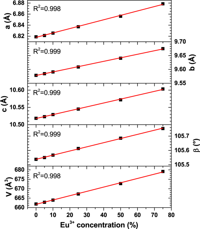
Y2Mo4O15:Eu3+ unit cell parameters as a function of Eu3+ concentration.
The recorded FTIR spectra (see Fig. s4) for undoped sample and samples doped with 10%, 25%, 50%, and 75% Eu3+ showed no significant differences between each other. Spectra consisted of several overlapping strong absorption bands in the range of 500–1000 cm−1. These bands can be attributed to the characteristic vibrations of [MoO4]2− and [Mo2O7]2− groups22,23,24.
The morphological features of undoped, 50% and 75% Eu3+ doped Y2Mo4O15 powder samples were investigated by taking high resolution SEM pictures, which are shown in Fig. 2. The obtained images clearly demonstrate that the particle size of the powders increases with increasing Eu3+ content in the structure. Particles are well shaped and their size distribution is rather broad. Moreover, another interesting feature is that there are very little dust particles on top of larger particles. This indicate high powder quality, since no washing or sieving of the obtained powders were performed.
Figure 2. SEM images.
(a) Y2Mo4O15, (b) Y2Mo4O15:50%Eu3+, and (c) Y2Mo4O15:75%Eu3+ under magnification of 5.0 k.
The body colour of undoped Y2Mo4O15 powder is white as shown in digital photo in Fig. 3b. White body colour indicates the absence of absorption in the visible range what is in good agreement with reflectance spectrum shown in Fig. 3a. The white body colour of undoped material gradually gained pale rose tint upon increasing Eu3+ concentration in the Y2Mo4O15 due to absorption in the violet-blue spectral range causing the luminescence of Eu3+ ions25. This is all in line with the reflection spectrum of Y2Mo4O15:75%Eu3+ shown in Fig. 3a, where typical Eu3+ absorption lines originating from the intraconfigurational 7F0 → 5DJ, 7F0 → 5LJ and 7F0 → 5GJ transitions26,27,28 are visible. Besides, it is evident that 7F1 and 7F2 levels are also thermally populated to some extent since absorption lines originating from the 7F2 → 5D0 (ca. 613 nm), 7F1 → 5D0 (ca. 590 nm), and 7F1 → 5D1 (ca. 527 nm) transitions are clearly visible in the reflection spectra29. The broad absorption band at short wavelengths (250–340 nm) can be attributed to the host lattice absorption.
Figure 3. Reflection spectra and digital photos.
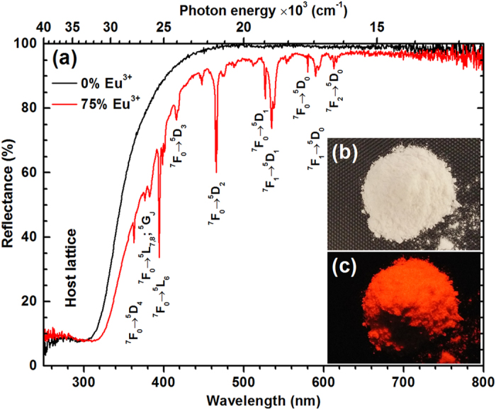
Reflection spectra of Y2Mo4O15 and Y2Mo4O15:75%Eu3+ (a), digital photos of Y2Mo4O15 at daylight (b) and Y2Mo4O15:75%Eu3+ under 254 nm excitation.
The optical band gap of the undoped, 10%, 25%, 50% and 75% Eu3+ materials was evaluated from absorption spectra. Absorption spectra F(R) were calculated from reflection spectra employing Kubelka-Munk function17,30:
 |
where R is reflectance, K is absorption coefficient, and S is scattering coefficient. It was found out that the energy of optical band gap decreases if the concentration of Eu3+ is increased. The band gap estimation is depicted in Fig. s5. The optical band gap energies for undoped, 10%, 25%, 50%, and 75% Eu3+ doped Y2Mo4O15 materials are 334 nm (3.71 eV), 336 nm (3.69 eV), 338 nm (3.67 eV), 340 nm (3.65 eV), and 344 nm (3.60 eV), respectively.
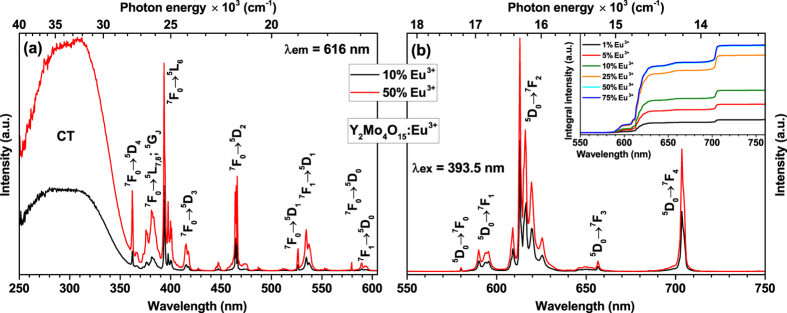
Figure 4a shows excitation spectra of 10% and 50% Eu3+ doped samples for 616 nm emission. The spectra consist of a broad band in the range of 250–360 nm and several sets of lines in the range of 360–600 nm. The broad excitation band can be attributed to the charge transfer (CT) transition from [MoO4]2− and [Mo2O7]2− groups to Eu3+ ions. It was also observed that the energy of charge transfer band slightly decreases (shifts towards longer wavelengths) with increasing Eu3+ concentration. This can be explained by the expansion of the lattice leading to the increased average distance between Eu3+ and surrounding anions31, thus easing the electron transfer from oxygen to europium. The sharp lines are associated with the intraconfigurational [Xe]4f6 → [Xe]4f6 transitions of Eu3+ ions. Excitation lines originating from 7F0 → 5L6 (≈394 nm) and 7F0 → 5D2 (≈465 nm) are very attractive for application as colour converters in LEDs since they overlap very well with the emission spectra of efficient near-UV and blue LEDs, respectively. All samples doped with Eu3+ ions showed bright red luminescence when excited with the UV lamp as indicated in Fig. 3c. The emission spectra of 10% and 50% Eu3+ doped samples under 393.5 nm excitation are given in Fig. 4b. The spectra consist of five sets of lines at around 580, 590, 615, 655, and 705 nm, which originate from 5D0 → 7F0, 5D0 → 7F1, 5D0 → 7F2, 5D0 → 7F3, and 5D0 → 7F4 optical transitions of Eu3+ ions, respectively. The intensity of 5D0 → 7F0 transition is the weakest since J = 0 ↔ J’ = 0 transitions are always forbidden32,33,34. The most intensive is an electric dipole (ED) 5D0 → 7F2 transition, whereas the intensity of a magnetic dipole (MD) 5D0 → 7F1 transition is at least by one order of magnitude lower. This indicates that Eu3+ ions occupy non-centrosymmetric sites in Y2Mo4O15 host matrix what is in good agreement with the crystallographic data. This is also confirmed by large value (ca. 7.8) of the asymmetry ratio (5D0 → 7F2)/(5D0 → 7F1), which gives a measure of distortion degree from the inversion symmetry of the local environment of Eu3+ ions in the lattice35,36. Another interesting feature of the emission spectra is oddly intensive emission peak at 705 nm. Such intensive 5D0 → 7F4 transitions lines of Eu3+ ions are usually observed in some phosphates and garnet structure materials37, however, in molybdates they are rather weak12,38. Nevertheless, the reason of such intensive 5D0 → 7F4 transition lines is still unclear and requires additional research.
Figure 4. Excitation and emission spectra.
(a) Excitation (λem = 616 nm) and (b) emission (λex = 393.5 nm) spectra of Y2Mo4O15:Eu3+ phosphors doped with 10% and 50% Eu3+. Inset shows emission (λex = 393.5 nm) integral intensity as a function of Eu3+ concentration.
It is also well known that the emission spectra of Eu3+ doped materials might contain the transitions that are overlapping. These transitions are 5D0 → 7F0 and 5D2 → 7F5, 5D0 → 7F2 and 5D1 → 7F4, 5D0 → 7F3 and 5D1 → 7F529. In order to check if any overlapping between the aforementioned transitions is occurring a sample doped with 50% Eu3+ was excited at four different wavelengths, namely, 393.5 nm (7F0 → 5L6), 465 nm (7F0 → 5D2), 534.5 nm (7F1 → 5D1), and 589.5 nm (7F1 → 5D0). With every increased excitation wavelength (decreased energy) lower and lower excited states of Eu3+ were populated. Therefore, if there were any emission from transitions starting at higher energy excited levels that are overlapping with the ones starting at 5D0 this would be visible in the emission spectra. However, no differences in emission spectra upon changing the excitation wavelength were observed as shown in Fig. s6a. Thus it can be concluded that the emission lines in the range of 580–720 nm originate from the lowest energy excited level 5D0. On the other hand, some weak emission lines originating from 5D1, 5D2 and 5D3 levels were observed when the 400–580 nm range was highly magnified. These lines are depicted in Fig. s6b,c. The following sets of lines were observed: 5D3 → 7F1 (ca. 416 nm), 5D3 → 7F2 (ca. 427 nm), 5D3 → 7F3 (ca. 446 nm), 5D3 → 7F4 and 5D2 → 7F0 (ca. 469 nm), 5D2 → 7F2 (ca. 486 nm), 5D2 → 7F3 (ca. 512 nm), 5D1 → 7F0 (ca. 526 nm), 5D1 → 7F1 (ca. 535 nm), 5D1 → 7F2 (ca. 555 nm). It also needs to be noted that the intensity of the first seven mentioned transitions is by four orders of magnitude lower if compared to the 5D0 → 7F2 transition, whereas the intensity of the latter two are by three orders of magnitude lower. This finding confirms that the radiationless decay from the higher energy levels to the 5D0 level is very effective29. The inset in Fig. 4b shows integrated emission spectra and conveys that the strongest emission is observed for the samples doped with 50% and 75% Eu3+ (overlapped).
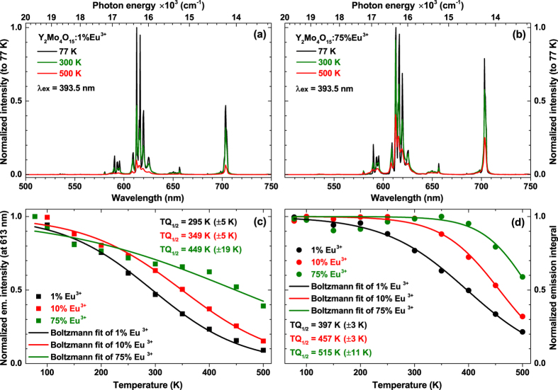
The emission spectra (λex = 393.5 nm) of samples doped with 1% and 75% Eu3+ as a function of temperature are depicted in Fig. 5a,b, respectively. It is evident that the emission intensity of sample doped with 1% Eu3+ decreases much faster upon temperature increase if compared to the 75% Eu3+ doped sample. It is also interesting to note that the intensity of 5D0 → 7F4 transition at 77 K temperature of 75% Eu3+ doped sample is much stronger than the one in 1% Eu3+ doped sample at the same temperature. In order to calculate the TQ1/2 (temperature at which phosphor loses half of its efficiency) values, the Boltzmann sigmoidal39 fit was performed to the normalized emission intensity data:
Figure 5. Temperature dependent emission spectra an TQ1/2 value evaluation.
Temperature dependent emission (λex = 393.5 nm) spectra of (a) Y2Mo4O15:1%Eu3+ and (b) Y2Mo4O15:75%Eu3+. Calculation of TQ1/2 values for the samples doped with 1%, 10% and 75% Eu3+ from normalized emission intensity (at 613 nm) (c) and normalized emission integral (d).
 |
Here y(x) is the normalized emission intensity value at a given x (temperature in K in this case), A1 and A2 are the initial value (left horizontal asymptote) and the final value (right horizontal asymptote), respectively. x0 (x0 = TQ1/2) is the centre of the sigmoid, and dx is the change in x corresponding to the most significant change in y(x) values. Since the fitting was performed on the normalized (divided by maximum intensity at 77 K for 613 nm line) emission intensity, the A1 and A2 values were set to 1 and 0, respectively. The obtained fitting lines and calculated TQ1/2 values for the samples doped with 1%, 10%, and 75% Eu3+ are given in Fig. 5c. The intensity of 613 nm line decreases gradually with increasing the temperature regardless the Eu3+ concentration. The calculated TQ1/2 values for 1%, 10%, and 75% Eu3+ doped samples are 295, 349, and 449 K, respectively. This shows that samples with low europium concentration suffers more from the thermal quenching. However, even though the emission intensity at elevated temperatures decreases, the emission lines of Eu3+ tend to broaden. Therefore, the total decrease of light output might be smaller than predicted from emission intensity change. Thus, the better way to calculate TQ1/2 values is by using emission integrals. The Boltzmann fit data of normalized emission integrals of 1%, 10%, and 75% Eu3+ doped samples is shown in Fig. 5d and the derived TQ1/2 values are 397, 457, and 515 K, respectively. It is obvious that the obtained TQ1/2 values are much higher than those calculated from emission intensity. Moreover, the emission integral method is also insensitive to the peak position change upon increasing temperature.
From the temperature dependent emission spectra the activation energy according to the single barrier quenching model can also be derived40,41,42:
 |
Here I(T) and I0 are temperature dependent emission integral and highest value of emission integral, respectively. EA is activation energy, k is Boltzmann constant (8.617342·10−5 eV/K)43, and T is temperature in K. B is the quenching frequency factor, which can also be expressed as Γ0/Γυ, where Γ0 is the attempt rate of the non-radiative process and Γυ is the radiative rate. The activation energies for 1%, 10%, and 75% Eu3+ doped samples are 0.15 ± 0.01 eV, 0.29 ± 0.02 eV, and 0.42 ± 0.12 eV, respectively. The obtained EA values are in good agreement with the respective TQ1/2 values, i.e. they both increase with increasing Eu3+ concentration in the Y2Mo4O15 lattice. The equation 4 can also be utilized to calculate the TQ1/2 values of the phosphor. One can easily derive that the TQ1/2 expression based on equation 4 is42:
 |
The EA values are given above and the B values for the samples doped with 1%, 10%, and 75% Eu3+ are 89, 1506, and 11742. Thus the calculated respective TQ1/2 values are 388, 460, and 520 K. These temperatures are very close to the ones obtained from the Boltzmann fit, therefore, both methods can be used for the TQ1/2 determination.
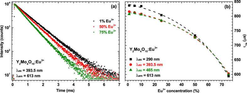
Figure 6a shows decay curves of 1%, 50%, and 75% Eu3+ doped samples when specimens were excited at 393.5 nm and emission monitored at 613 nm. Decay curves for all Eu3+ concentrations are linear implying only one depopulation mechanism of 5D0 level. The increasing europium concentration in the Y2Mo4O15 host lattice resulted in steeper decay curves indicating the decrease of luminescence lifetimes. All decay curves were fitted employing a single exponential decay function:
Figure 6. Photoluminescence decay curves and calculated decay constants.
(a) Photoluminescence decay (λex = 393.5 nm, λem = 613 nm) curves of Y2Mo4O15:Eu3+ samples doped with 1%, 50% and 75% Eu3+. (b) Decay constants of Y2Mo4O15:Eu3+ phosphors as a function of Eu3+ concentration and excitation wavelength.
 |
where I(t) is the intensity at a given time t, I0 is initial intensity and τ is a decay constant (lifetime)35. The obtained decay constants as a function of Eu3+ concentration and excitation wavelength are depicted in Fig. 6b. Excitation of samples at 393.5 nm (7F0 → 5L6 transition) and 465 nm (7F0 → 5D2 transition) yields the same decay constants regardless the Eu3+ concentration, what shows that radiationless decay from the higher energy levels (5L6 and 5D2) to the 5D0 level, from which the luminescence occurs, is very fast and of the same order. Excitation of samples through CT band gives slightly larger values of decay constant at Eu3+ concentrations up to 25%. However, at higher activator concentrations decay constants levels. The exact calculated photoluminescence decay values with standard deviations as a function of Eu3+ concentration and excitation wavelength are tabulated in Table s2.
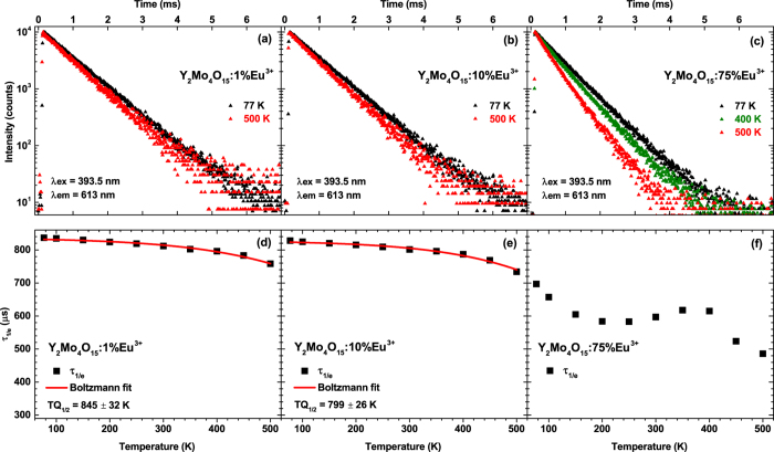
A better insight to the thermal quenching processes might be gained by measuring temperature dependent decay curves. Such curves for samples doped with 1%, 10%, and 75% Eu3+ are shown in Fig. 7a–c, respectively. The decay curves for all samples become steeper with increasing temperature, thus the photoluminescence lifetime gets shorter. However, the change of decay constants of samples doped with 1% and 10% Eu3+ is rather small, whereas for the 75% Eu3+ doped sample this change is quite substantial. The calculated photoluminescence lifetimes as a function of temperature were also fitted with Boltzmann function in order to calculate the TQ1/2. The obtained results are shown in Fig. 7d–f. Unfortunately, it was not possible to fit the data obtained for the sample doped with 75% Eu3+, since the lifetimes as a function of temperature distributes in rather bizarre way and the Boltzmann fit did not converge. The calculated TQ1/2 values for 1% and 10% Eu3+ doped samples are 845 ± 32 K and 799 ± 26 K, respectively. The relatively high standard deviation values are obtained due to the fact, that the fitting function do not reach the turning point. It is also clear that the calculated TQ1/2 values from the temperature dependent lifetime data are much larger if compared to ones obtained from temperature dependent emission integrals. Moreover, the smaller decrease of lifetime values upon temperature increase is evident. This implies that the internal quantum yield (IQY) decreases slower than the external quantum yield (EQY). The internal quantum yield of the phosphor can be expressed as25:
Figure 7. Temperature dependent photoluminescence decay curves and calculated decay constants.
Decay (λex = 393.5 nm, λem = 613 nm) curves of (a) Y2Mo4O15:1%Eu3+, (b) Y2Mo4O15:10%Eu3+, (c) Y2Mo4O15:75%Eu3+. Calculation of TQ1/2 values for the samples doped with (d) 1% Eu3+ and (e) 10% Eu3+. (f) Temperature dependent decay constants of 75% Eu3+ doped sample.
 |
where Wr and Wnr are the probabilities of radiative and non-radiative transitions, respectively. The sum of radiative and non-radiative probabilities is unity, therefore, the decreasing IQY is related to the increased probability of Wnr. The internal quantum yield is dependent only on the activator ion and its local surrounding. The external quantum yield can be expressed as25:
 |
where ηesc is the escape efficiency of the photons from the phosphor particle. The faster decrease of EQY in our case is related to the decreasing photon escape efficiency. All calculated photoluminescence decay values with standard deviations as a function of temperature are listed in Table s3.
The calculated external quantum yields for Y2Mo4O15:Eu3+ phosphor samples under the three excitation wavelengths are depicted in Fig. 8. The quantum yields reaches top values when 50% of Eu3+ is introduced into the host lattice. Further increase of Eu3+ concentration leads to the decrease of quantum yield. This decrease is probably caused by concentration quenching. Since the quenching concentration is rather high it can be concluded that [MoO4]2− and [Mo2O7]2− groups that are present in the structure effectively shields Eu3+ ions from each other, thus, preventing the energy transfer between Eu3+ pairs. The same pattern was observed for all three excitation wavelengths. Moreover, the efficiency increases with increasing excitation wavelength (decreasing energy). The maximum efficiency (ca. 85%) was obtained for the sample doped with 50% of Eu3+ and excited at 465 nm. The mentioned energy transfer between Eu3+ pairs is strongly dependent on the average distance (Rc) between the Eu3+ ions. Therefore, knowing the critical concentration of Eu3+ it is possible to calculate this distance44:
Figure 8. Quantum yields of Y2Mo4O15:Eu3+ phosphors as a function of Eu3+ concentration and excitation wavelength.
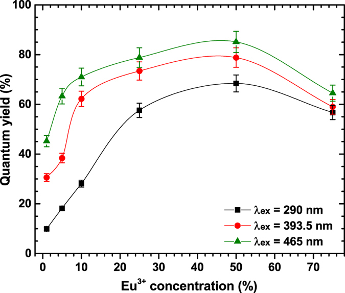
The lines are drawn to guide the eye.
 |
where V is the volume of the crystallographic unit cell, xc is the critical concentration and N is the number of lattice sites that can be occupied by activator ions. In our case V = 672.74 Å3 (see Table s1), xc is 75%, i.e. 0.75 and N = 4, thus the obtained Rc value is equal to 7.54 Å.
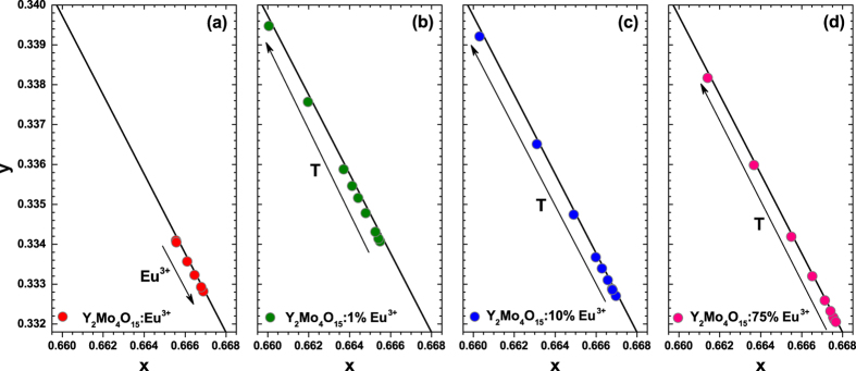
In order to demonstrate the emission colour dependency of the synthesized phosphors, the CIE chromaticity coordinates in 1931 and 1976 colour space diagrams were calculated. Table s4 shows the calculated colour coordinates as a function of Eu3+ concentration and excitation wavelength, whereas colour coordinates as a function of temperature of samples doped with 1%, 10%, and 75% Eu3+ are given in Table s5. Besides the colour coordinates, the luminous efficacy (LE) (a parameter describing how bright the radiation is perceived by the average human eye) values were also calculated from the respective emission spectra by using the following equation4:
 |
Here I(λ) is the emission spectrum of the phosphor and V(λ) is the human eye sensitivity curve. The human eye sensitivity curve possesses a maxima at 555 nm, therefore, the highest possible LE value (683 lm/Wopt) is obtained for monochromatic green radiation at 555 nm. The obtained LE values were around 242 lm/Wopt for all Eu3+ concentrations regardless the excitation wavelength. These values are rather high and can be compared to such well known red emitting phosphors as Sr2Si5N8:Eu2+ (λem = 620 nm, LE = 240 lm/Wopt), CaAlSiN3:Eu2+ (λem = 650 nm, LE = 150 lm/Wopt), CaS:Eu2+ (λem = 650 nm, LE = 85 lm/Wopt)45. The LE values increased from 231 to around 242 lm/Wopt when the temperature was increased from 77 to 500 K. This shift goes hand in hand with the shift of colour coordinates (see Fig. 9b–d) to the direction of orange region when temperature is increased. The so-called blue shift of emission results in higher overlap between emission spectra and human eye sensitivity curve thus leading to larger LE values.
Figure 9. Fragments of the CIE 1931 colour diagram.
The colour points of (a) Y2Mo4O15:Eu3+ as a function of Eu3+ concentration and as a function of temperature of (b) 1%, (c) 10%, and (d) 75% Eu3+ doped samples. All samples were excited at 393.5 nm.
Fragments of the CIE 1931 colour space diagrams with the colour coordinates of Y2Mo4O15:Eu3+ phosphors as a function of Eu3+ concentration are shown in Fig. 9a. The colour coordinates are relatively stable with respect to Eu3+ concentration, however, the tendency is that they slightly shift towards deeper red region if the Eu3+ concentration increases. Colour coordinates are also very close to the edge of the colour space diagram indicating the high colour purity of the prepared phosphors.
Due to the spin and parity forbidden character of their [Xe]4fn → [Xe]4fn transitions, lanthanide ions often suffers from weak absorption5,45,46. One way to increase the absorption strength of the phosphor is preparing the ceramics. In such way the pathway of the incident photons increases, thus increasing the probability of absorption of these photons. The ceramic disks of Y2Mo4O15:75%Eu3+ sample with the thickness of 0.71 and 0.98 mm (ρ = 2.93 g/cm3) were prepared and their optical properties investigated. These disks were placed on the top of three LEDs with the emission maximum at 375, 400, and 455 nm, i.e. the emission spectra were recorded in the transmission mode. The respective emission spectra of mentioned LEDs are shown in Fig. 10a–c. the emission spectra of ceramic disks excited by LEDs are shown in Fig. 10d–f. It is obvious that ceramic disks of both thicknesses absorb all radiation emitted by 375 and 400 nm LEDs and convert it into the red light. Therefore, even thinner ceramics could be used. On the other hand, preparation of thinner ceramic disks with available equipment was rather complicated. This strong absorption in the mentioned spectral regions is possible due to the abundance of Eu3+ absorption lines in these spectral regions. The situation, however, is different if disks are excited with 455 nm LED. In this spectral region there is only one 7F0 → 5D2 transition with narrow absorption lines at around 465 nm. Thus, a large portion of the blue light emitted by LED passes through ceramic disk without being absorbed. This is visible in emission spectra depicted in Fig. 10f, where absorption lines of Eu3+ due 7F0 → 5D2 transition are visible in the region of emission spectrum of 455 nm LED. Obviously, the 455 nm LED is not suitable for gaining red light, however, various tints of pink light might be obtained as shown in Fig. 11. It is also interesting to note that the emission spectra of ceramic disks are a little bit different if compared to the powder samples, namely, the five narrow peaks of 5D0 → 7F2 transition merge into the three broader peaks and the emission line originating from 5D0 → 7F4 transition become even stronger. The latter also slightly increases with the increase of ceramic disk thickness. Considering this ceramics for colour conversion, however, such increase in emission intensity at around 700 nm is undesirable, since the human eye sensitivity at this wavelength is very low and this would reduce the luminous efficacy of the prepared light source.
Figure 10. Emission spectra of LEDs and ceramics.
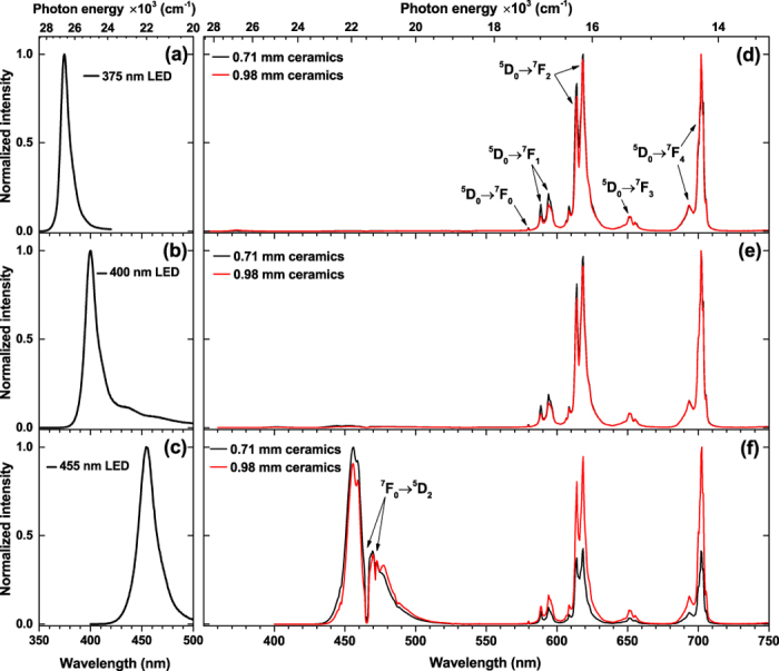
(a) 375 nm LED, (b) 400 nm LED, and (c) 455 nm LED. Emission spectra of Y2Mo4O15:75%Eu3+ ceramic disks excited with (d) 375 nm LED, (e) 400 nm LED, and (f) 455 nm LED.
Figure 11. Fragment of the CIE 1931 colour diagram.
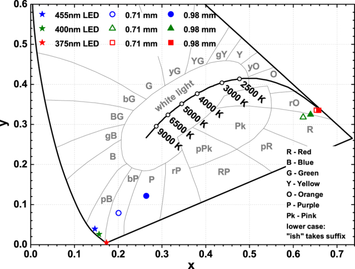
The colour points of Y2Mo4O15:75Eu3+ ceramics combined with 375, 400, and 455 nm LEDs. Stars, open, and filled symbols denote colour points of LEDs, LEDs with 0.71 mm ceramic disk, and LEDs with 0.98 mm ceramic disk, respectively.
The calculated colour coordinates in CIE 1931 colour space of light sources obtained by combining 375, 400, and 455 nm LEDs and Y2Mo4O15:75%Eu3+ ceramic disks of both thicknesses are depicted in Fig. 11. The colour points of LEDs are also included for the reference. The exact values are tabulated in Table s6 together with calculated luminous efficacies. The colour coordinates of light source obtained from 375 nm LED and Y2Mo4O15:75%Eu3+ ceramics are close to the edge of the colour space diagram, therefore, the colour purity is high. However, the colour purity decreases if 400 nm LED is used as an excitation source. It is possible that small amount of violet light emitted by LED passes through the ceramic disks thus reducing the colour purity. As was already mentioned the combination of 455 nm LED and prepared ceramic disks have not yielded the red colour. On the other hand, one combining this LED with ceramic disks could obtain the light source with the colours ranging from bluish purple to purple or even reddish purple depending on the thickness of ceramics. The calculated luminous efficacy values were in the range of 200–184 lm/Wopt for light sources prepared employing 375 and 400 nm LEDs. The slightly lower values if compared to the powder samples are obtained due to the increased emission intensity in deep red spectral region at around 700 nm. Using 455 nm LED as an excitation source yielded substantially lover LE values in the range of 94–121 lm/Wopt. This value increase with increasing thickness of the ceramics due to decreasing intensity of blue light passing through the disk unabsorbed.
Conclusions
Single phase Y2Mo4O15:Eu3+ phosphors with Eu3+ concentration in the range of 1–75% were prepared by high temperature solid state reaction. Samples showed bright red luminescence under excitation of near-UV and blue radiation. These phosphors possess high colour purity, excellent thermal stability and high quantum yields. Temperature dependent photoluminescence decay measurements indicated that internal quantum yield decreases much slower than the external one what was related to the decreasing photon escape efficiency from the particle upon increasing the temperature. The combination of Y2Mo4O15:75%Eu3+ ceramic disks with 375, and 400 nm LEDs yielded red light sources and with 455 nm LED resulted in various tints of purple colour.
Additional Information
How to cite this article: Janulevicius, M. et al. Luminescence and luminescence quenching of highly efficient Y2Mo4O15:Eu3+ phosphors and ceramics. Sci. Rep. 6, 26098; doi: 10.1038/srep26098 (2016).
Supplementary Material
Footnotes
Author Contributions M.J. and P.M. prepared the sample powders and ceramics. M.M. and A.K. performed XRD analysis and Rietveld refinement. L.M. performed luminescence measurements. J.G. performed temperature dependent measurements. S.S. and A.K. initialized the study and wrote the manuscript. All authors discussed the results and reviewed the manuscript.
References
- Nakamura S. GaN growth using GaN buffer layer. Jpn. J. Appl. Phys. 30, L1705–L1707 (1991). [Google Scholar]
- Bachmann V., Ronda C., Oeckler O., Schnick W. & Meijerink A. Color Point Tuning for (Sr,Ca,Ba)Si2O2N2:Eu2+ for white light LEDs. Chem. Mater. 21, 316–325 (2009). [Google Scholar]
- Lim M. A., Park J. K., Kim C. H., Park H. D. & Han M. W. Luminescence characteristics of green light emitting Ba2SiO4:Eu2+ phosphor. J. Mater. Sci. Lett. 22, 1351–1353 (2003). [Google Scholar]
- Smet P. F., Parmentier A. B. & Poelman D. Selecting conversion phosphors for white light-emitting diodes. J. Electrochem. Soc. 158, R37–R54 (2011). [Google Scholar]
- Blasse G. & Grabmaier B. C. Luminescent Materials (Springer-Verlag, 1994). [Google Scholar]
- Gavrilović T. V., Jovanović D. J., Lojpur V. & Dramićanin M. D. Multifunctional Eu3+- and Er3+/Yb3+-doped GdVO4 nanoparticles synthesized by reverse micelle method. Sci. Rep. 4, 4209 (2014). [DOI] [PMC free article] [PubMed] [Google Scholar]
- Roof I. P., Jagau T.-C., Zeier W. G., Smith M. D. & zur Loye H.-C. Crystal growth of a new series of complex niobates, LnKNaNbO5 (Ln = La, Pr, Nd, Sm, Eu, Gd, and Tb): structural properties and photoluminescence. Chem. Mater. 21, 1955–1961 (2009). [Google Scholar]
- Chang Y.-S., Shi Z.-R., Tsai Y.-Y., Wu S. & Chen H.-L. The effect of Eu3+-activated InVO4 phosphors prepared by sol–gel method. Opt. Mater. 33, 375–380 (2011). [Google Scholar]
- Han X., Lahera D. E., Serrano M. D., Cascales C. & Zaldo C. Ultraviolet to infrared refractive indices of tetragonal double tungstate and double molybdate laser crystals. Applied Physics B 108, 509–514 (2012). [Google Scholar]
- Nagornaya L. L. et al. Tungstate and molybdate scintillators to search for dark matter and double beta decay. IEEE Trans. Nucl. Sci. 56, 2513–2518 (2009). [Google Scholar]
- Wang H.-Q. et al. A new crystal phase molybdate Yb2Mo4O15: the synthesis and upconversion properties. Particle & Particle Systems Characterization 32, 340–346 (2015). [Google Scholar]
- Yang M. et al. Multifunctional luminescent nanomaterials from NaLa(MoO4)2:Eu3+/Tb3+ with tunable decay lifetimes, emission colors, and enhanced cell viability. Sci. Rep. 5, 11844 (2015). [DOI] [PMC free article] [PubMed] [Google Scholar]
- Hartenbach I., Hoppe H. A., Kazmierczak K. & Schleid T. Synthesis, crystal structure and spectroscopic properties of a novel yttrium(iii) fluoride dimolybdate(vi): YFMo2O7. Dalton Trans. 43, 14016–14021 (2014). [DOI] [PubMed] [Google Scholar]
- Baur F., Katelnikovas A., Sakirzanovas S., Petry R. & Justel T. Synthesis and optical properties of Li3Ba2La3(MoO4)8:Sm3+ powders for pcLEDs. Z. Naturforsch. B 69, 183–192 (2014). [Google Scholar]
- Efremov V. A., Davydova N. N., Gokhman L. Z., Evdokimov A. A. & Trunov V. K. Crystal structure of Ho2Mo4O15 holmium tetramolybdate. Zh. Neorg. Khim. 33, 3005–3010 (1988). [Google Scholar]
- Laufer S. et al. Yttrium(III) oxomolybdates(VI) as potential host materials for luminescence applications: an investigation of Eu3+-doped Y2[MoO4]3 and Y2[MoO4]2[Mo2O7]. New J. Chem. 37, 1919–1926 (2013). [Google Scholar]
- Deng H. et al. Photoluminescence properties of a new orange–red emitting Sm3+-doped Y2Mo4O15 phosphor. J. Solid State Chem. 228, 110–116 (2015). [Google Scholar]
- Wang H., Peng J., Chen D. F. & Hu Z. Structure and thermal expansion behavior of Y2−xLnxMo4O15 (Ln = Nd, Dy). Solid State Sci. 8, 1144–1151 (2006). [Google Scholar]
- Wang H. et al. Structural and thermal expansion properties of solid solution Y2−xSmxMo4O15 (x = 0.0 − 0.8). Physica B 388, 278–284 (2007). [Google Scholar]
- Shannon R. D. Revised effective ionic radii and systematic studies of interatomic distances in halides and chalcogenides. Acta Crystallogr. A32, 751–767 (1976). [Google Scholar]
- Ropp R. C. Luminescence and the solid state 2nd edn (Elsevier, 2004). [Google Scholar]
- Nyquist R. A. & Kagel R. O. Infrared spectra of inorganic compounds (3800-45cm−1) (Academic Press, 1971). [Google Scholar]
- Nyquist R. A., Putzig C. L., Leugers M. A., Kagel R. O. & Nyquist R. A. The handbook of infrared and Raman spectra of inorganic compounds and organic salts (Academic Press, 1997). [Google Scholar]
- Maczka M. et al. Structural and vibrational properties of K3Fe(MoO4)2(Mo2O7)-a novel layered molybdate. Journal of Physics-Condensed Matter 21, 095402 (2009). [DOI] [PubMed] [Google Scholar]
- Katelnikovas A. et al. Synthesis and optical properties of Li3Ba2La3(MoO4)8:Eu3+ powders and ceramics for pcLEDs. J. Mater. Chem. 22, 22126–22134 (2012). [Google Scholar]
- Yi L. et al. A potential red phosphor LiGd(MoO4)2:Eu3+ for light-emitting diode application. J. Lumin. 130, 1113–1117 (2010). [Google Scholar]
- Lee G. H. & Kang S. Studies in crystal structure and luminescence properties of Eu3+-doped metal tungstate phosphors for white LEDs. J. Lumin. 131, 2606–2611 (2011). [Google Scholar]
- Carnall W. T., Crosswhite H. & Crosswhite H. M. Energy level structure and transition probabilities in the spectra of the trivalent lanthanides in LaF3. Argonne National Laboratory Report Unnumbered (1977). [Google Scholar]
- Binnemans K. A comparative spectroscopic study of Eu3+ in crystalline host matrices. Bull. Soc. Chim. Belg. 105, 793–798 (1996). [Google Scholar]
- Kubelka P. New contributions to the optics of intensely light-scattering materials. Part I. J. Opt. Soc. Am. 38, 448–457 (1948). [DOI] [PubMed] [Google Scholar]
- Dorenbos P. The Eu3+ charge transfer energy and the relation with the band gap of compounds. J. Lumin. 111, 89–104 (2005). [Google Scholar]
- Walsh B. M. Judd-Ofelt theory: principles and practices. 403–433 (Springer, 2006).
- Eliseeva S. V. & Bunzli J.-C. G. Lanthanide luminescence for functional materials and bio-sciences. Chem. Soc. Rev. 39, 189–227 (2010). [DOI] [PubMed] [Google Scholar]
- Görller-Walrand C. & Binnemans K. In Handbook on the physics and chemistry of rare earths (eds Gschneidner K. A. & Eyring L.) Ch. 167, 101–264 (Elsevier Science B.V., 1998). [Google Scholar]
- Chang Y. C., Liang C. H., Yan S. A. & Chang Y. S. Synthesis and photoluminescence characteristics of high color purity and brightness Li3Ba2Gd3(MoO4)8:Eu3+ red phosphors. J. Phys. Chem. C 114, 3645–3652 (2010). [Google Scholar]
- Tsai B.-S., Chang Y.-H. & Chen Y.-C. Synthesis and luminescent properties of MgIn2-xGaxO4:Eu3+ phosphors. Electrochem. Solid-State Lett. 8, H55–H57 (2005). [Google Scholar]
- Skaudzius R., Katelnikovas A., Enseling D., Kareiva A. & Justel T. Dependence of the 5D0 → 7F4 transitions of Eu3+ on the local environment in phosphates and garnets. J. Lumin. 147, 290–294 (2014). [Google Scholar]
- Uhlich D. Kristallographische und spektroskopische Untersuchungen an Eu3+-dotierten Molybdaten als potentielle Konverter für LEDs, PhD thesis, Universität Osnabrück, (2009).
- Aguiar J., Carpena P., Molina-Bolívar J. A. & Carnero Ruiz C. On the determination of the critical micelle concentration by the pyrene 1:3 ratio method. J. Colloid Interface Sci. 258, 116–122 (2003). [Google Scholar]
- Baur F., Glocker F. & Justel T. Photoluminescence and energy transfer rates and efficiencies in Eu3+ activated Tb2Mo3O12. J. Mater. Chem. C 3, 2054–2064 (2015). [Google Scholar]
- Ueda J., Dorenbos P., Bos A. J. J., Meijerink A. & Tanabe S. Insight into the thermal quenching mechanism for Y3Al5O12:Ce3+ through thermo luminescence excitation spectroscopy. J. Phys. Chem. C 119, 25003–25008 (2015). [Google Scholar]
- Muller M., Fischer S. & Justel T. Luminescence and energy transfer of co-doped Sr5MgLa2(BO3)6:Ce3+,Mn2 +. RSC Adv. 5, 67979–67987 (2015). [Google Scholar]
- CRC handbook of chemistry and physics 90th edn, (CD-ROM Version 2010) (CRC Press/Taylor and Francis, 2010). [Google Scholar]
- Blasse G. Energy transfer in oxidic phosphors. Physics Letters 28A, 444–445 (1968). [Google Scholar]
- Ronda C. R. Luminescence: from theory to applications (Wiley-VCH, 2008). [Google Scholar]
- Yen W. M., Shionoya S. & Yamamoto H. Fundamentals of phosphors (CRC Press, 2007). [Google Scholar]
Associated Data
This section collects any data citations, data availability statements, or supplementary materials included in this article.


