Abstract
[Purpose] The biomechanical effects of foot orthoses on malalignment syndrome have not been fully clarified. This experimental investigation was conducted to evaluate the effects of orthoses on the gait patterns of patients with malalignment syndrome. [Subjects and Methods] Ten patients with malalignment syndrome were recruited. For each participant, kinematic and kinetic data were collected under three test conditions: walking barefoot, walking with flat insoles in shoes, and walking with a biomechanical foot orthosis (BFO) in shoes. Gait patterns were analyzed using a motion analysis system. [Results] Spatiotemporal data showed the step and stride lengths when wearing shoes with flat insoles or BFO were significantly greater than when barefoot, and that the walking speed when wearing shoes with BFO was significantly faster than when walking barefoot or with shoes with flat insoles. Kinetic data, showed peak pelvic tilt and obliquity angle were significantly greater when wearing BFO in shoes than when barefoot, and that peak hip flexion/extension angle and peak knee flexion/extension and rotation angles were significantly greater when wearing BFO and flat insoles in shoes than when barefoot. [Conclusion] BFOs can correct pelvic asymmetry while walking.
Key words: Three-dimensional gait analysis, Biomechanical foot orthosis, Malalignment syndrome
INTRODUCTION
Malalignment syndrome was defined by Wolf Schamberger as a set of clinical orthopaedic symptoms, as follows: malalignment associated biomechanical changes, especially a shift in weight bearing and asymmetries of muscle tension and strength, and joint ranges of motion affecting soft tissues, joints, and organ systems throughout the body. In particular, the pelvis transfers loads generated by body weight and gravity during standing, walking and sitting1, 2), and acts as a basis for the axial system. Thus, pelvic alignment influences spinal posture and stability, and pelvic malalignment is a common cause of lower back, hip, and leg pain in both the general public and in athletes. The symptoms and signs of malalignment syndrome include persistent foot, leg, or low back pain that is activity dependent, curvature of the spine, asymmetrical muscle bulk, or strength or inability to turn the body as much in a particular direction. According to Schamberger, there are three common presentations of pelvic malalignment, rotational malalignment, upslip of the sacroiliac joint, and inflare/outflare, and all three cause some form of asymmetry.
Malalignment syndrome is commonly treated using some form of orthotic device, and biomechanical foot orthosis (BFO) treatment is the only form of therapy that addresses the correction of biomechanical malalignments in the lower extremity kinetic chain. The goal of treatment is the restoration of normal structure and function of the spine and pelvis; therefore, the devices used are designed to realign the body. These orthotics increase foot stability by providing contact for weight bearing across a larger part of the sole and decrease the tendency of the feet to roll inwards or outwards after alignment has been achieved, and may decrease torque forces on legs. Orthotics increase sensory input from the sole surface, and the stimulation of proprioceptive receptors has been shown to help control pain. A reduced perception of pain may also elicit reflex relaxation of muscles, which may help to reduce muscle tension asymmetry (a chronic tension myalgia caused by a constant state of compensatory muscle contraction). Orthotic intervention is believed to influence the pattern of lower extremity movement through a combination of mechanical control and biofeedback.
Foot orthoses have been widely used to successfully treat a range of pathologies related to biomechanical dysfunction of the lower limb by altering impact forces and kinematic variables3,4,5,6,7). Orthosis inserts are widely prescribed in the belief that they can alter lower extremity joint alignment and movement8,9,10,11), but the biomechanical effects of the orthoses used for the clinical treatment of malalignment syndrome are not completely understood. Previous studies have focused primarily on the effects of orthotic devices on foot structure rather than on the pelvis or lower limbs, and the wider biomedical implications of orthoses remain unclear. Furthermore, recent reports have seriously questioned the reliabilities of clinical measurements, and the validities of static measurements with respect to predicting dynamic foot functional behavior12,13,14). This study investigated the effects of BFO on malalignment syndrome using three-dimensional gait analysis, focusing on the correction of asymmetry.
SUBJECTS AND METHODS
Ten patients (two males and eight females; mean±SD, age 42.2±13.04 years, range 20 to 57 years, height 163.1±5.76 cm, weight 55.5±8.03 kg) were recruited. No patient had a previous history of a neurologic or psychiatric problem, joint abnormalities of the lower limbs or operative intervention, pregnancy, vascular insufficiency, or a systemic problem (e.g., a cancerous, cardiovascular, or endocrinologic disease) that affected ability to walk. Patients were selected using the following criteria: pelvic rotational malalignment with or without low back pain, that showed more posterior rotation on one side during walking on the spot as measured by a pelvic angulometer (Biomechanics Co., Bundang, Korea); an age of 20–60 years; and varus or valgus abnormalities of the rearfoot. All participants provided their written consent prior to participating in the study, which was approved by the institutional review board of Yeungnam University Hospital.
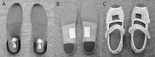
Three types of walking conditions were used in this study: walking barefoot (barefoot), walking with a flat insole in shoes (shoes), or walking with a BFO in shoes (BFO). A flat, full-length insole was added to control the height of the BFO, and was made without inclination because heel lift has been demonstrated to alter pelvic position and tilt during the stance period of the gait cycle15, 16). All the shoes and insoles were made by Biomechanics Co. (Bundang, Korea) and GO Meditech (Daegu, Korea). BFOs were custom-made and the sizes and heights of the insoles were adjusted to fit each subject. The shoes were made with exposures for markers. The bottoms of the shoes and insoles were composed of ethylene vinyl acetate, whereas the BFOs were of the standard full-length type comprising a polypropylene shell, metatarsal base assistance dome, heel lift, and full length lift, with an accessory posting made of ethylene vinyl acetate covered with cork (Fig. 1).
Fig. 1.
The biomechanical foot orthosis (BFO) and shoes
(A) Front side of custom-made BFO and (B) back side
(C) Shoes showing exposure for foot markers; BFOs or flat insoles were inserted in the shoes
Three-dimensional gait analysis was conducted using the 12-camera VICON motion analysis system (Oxford Metrics, Oxford, UK) with two force plates (AMTI OR6 series) to capture ground reaction forces and identify gait cycle events. Sixteen 14-mm-diameter reflective markers were placed over the following anatomic locations of the pelvis and lower extremities: anterior superior iliac spines (ASIS), posterior superior iliac spine (PSIS), lateral epicondyle of the knee joints, lateral malleoli, heels, second metatarsals, and lateral aspects of the thigh and calf segments. The reliability and validity of gait analysis using the VICON motion analysis system are well-established17,18,19).
Before beginning the gait analysis experiments, subjects were instructed to walk a number of times along an 8 m walkway at a normal comfortable walking speed, and a static trial was conducted to establish relationships between the markers in a static position for each subject in the initial anatomical position. Data were then collected under the three specific test conditions: walking barefoot (barefoot), with flat insoles in shoes (shoes), or with BFO in shoes (BFO). Each subject walked barefoot, with a flat insole in the shoes, and with a BFO in the shoes, in that order. For each condition, five trials of gait data were acquired for each subject walking at a self-selected speed. Mean values were used in the subsequent analysis. No patient experienced a problem on the gait walkway during the experiments.
Motion capture and modeling of all trials were performed using VICON Nexus1.4 software. Kinetic and kinematic data were then computed using POLYGON software. Gait patterns were obtained by averaging the data of five different cycles of gait for each participant. The results are presented as mean±SDs.
All statistical analysis was performed using SPSS for Windows (SPSS version 12.0 K, SPSS Korea). Repeated-measures ANOVA was used to compare the three different test conditions. The difference between equivalent measurements was interpreted to be significant when the corresponding p values were less than 0.05.
RESULTS
The spatiotemporal data of each test condition are shown in Table 1. Step and stride lengths in shoes and BFO were significantly longer than when barefoot (step length: shoes vs. barefoot, p<0.001; BFO vs. barefoot, p<0.001, stride length: shoes vs. barefoot, p=0.001; BFO vs barefoot, p<0.001; BFO vs shoes, p=0.018). Also, walking speed of BFO was significantly faster than walking barefoot or in shoes (shoes vs. barefoot, p=0.034; BFO vs. barefoot, p=0.01; BFO vs. shoes, p=0.028). However, the other gait parameters were not significantly different.
Table 1. Spatiotemporal gait parameters.
| Barefoot | With shoes | With BFO | ||||
|---|---|---|---|---|---|---|
| Mean | SD | Mean | SD | Mean | SD | |
| Cadence (steps/min) | 105.57 | 6.92 | 104.63 | 5.83 | 106.52 | 5.50 |
| Step length (m) | 0.55 | 0.05 | 0.59* | 0.04 | 0.60* | 0.04 |
| Step time (s) | 0.57 | 0.03 | 0.57 | 0.03 | 0.57 | 0.03 |
| Step width (m) | 0.14 | 0.05 | 0.15 | 0.04 | 0.24 | 0.31 |
| Stride length (m) | 1.10 | 0.10 | 1.18* | 0.07 | 1.20*† | 0.08 |
| Stride time (s) | 1.14 | 0.07 | 1.14 | 0.07 | 1.13 | 0.06 |
| Speed (m/s) | 0.97 | 0.14 | 1.03* | 0.11 | 1.07*† | 0.11 |
*p<0.05, vs. barefoot; †p<0.05, vs. shoes; SD: standard deviation; BFO: biomechanical foot orthosis
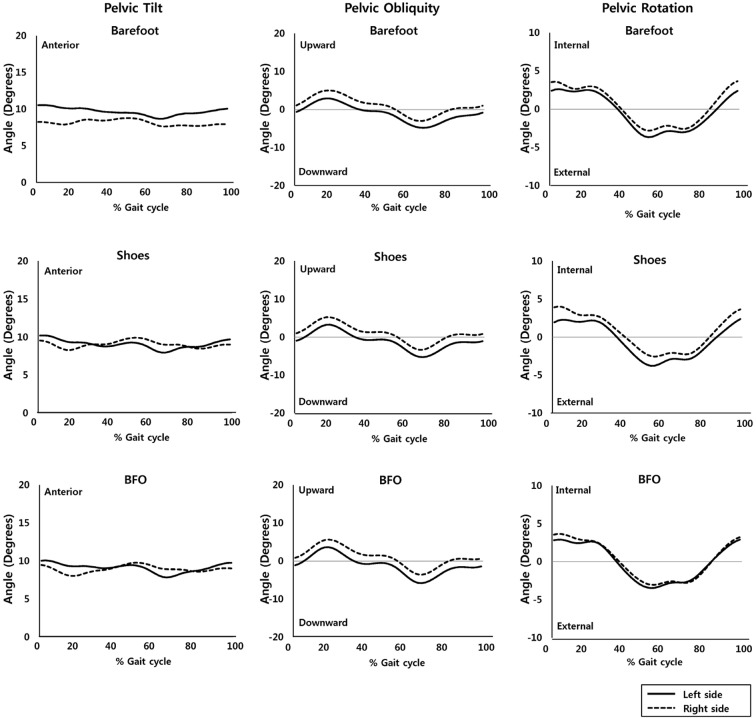
Peak pelvic tilt and obliquity angle were significantly higher for BFO than for barefoot (pelvic tilt p=0.037; pelvic obliquity p=0.02). Furthermore, peak hip flexion/extension angle (shoes vs. barefoot, p=0.002; BFO vs. barefoot, p<0.001), peak knee flexion/extension (shoes vs. barefoot, p<0.001; BFO vs. barefoot, p<0.001), and rotation angle (shoes vs. barefoot, p=0.013; BFO vs. barefoot, p=0.003) were significantly higher for the BFO and shoes conditions than for the barefoot condition. However, a significant difference was not found between BFO and shoes. Peak pelvic rotation angle was non-significantly lower for BFO than for shoes or barefoot (Table 2). However, BFO reduced the difference of the pelvic rotation between the left and right sides significantly more than the other conditions (BFO vs. barefoot, p=0.003; BFO vs. shoes, p=0.001) (Table 3, Fig. 2).
Table 2. Kinematic and kinetic data.
| Barefoot | With shoes | With BFO | ||||
|---|---|---|---|---|---|---|
| Mean | SD | Mean | SD | Mean | SD | |
| Peak pelvic tilt angle (°) | 9.02 | 4.04 | 10.36 | 5.58 | 10.48* | 5.58 |
| Peak pelvic obliquity angle (°) | 4.43 | 1.23 | 4.68 | 1.25 | 4.92* | 1.37 |
| Peak pelvic rotation angle (°) | 4.43 | 1.63 | 3.99 | 1.43 | 3.98 | 1.19 |
| Peak hip F/E angle (°) | 35.54 | 7.88 | 37.11* | 8.40 | 37.55* | 8.10 |
| Peak hip Ab/Ad angle (°) | 7.09 | 2.70 | 7.25 | 2.74 | 7.42 | 2.91 |
| Peak hip rotation angle (°) | 7.32 | 20.52 | 9.37* | 19.86 | 9.04 | 19.68 |
| Peak knee F/E angle (°) | 61.85 | 5.12 | 70.38* | 4.61 | 69.15*† | 4.82 |
| Peak knee Ab/Ad angle (°) | 12.38 | 16.79 | 13.40* | 17.12 | 13.03 | 16.68 |
| Peak knee rotation angle (°) | 21.42 | 8.29 | 24.16* | 7.27 | 24.47* | 7.63 |
| Peak ankle DF/PF angle (°) | 18.82 | 4.24 | 18.10 | 5.90 | 17.63 | 5.41 |
*p<0.05, vs. barefoot; †p<0.05, vs. shoes; SD: standard deviation; BFO: biomechanical foot orthosis; Ab: abduction; Ad: adduction; F: flexion; E: extension; DF: dorsiflexion; PF: plantarflexion
Table 3. Kinematic data of left-to-right differences.
| Difference | Barefoot | With shoes | With BFO | |||
|---|---|---|---|---|---|---|
| Mean | SD | Mean | SD | Mean | SD | |
| Pelvis tilt (°) | 1.03 | 0.48 | 0.97 | 0.32 | 0.92 | 0.42 |
| Pelvis obliquity (°) | 3.72 | 3.74 | 3.87 | 3.67 | 4.04 | 3.87 |
| Pelvis rotation (°) | 4.46 | 3.08 | 3.89* | 2.78 | 2.79*† | 2.20 |
| Hip F/E (°) | 2.72 | 1.24 | 1.86 | 0.49 | 1.83 | 0.43 |
| Hip Ab/Ad (°) | 3.69 | 2.59 | 3.76 | 2.60 | 2.78 | 2.36 |
| Hip rotation (°) | 6.55 | 3.20 | 6.56 | 3.63 | 6.37 | 3.83 |
| Knee F/E (°) | 3.50 | 1.51 | 2.56 | 0.86 | 2.23* | 0.84 |
| Knee Ab/Ad (°) | 3.20 | 2.31 | 3.38 | 2.27 | 3.06 | 2.25 |
| Knee rotation (°) | 5.98 | 4.83 | 6.32 | 5.02 | 6.28 | 4.80 |
| Ankle DF/PF (°) | 3.33 | 1.03 | 2.37* | 1.03 | 2.52* | 1.31 |
*p<0.05, vs. barefoot; †p<0.05, vs. shoes; SD: standard deviation; BFO: biomechanical foot orthosis; Ab: abduction; Ad: adduction; F: flexion; E: extension; DF: dorsiflexion; PF: plantarflexion
Fig. 2.
Plot of pelvic angle
Angles of left (black line) and right (dotted line) sides. Right- and left-sided pelvic rotation was significantly different among the barefoot, shoes, and BFO conditions (BFO vs. barefoot, p=0.003; BFO vs. shoes, p=0.001).
DISCUSSION
Many studies have reported the effects of foot orthoses or insoles on the biomechanics of the lower extremity joints, but the majority focused on the biomechanics of the knee or ankle joints. Furthermore, it has been suggested by several electromyographic (EMG) studies of standardized and medial wedge foot orthoses, that orthoses significantly alter the onset and amplitude of low back and pelvic muscle activities20,21,22,23,24,25). Rothbart et al.26) demonstrated that foot orthoses with a large medial forefoot wedge can change the body posture during walking of patients with chronic low back pain. Therefore, it is accepted that foot orthoses affect the biomechanics of the proximal lower extremities, including the pelvis and low back. However, little is known about the clinical usefulness of BFO for the pelvic alignment of patients with malalignment syndrome.
The present study described the immediate influence of BFO on malalignment syndrome, especially with respect to changes in pelvic asymmetry. According to the pelvic kinematic data of right-to-left differences, both shoes and BFO were found to reduce pelvic-tilt sidedness, and pelvic rotation angle asymmetry was significantly decreased in the BFO condition, compared to the barefoot and shoes conditions, which implies that BFO contributes to the correction of pelvic rotational asymmetry. An alternative explanation is that BFO reduces body alignment asymmetry thereby reducing muscle activities1, 2, 27). During walking, motion in the plane of progression is stimulated by a change in momentum induced by foot-floor contact and the height of the body’s center of gravity. Bendovā et al. reported a change in load distribution was detected under the feet when pelvic floor muscles were unilaterally activated, and suggested tension asymmetry of the pelvic floor affected the relative positioning of the pelvis leading to pelvic malalignment27). This result suggests that an equal redistribution of load on the rearfoot improves pelvic asymmetry. The pelvis is a mobile link between the two lower limbs, and serves as the bottom segment of the unit from the head to trunk that rides on the hip joint. For these reasons, the pelvis is an important part of the body alignment and energy conservation system.
It has been established that the axial alignment at the knee is related to maximal knee adduction moment and motion in the coronal plane, and that it facilitates vertical balance over the limb. Nester and Chen28, 29) reported that medially wedged foot orthoses increase knee adduction moment but have no remarkable effect on knee adduction kinematics in normal feet. In the present study, increases in knee adduction moment and angle were observed under the BFO and shoes conditions, but no significant differences, other than that of knee adduction angle, were observed between the shoes and barefoot conditions. In a previous study, it was suggested that the effect of orthoses on knee adduction moment might produce a change in knee adduction motion in the long-term. The present result can be understood in the same context.
In conclusion, the present study showed BFO can correct pelvic asymmetry. This is the first study to report a relation between asymmetry during walking and the efficacy of BFO in malalignment syndrome. However, this study was limited by its small sample size and the lack of a follow-up study. In addition, the study participants had moderate to severe pelvic asymmetry, and it remains undetermined whether BFO is effective for patients with mild pelvic asymmetry. Further complementary large-scale studies with long-term follow-up are warranted, and combined studies of EMG of lumbosacral muscles are needed in order to confirm the effect of BFO on the correction of pelvic alignment.
Acknowledgments
This study was supported by a grant of the Korea Health care technology R&D Project, Ministry for Health, Welfare & Family Affairs, Republic of Korea (A084177).
REFERENCES
- 1.Snijders CJ, Vleeming A, Stoeckart R: Transfer of lumbosacral load to iliac bones and legs Part 2: loading of the sacroiliac joints when lifting in a stooped posture. Clin Biomech (Bristol, Avon), 1993, 8: 295–301. [DOI] [PubMed] [Google Scholar]
- 2.Pool-Goudzwaard A, van Dijke GH, van Gurp M, et al. : Contribution of pelvic floor muscles to stiffness of the pelvic ring. Clin Biomech (Bristol, Avon), 2004, 19: 564–571. [DOI] [PubMed] [Google Scholar]
- 3.Landorf KB, Keenan AM: Efficacy of foot orthoses. What does the literature tell us? 2000, 90: 149–158. [DOI] [PubMed] [Google Scholar]
- 4.Payne C, Chuter V: The clash between theory and science on the kinematic effectiveness of foot orthoses. Clin Podiatr Med Surg, 2001, 18: 705–713, vi. [PubMed] [Google Scholar]
- 5.Finestone A, Novack V, Farfel A, et al. : A prospective study of the effect of foot orthoses composition and fabrication on comfort and the incidence of overuse injuries. Foot Ankle Int, 2004, 25: 462–466. [DOI] [PubMed] [Google Scholar]
- 6.Landorf KB, Keenan AM, Herbert RD: Effectiveness of foot orthoses to treat plantar fasciitis: a randomized trial. Arch Intern Med, 2006, 166: 1305–1310. [DOI] [PubMed] [Google Scholar]
- 7.Christovão TC, Neto HP, Grecco LA, et al. : Effect of different insoles on postural balance: a systematic review. J Phys Ther Sci, 2013, 25: 1353–1356. [DOI] [PMC free article] [PubMed] [Google Scholar]
- 8.Seo KC, Park KY: The effects of foot orthosis on the gait ability of college students in their 20s with flat feet. J Phys Ther Sci, 2014, 26: 1567–1569. [DOI] [PMC free article] [PubMed] [Google Scholar]
- 9.Park K, Seo K: Effects of a functional foot orthosis on the knee angle in the sagittal plane of college students in their 20s with flatfoot. J Phys Ther Sci, 2015, 27: 1211–1213. [DOI] [PMC free article] [PubMed] [Google Scholar]
- 10.Kim H, Shim J: The effect of insole height on foot pressure of adult males in twenties. J Phys Ther Sci, 2011, 23: 761–763. [Google Scholar]
- 11.Ko DY, Lee HS: The changes of COP and foot pressure after one hour’s walking wearing high-heeled and flat shoes. J Phys Ther Sci, 2013, 25: 1309–1312. [DOI] [PMC free article] [PubMed] [Google Scholar]
- 12.Hamill J, Bates BT, Knutzen KM, et al. : Relationship between selected static an dynamic lower extremity measures. Clin Biomech (Bristol, Avon), 1989, 4: 217–225. [Google Scholar]
- 13.Menz HB: Clinical hindfoot measurement: a critical review of the literature. Foot, 1995, 5: 57–64. [Google Scholar]
- 14.Knutzen KM, Price A: Lower extremity static and dynamic relationships with rearfoot motion in gait. J Am Podiatr Med Assoc, 1994, 84: 171–180. [DOI] [PubMed] [Google Scholar]
- 15.Zabjek KF, Leroux MA, Coillard C, et al. : Acute postural adaptations induced by a shoe lift in idiopathic scoliosis patients. Eur Spine J, 2001, 10: 107–113. [DOI] [PMC free article] [PubMed] [Google Scholar]
- 16.Shimizu M, Andrew PD: Effect of heel height on the foot in unilateral standing. J Phys Ther Sci, 1999, 11: 95–100. [Google Scholar]
- 17.Chambers C, Goode B: Variability in gait measurements across multiple sites. Gait Posture, 1996, 4: 167. [Google Scholar]
- 18.Greenberg MB, Gronley JA, Greenberg MB, et al. : Concurrent validity of observational gait analysis using the Vicon motion analysis system. Gait Posture, 1996, 4: 167–168. [Google Scholar]
- 19.Stoui JL, Starr R, Schutte L: The effect of variability of placement of the knee alignment device on kinematic data. Gait Posture, 1996, 4: 168. [Google Scholar]
- 20.Bird AR, Bendrups AP, Payne CB: The effect of foot wedging on electromyographic activity in the erector spinae and gluteus medius muscles during walking. Gait Posture, 2003, 18: 81–91. [DOI] [PubMed] [Google Scholar]
- 21.Mündermann A, Nigg BM, Humble RN, et al. : Orthotic comfort is related to kinematics, kinetics, and EMG in recreational runners. Med Sci Sports Exerc, 2003, 35: 1710–1719. [DOI] [PubMed] [Google Scholar]
- 22.Nawoczenski DA, Ludewig PM: Electromyographic effects of foot orthotics on selected lower extremity muscles during running. Arch Phys Med Rehabil, 1999, 80: 540–544. [DOI] [PubMed] [Google Scholar]
- 23.O’Connor KM, Hamill J: The role of selected extrinsic foot muscles during running. Clin Biomech (Bristol, Avon), 2004, 19: 71–77. [DOI] [PubMed] [Google Scholar]
- 24.Tomaro J, Burdett RG: The effects of foot orthotics on the EMG activity of selected leg muscles during gait. J Orthop Sports Phys Ther, 1993, 18: 532–536. [DOI] [PubMed] [Google Scholar]
- 25.Murley GS, Bird AR: The effect of three levels of foot orthotic wedging on the surface electromyographic activity of selected lower limb muscles during gait. Clin Biomech (Bristol, Avon), 2006, 21: 1074–1080. [DOI] [PubMed] [Google Scholar]
- 26.Rothbart BA, Hansen K, Liley P, et al. : Resolving chronic low back pain: the foot connection. Am J Pain Manage, 1995, 5: 84–90. [Google Scholar]
- 27.Bendová P, Růzicka P, Peterová V, et al. : MRI-based registration of pelvic alignment affected by altered pelvic floor muscle characteristics. Clin Biomech (Bristol, Avon), 2007, 22: 980–987. [DOI] [PubMed] [Google Scholar]
- 28.Nester CJ, van der Linden ML, Bowker P: Effect of foot orthoses on the kinematics and kinetics of normal walking gait. Gait Posture, 2003, 17: 180–187. [DOI] [PubMed] [Google Scholar]
- 29.Chen YC, Lou SZ, Huang CY, et al. : Effects of foot orthoses on gait patterns of flat feet patients. Clin Biomech (Bristol, Avon), 2010, 25: 265–270. [DOI] [PubMed] [Google Scholar]


