Abstract
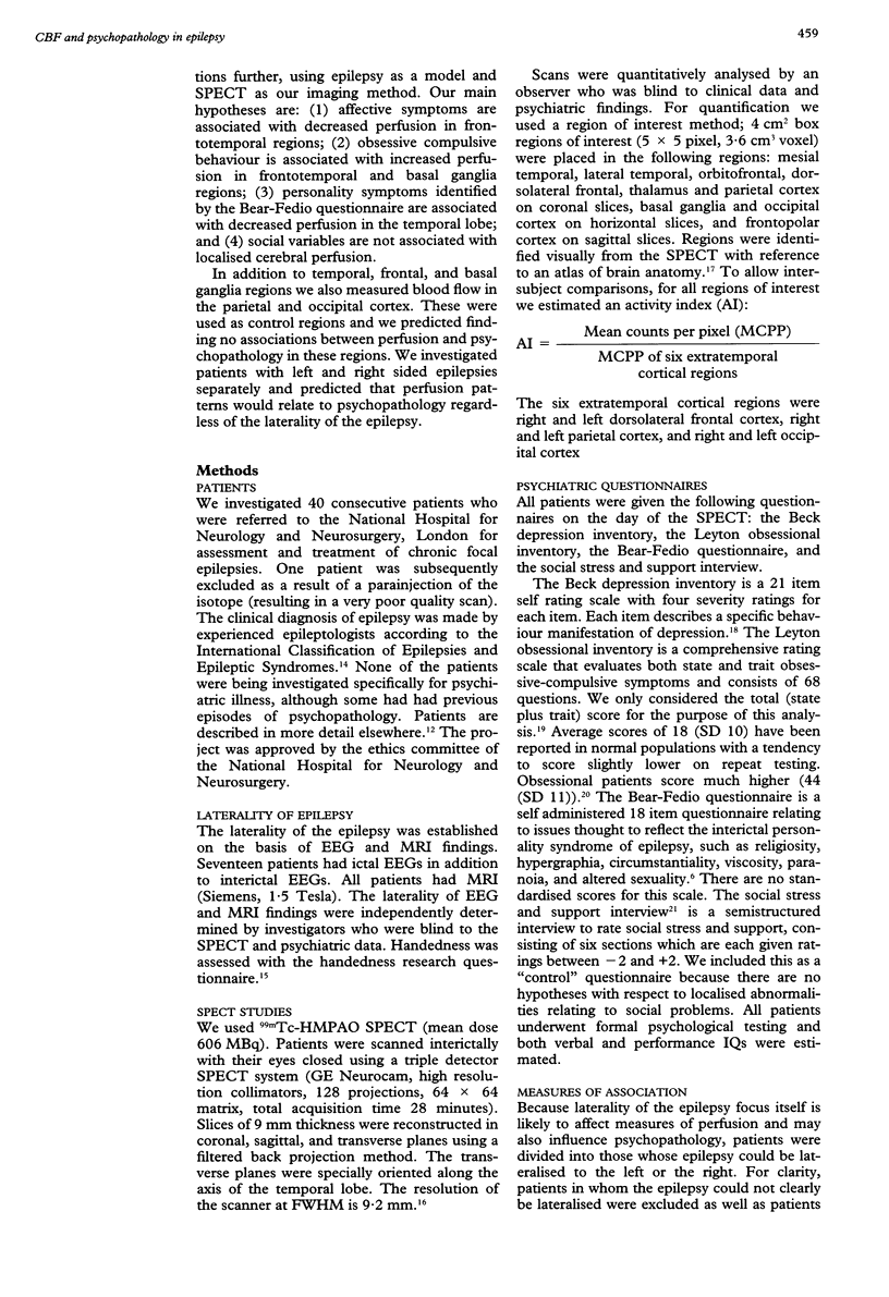
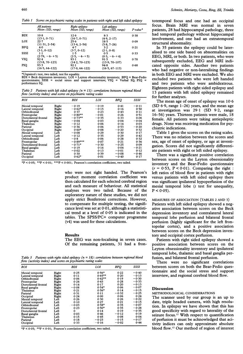
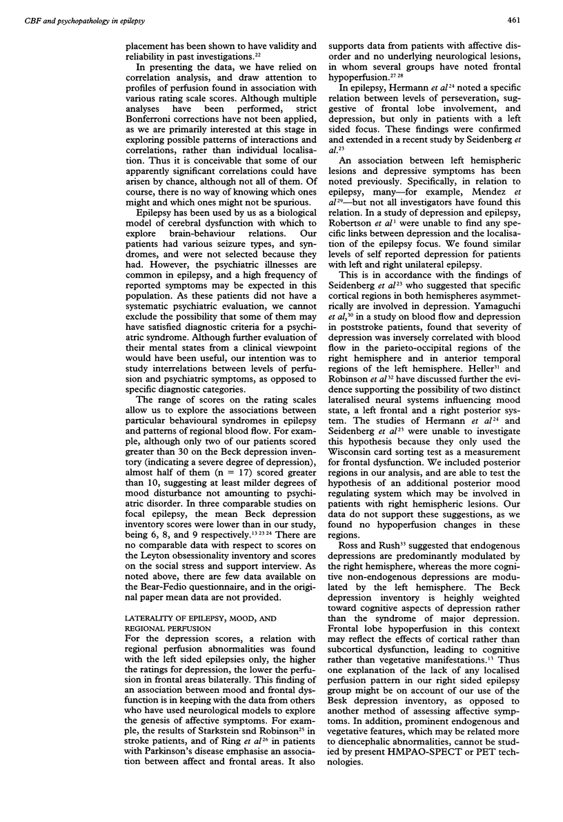
OBJECTIVES: In a study of patients with focal epilepsy the hypothesis was explored that different measurements of psychopathology are related to specific distributions of cerebral perfusion. METHODS: Forty patients had SPECT performed with (99m)Tc-HMPAO. In addition, patients received a psychiatric evaluation with the following psychiatric questionnaires: the Beck depression inventory, the Leyton obsessionality inventory, the Bear-Fedio questionnaire, and the social stress and support interview. Patients were analysed in two groups according to the laterality of the epilepsy. Nine patients were excluded based on poor quality scans (n = 1), unlateralised epilepsy (n = 4), and left or ambidextrous handedness (n = 4). RESULTS: There were no overall differences between the left and right epilepsy groups on measures of psychopathology. Associations were found between scores on some of the rating scales and regional cerebral blood flow. Specifically, for patients with left sided epilepsy, higher scores on the Beck depression inventory were associated with lower contralateral temporal and bilateral frontal perfusion, and higher occipital perfusion. For patients with right sided epilepsy higher scores on the Leyton obsessionality inventory were associated with increased perfusion in ipsilateral temporal, thalamic, and basal ganglia regions and bilateral frontal regions. CONCLUSION: The results do not support the notion that lateralised epileptogenic lesions are associated with different levels of depression, obsessionality, or personality traits. They support the view that certain psychopathological symptom patterns are related to specific regional dysfunctions depending on the laterality of a hemispheric lesion.
Full text
PDF
Selected References
These references are in PubMed. This may not be the complete list of references from this article.
- Annett M. The binomial distribution of right, mixed and left handedness. Q J Exp Psychol. 1967 Nov;19(4):327–333. doi: 10.1080/14640746708400109. [DOI] [PubMed] [Google Scholar]
- BECK A. T., WARD C. H., MENDELSON M., MOCK J., ERBAUGH J. An inventory for measuring depression. Arch Gen Psychiatry. 1961 Jun;4:561–571. doi: 10.1001/archpsyc.1961.01710120031004. [DOI] [PubMed] [Google Scholar]
- Baxter L. R., Jr, Schwartz J. M., Bergman K. S., Szuba M. P., Guze B. H., Mazziotta J. C., Alazraki A., Selin C. E., Ferng H. K., Munford P. Caudate glucose metabolic rate changes with both drug and behavior therapy for obsessive-compulsive disorder. Arch Gen Psychiatry. 1992 Sep;49(9):681–689. doi: 10.1001/archpsyc.1992.01820090009002. [DOI] [PubMed] [Google Scholar]
- Baxter L. R., Jr, Schwartz J. M., Mazziotta J. C., Phelps M. E., Pahl J. J., Guze B. H., Fairbanks L. Cerebral glucose metabolic rates in nondepressed patients with obsessive-compulsive disorder. Am J Psychiatry. 1988 Dec;145(12):1560–1563. doi: 10.1176/ajp.145.12.1560. [DOI] [PubMed] [Google Scholar]
- Baxter L. R., Jr, Schwartz J. M., Phelps M. E., Mazziotta J. C., Guze B. H., Selin C. E., Gerner R. H., Sumida R. M. Reduction of prefrontal cortex glucose metabolism common to three types of depression. Arch Gen Psychiatry. 1989 Mar;46(3):243–250. doi: 10.1001/archpsyc.1989.01810030049007. [DOI] [PubMed] [Google Scholar]
- Bear D. M., Fedio P. Quantitative analysis of interictal behavior in temporal lobe epilepsy. Arch Neurol. 1977 Aug;34(8):454–467. doi: 10.1001/archneur.1977.00500200014003. [DOI] [PubMed] [Google Scholar]
- Bench C. J., Friston K. J., Brown R. G., Frackowiak R. S., Dolan R. J. Regional cerebral blood flow in depression measured by positron emission tomography: the relationship with clinical dimensions. Psychol Med. 1993 Aug;23(3):579–590. doi: 10.1017/s0033291700025368. [DOI] [PubMed] [Google Scholar]
- Bench C. J., Friston K. J., Brown R. G., Scott L. C., Frackowiak R. S., Dolan R. J. The anatomy of melancholia--focal abnormalities of cerebral blood flow in major depression. Psychol Med. 1992 Aug;22(3):607–615. doi: 10.1017/s003329170003806x. [DOI] [PubMed] [Google Scholar]
- Bromfield E. B., Altshuler L., Leiderman D. B., Balish M., Ketter T. A., Devinsky O., Post R. M., Theodore W. H. Cerebral metabolism and depression in patients with complex partial seizures. Arch Neurol. 1992 Jun;49(6):617–623. doi: 10.1001/archneur.1992.00530300049010. [DOI] [PubMed] [Google Scholar]
- Cooper J. The Leyton obsessional inventory. Psychol Med. 1970 Nov;1(1):48–64. doi: 10.1017/s0033291700040010. [DOI] [PubMed] [Google Scholar]
- Cummings J. L. Frontal-subcortical circuits and human behavior. Arch Neurol. 1993 Aug;50(8):873–880. doi: 10.1001/archneur.1993.00540080076020. [DOI] [PubMed] [Google Scholar]
- Duncan R., Patterson J., Hadley D. M., Macpherson P., Brodie M. J., Bone I., McGeorge A. P., Wyper D. J. CT, MR and SPECT imaging in temporal lobe epilepsy. J Neurol Neurosurg Psychiatry. 1990 Jan;53(1):11–15. doi: 10.1136/jnnp.53.1.11. [DOI] [PMC free article] [PubMed] [Google Scholar]
- Hermann B. P., Seidenberg M., Haltiner A., Wyler A. R. Mood state in unilateral temporal lobe epilepsy. Biol Psychiatry. 1991 Dec 15;30(12):1205–1218. doi: 10.1016/0006-3223(91)90157-h. [DOI] [PubMed] [Google Scholar]
- Hoehn-Saric R., Pearlson G. D., Harris G. J., Machlin S. R., Camargo E. E. Effects of fluoxetine on regional cerebral blood flow in obsessive-compulsive patients. Am J Psychiatry. 1991 Sep;148(9):1243–1245. doi: 10.1176/ajp.148.9.1243. [DOI] [PubMed] [Google Scholar]
- Jenkins R., Mann A. H., Belsey E. The background, design and use of a short interview to assess social stress and support in research and clinical settings. Soc Sci Med E. 1981 Aug;15(3):195–203. doi: 10.1016/0271-5384(81)90013-2. [DOI] [PubMed] [Google Scholar]
- Kettl P. A., Marks I. M. Neurological factors in obsessive compulsive disorder. Two case reports and a review of the literature. Br J Psychiatry. 1986 Sep;149:315–319. doi: 10.1192/bjp.149.3.315. [DOI] [PubMed] [Google Scholar]
- Kouris K., Jarritt P. H., Costa D. C., Ell P. J. Physical assessment of the GE/CGR Neurocam and comparison with a single rotating gamma-camera. Eur J Nucl Med. 1992;19(4):236–242. doi: 10.1007/BF00175135. [DOI] [PubMed] [Google Scholar]
- Kroll L., Drummond L. M. Temporal lobe epilepsy and obsessive-compulsive symptoms. J Nerv Ment Dis. 1993 Jul;181(7):457–458. doi: 10.1097/00005053-199307000-00011. [DOI] [PubMed] [Google Scholar]
- McGuire P. K., Bench C. J., Frith C. D., Marks I. M., Frackowiak R. S., Dolan R. J. Functional anatomy of obsessive-compulsive phenomena. Br J Psychiatry. 1994 Apr;164(4):459–468. doi: 10.1192/bjp.164.4.459. [DOI] [PubMed] [Google Scholar]
- Mendez M. F., Taylor J. L., Doss R. C., Salguero P. Depression in secondary epilepsy: relation to lesion laterality. J Neurol Neurosurg Psychiatry. 1994 Feb;57(2):232–233. doi: 10.1136/jnnp.57.2.232. [DOI] [PMC free article] [PubMed] [Google Scholar]
- Mungas D. Interictal behavior abnormality in temporal lobe epilepsy. A specific syndrome or nonspecific psychopathology? Arch Gen Psychiatry. 1982 Jan;39(1):108–111. doi: 10.1001/archpsyc.1982.04290010080014. [DOI] [PubMed] [Google Scholar]
- Rauch S. L., Jenike M. A., Alpert N. M., Baer L., Breiter H. C., Savage C. R., Fischman A. J. Regional cerebral blood flow measured during symptom provocation in obsessive-compulsive disorder using oxygen 15-labeled carbon dioxide and positron emission tomography. Arch Gen Psychiatry. 1994 Jan;51(1):62–70. doi: 10.1001/archpsyc.1994.03950010062008. [DOI] [PubMed] [Google Scholar]
- Ring H. A., Bench C. J., Trimble M. R., Brooks D. J., Frackowiak R. S., Dolan R. J. Depression in Parkinson's disease. A positron emission study. Br J Psychiatry. 1994 Sep;165(3):333–339. doi: 10.1192/bjp.165.3.333. [DOI] [PubMed] [Google Scholar]
- Robinson R. G., Starr L. B., Price T. R. A two year longitudinal study of mood disorders following stroke. Prevalence and duration at six months follow-up. Br J Psychiatry. 1984 Mar;144:256–262. doi: 10.1192/bjp.144.3.256. [DOI] [PubMed] [Google Scholar]
- Rodin E., Schmaltz S. The Bear-Fedio personality inventory and temporal lobe epilepsy. Neurology. 1984 May;34(5):591–596. doi: 10.1212/wnl.34.5.591. [DOI] [PubMed] [Google Scholar]
- Ross E. D., Rush A. J. Diagnosis and neuroanatomical correlates of depression in brain-damaged patients. Implications for a neurology of depression. Arch Gen Psychiatry. 1981 Dec;38(12):1344–1354. doi: 10.1001/archpsyc.1981.01780370046005. [DOI] [PubMed] [Google Scholar]
- Schmitz E. B., Costa D. C., Jackson G. D., Moriarty J., Duncan J. S., Trimble M. R., Ell P. J. Optimised interictal HMPAO-SPECT in the evaluation of partial epilepsies. Epilepsy Res. 1995 Jun;21(2):159–167. doi: 10.1016/0920-1211(95)00020-b. [DOI] [PubMed] [Google Scholar]
- Snowdon J. A comparison of written and postbox forms of the Leyton Obsessional Inventory. Psychol Med. 1980 Feb;10(1):165–170. doi: 10.1017/s0033291700039714. [DOI] [PubMed] [Google Scholar]
- Starkstein S. E., Robinson R. G. Affective disorders and cerebral vascular disease. Br J Psychiatry. 1989 Feb;154:170–182. doi: 10.1192/bjp.154.2.170. [DOI] [PubMed] [Google Scholar]
- Yamaguchi S., Kobayashi S., Koide H., Tsunematsu T. Longitudinal study of regional cerebral blood flow changes in depression after stroke. Stroke. 1992 Dec;23(12):1716–1722. doi: 10.1161/01.str.23.12.1716. [DOI] [PubMed] [Google Scholar]


