

Because the dawning of the era in which interventions for congenital heart disease (CHD) became possible, the primary intellectual framework undergirding pediatric cardiology and cardiothoracic surgery has been based on morphology and hemodynamics (henceforth, the M/H model). This model has been wildly successful, enabling practitioners to devise creative interventions for even the most complex forms of CHD.
Today, we are confronting new challenges as we have awakened to the realization that longer-term outcomes for CHD, both cardiac and extracardiac, are well below earlier expectations.1 Relying on the M/H model that has permitted us to discover the unnatural history of CHD, our field is bringing scientific rigor to examining those outcomes and developing interventions to improve them.
Although this M/H model–based approach to improving outcomes is eminently reasonable, sole reliance on it is ultimately limiting. Use of a model based on causes might enable us to envision other therapies for CHD. For example, the principal risk associated with bicuspid aortic valve is valve calcification in later adulthood. Using the M/H model, we have attributed bicuspid aortic valve–associated valve calcification to hemodynamic stresses and used surveillance of valve function to determine the timing of surgical intervention. Operating under the causation model, it was discovered that a small percentage of patients with bicuspid aortic valve harbor NOTCH1 mutations, which are associated with early valve calcification.2 Aside from improving prognostication, the cause-based understanding of this form of bicuspid aortic valve suggests that therapies altering NOTCH signaling might prevent the deterioration of aortic valve function.
The causation model once occupied a larger role among CHD thought leaders. This period preceded the development of heart surgery and catheterization, when the only viable route seemed to be preventative strategies. Early thinkers emphasized environmental factors and, less importantly, heredity. Today, we still point to those 2 factors but think that their relative importance is reversed. Of note, the widespread belief in the role of genetics for CHD causation arose despite the facts that CHD usually arises sporadically and specific genetic defects can be identified for few affected individuals. In this review, I will explore the history of the causation model of CHD, emphasizing how we arrived at our current understanding, particularly with respect to genetics. I will then discuss how the causation model might contribute to CHD care in the future.
Preinterventional Era
Thomas Bevill Peacock
Thomas Bevill Peacock (Figure 1) was a physician who practiced in the 1800s at St Thomas’ Hospital in London.3,4 Aside from publishing numerous cases reports, focusing especially on cardiovascular diseases, Peacock authored an important monograph in 1858 entitled On Malformations of the Human Heart.5 Maude Abbott credited Peacock as the first to present CHD knowledge in an organized fashion.6 Unlike many of his contemporaries who wrote about CHD cases in haphazard fashion because of a lack of pathophysiologic principals, Peacock combined anatomy and embryology to classify CHD into 4 categories: misplacements of the heart, pericardial abnormalities, cardiac malformations, and irregularities of the primary vessels. In the monograph’s final section, Peacock wrote about the causes of CHD, attributing most forms to abnormalities in embryonic development, particularly growth arrest. He posited that milder defects were likely to have arisen later in development. With respect to the root causes, Peacock offered this: “The occurrence of accidents and strong impressions upon the mind of the mother are also supposed to conduce to the irregular development of the offspring, and in many cases such causes appear to have operated. In several instances which have fallen under my notice, the mothers of children laboring under malformations of the heart have assigned the defect in the children to strong mental impressions or shocks which they sustained during pregnancy; and there seems reason to think that such causes, by deranging the fetal circulation, might produce the effects.”
Figure 1.
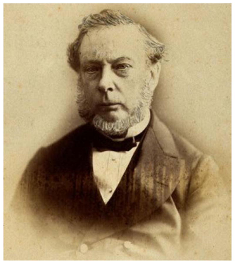
Thomas Bevill Peacock. Reprinted with permission from the Wellcome Library no. 13379i, London; http://catalogue.wellcomelibrary.org.
Aside from these environmental causes of CHD, Peacock noted instances in which parents had >1 child with CHD, which he termed an hereditary predisposition to defective development of the heart. Of note, Peacock’s monograph was printed a few years before the publication of Gregor Mendel’s masterpiece on the laws of inheritance, which was based on studies with sweet peas.7 Thus, Peacock had no scientific framework with which to think about CHD genetics.
Maude Abbott
Maude Abbott, the renowned pathologist at McGill University, worked primarily in the first decades of the 1900s. Her fascinating history has been well documented8 so will not be revisited extensively. She was encouraged by William Osler as she sought to reorganize the McGill pathological museum. Abbott rediscovered the original Holmes heart, resulting in her 1901 publication about that form of CHD9 and Osler’s subsequent invitation to write a chapter on CHD for his textbook Modern Medicine.6 Of note, Peacock had also cited Holmes’ report of that heart.5 Because Abbott knew that,6 we can assume that she was aware of Peacock’s views on CHD causality.
In Abbott’s chapter “Congenital cardiac disease” in Osler’s Modern Medicine, published in 1908,6 she sought to address a few burning questions about CHD including: what is the cause of the defect? Is it developmental or because of intra-uterine disease? Largely rejecting fetal diseases, such as acute endocarditis as causal, Abbott, like Peacock, pointed to arrest of development as the primary cause of CHD. She noted the vastly increased frequency of associated extracardiac anomalies, including neurocognitive issues, among those with CHD. She also observed that familial recurrence of CHD was most often in sibships, leading her to conclude that environmental factors were at play. Abbott specifically mentioned “baneful influences acting on the mother during the early weeks of pregnancy,” among which she included great trouble and fright.
Abbott understood that genetics was relevant for CHD. She wrote “Heredity, although not so clear or constant a factor in cardiac defects as some other anomalies (eg, polydactylism), certainly bears some part,” and then she cited families with multigenerational recurrence of CHD and cases of CHD with polydactyly. Like Peacock, Abbott was probably unaware of Mendelian genetics. Mendel’s magnum opus, published in 1866,7 was lost to the scientific community until 1900 and then took time to penetrate medical thinking. Thus, Abbott was operating from the same pre-Mendelian mindset as Peacock.
Abbott used the co-occurrence of CHD and polydactyly to bolster her argument that heredity was relevant for CHD. That association is rare, making her recitation of it striking. In Abbott’s time, polydactyly was considered the example par excellence of an hereditary trait. This was based on Pierre Louis de Maupertius’s description in 1753 of a 4-generation German family who had inherited polydactyly in an autosomal dominant pattern.10 The occurrence of polydactyly, a rare anomaly, in successive generations provided the first clear example of inheritance of a genetic trait in humans. By highlighting the association of CHD with polydactyly, Abbott was signaling that CHD could also be inherited.
Because of the primitive state of human genetics, Abbott missed the significance of several observations that she cited. She recounted the association between Down syndrome and CHD. Although she recognized the pathogenic role of advanced parental age, chromosomes and trisomies were as yet unknown so Abbott incorrectly attributed the mechanism to an exhaustion of the reproductive organs.6,11 She also noted several instances of isolated CHD in offspring born to parents in advanced middle age, unaware of the role of paternal age in de novo mutagenesis.11 Finally, Abbott cited consanguinity among parents as a factor for CHD but did not understand its implication vis-à-vis autosomal recessive inheritance.6,11
Interventional Era
Helen Brooke Taussig
Helen Brooke Taussig, arguably the most famous pediatric cardiologist of all time, wrote the first comprehensive textbook devoted to CHD, Congenital Malformations of the Heart.12 This book, published in 1947 but begun in the late 1930s, stands astride the preinterventional and postinterventional eras. Recall that Robert Gross’s first ligation of a patent ductus arteriosus was performed in 1939 and the first Blalock–Taussig–Thomas shunt was performed in 1944.13,14 Although Helen Taussig’s exemplary career propelled the M/H model forward, she never stopped thinking about the origins of CHD.15
Taussig attributed CHD to intrinsic and extrinsic factors.12 For the intrinsic factors, she deemed gene defects as of “real importance” but noted that the hereditary nature of CHD was often obscure. Among the extrinsic factors, Taussig cited vitamin deficiencies, skeletal abnormalities, and viral infections.
Taussig’s views shifted over time.16 She ultimately rejected teratogens as being of much importance. Her logic was that exposure to environmental toxicants resulted in widespread damage to the fetus. Thus, Taussig reasoned that teratogens were unlikely to underlie isolated CHD. In addition, she concluded that the relative constancy of CHD epidemiology around the world made a large role for exposures less likely because the environment differs so much geographically.
In the end, Taussig rejected the idea that CHD resulted from developmental errors as Abbott had maintained. Abbott had cited the work of Meckel,17 who had noted the similarity of certain forms of CHD to the hearts of more primitive animals, and the comparative evolutionary studies of Rokitansky and Spitzer.18,19 This led to comparisons, still heard today, between certain forms of CHD and snake, frog, fish and bird hearts. In this view, human CHD results from arrest during development, a failure along the ontogeny recapitulates phylogeny path. Taussig, in contrast, was struck by the presence of CHD in animals. She emphasized several breeds of dogs inheriting specific forms of CHD,16 and her last peer-reviewed article, published posthumously, was about CHD in birds.20 Because forms of CHD similar to those observed in patients are found widely in the animal kingdom, Taussig concluded that the genetic variation causing CHD must be ancient, making hearts with CHD evolutionary remnants, not developmental errors. Although this idiosyncratic view has not gained acceptance, Taussig’s emphasis on naturally occurring CHD in other species provided a strong rationale for understanding cardiac development in animals to gain insights into CHD pathogenesis in humans.
John Maurice Hardman Campbell
John Maurice Hardman Campbell (Figure 2) was a cardiologist who practiced at Guy’s Hospital in London.21 He was a founding member of the Cardiac Society of Great Britain and Ireland, which later sponsored the British Heart Journal, of which Campbell was the first editor. Campbell’s interests were far-ranging; he was an expert on ornithology and on Sherlock Holmes, even appearing as Doctor Watson in a re-enactment.
Figure 2.
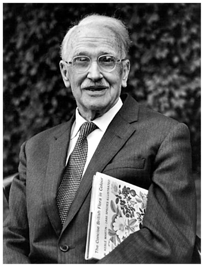
Maurice Campbell. Reprinted with permission from Horst Kolo, London.
In a career-altering event, Campbell helped to host a visit from Blalock in 1947, during which the first shunt procedures for CHD were performed in Great Britain. Fascinated, Campbell went on to collaborate with Russell Brock, providing the first patients for surgical pulmonary valvotomy.22 His interest in CHD continued for the duration of his productive career.
Campbell became interested in heredity as a teenager and undertook his first genetics research early in his medical career. During his research fellowship, he published an article about the heredity of hereditary spherocytosis.23 In it, he revealed that he was fully apprised about the exciting developments in human genetics. He reviewed Mendelian genetics at length. More impressively, he understood concepts about gene–environment interaction based on work with Drosophila by Thomas Morgan and others, which he needed to explain why hereditary spherocytosis could be a simple genetic trait despite the fact that the number of at-risk individuals manifesting jaundice was < 50% expected from Mendelian theory.
Campbell’s deep understanding of genetics arose fortuitously. As a boy, he attended a preparatory school in Oxford, where many of the university’s professors sent their children (M. Campbell, “Through changing years: A physician’s autobiography, unpublished article, communicated by Donald Campbell). Among those was J.B.S. Haldane, who was a year younger but supplanted Campbell as the best student in mathematics. The 2 became good friends. Years later, Campbell and Haldane attended Oxford University at the same time, and both studied oxygen physiology with Haldane’s father, resulting in each of their first scientific articles.24,25 After serving in the Royal Army Medical Corps during World War I, Campbell renewed his studies of oxygen, spending 3 years in the early 1920s in the Department of Physiology at Guy’s Hospital under the mentorship of Marcus S. Pembrey, who had also worked at Oxford with the elder Haldane.21,26 By the mid-1920s when Campbell was studying hereditary spherocytosis, J.B.S. Haldane, a polymath, had become an intellectual leader in genetics. Haldane had documented the first example of genetic linkage in mammals in 1915 and was then developing mathematical ideas underpinning population genetics.27 Campbell credited Haldane for drawing his attention to the ideas about gene–environment interaction, which informed his analysis of hereditary spherocytosis but also influenced his subsequent thinking about the genetics of CHD.23
Campbell married his robust understanding of human genetics with his interest in CHD, publishing numerous articles about this topic. In his first publication in 1949, he reported his limited conclusions from studying 300 subjects.28 Campbell confirmed the role of in utero rubella infection for a small percentage of his CHD cases but concluded that “(n)o decisive evidence of the importance of other environmental factors has been found.” Although he could identify Mendelian inheritance in only a small proportion, he used a process of elimination to conclude that “(t)he causes of CHD are mainly genetic.” This claim, bold in comparison with Taussig’s contemporaneous assessment that genetic factors were of real importance, has stood the test of time.
In Campbell’s last article about CHD cause, published in 1965 after his retirement, he summarized his seminal observations from his cohort, which had grown to >1200.29 Like others, he observed an increased risk of CHD among siblings but also noted the high rate of lesion concurrence when 2 siblings were affected. Campbell concluded that this provided strong evidence for genetic factors. He also searched for recurrence among parents and offspring. He found it among the parents of children with atrial septal defects but recognized that his cohort was biased by the low survival and reproductive fitness for CHD generally. Campbell felt that CHD was increased among the few offspring of individuals with CHD he had and deemed this “worth investigation on a larger scale.” Campbell, like others, found strong evidence for parental consanguinity, an indicator of autosome recessive inheritance, among those with situs inversus totalis and heterotaxy, and observed less striking increases over population rates for several other forms of CHD.
Campbell studied his cohort for parental age, looking for evidence that fathers of children with CHD were older because the paternal age effect had already been described for other traits.30 He found that the fathers of children with CHD were 3.3 years older than the mothers, a gap >0.5 year wider than observed in the general population. Noting that Maurice Lamy had observed something similar,31 Campbell concluded that the paternal age effect was operative for CHD.
On the basis of his extensive studies, Campbell decided that neither simple genetic factors nor environmental factors acting alone principally cause CHD. He posited that both were likely at play and favored genetic complexity. This was echoed by others who undertook retrospective cohort studies of CHD cause, most notably Lamy and Fuhrman.31,32 In the end, however, Campbell specified no genetic model for CHD cause.
James W. Nora
James W. Nora (Figure 3) was a pediatric cardiologist who wrote about genetic and environmental factors causing CHD throughout his career, often with his wife Audrey Nora. Nora’s purpose was to develop a coherent model incorporating those 2 factors, which could be used to understand CHD and to inform counseling affected families. In its earliest iteration, Nora termed his approach the multifactorial inheritance model.33 In that 1968 article, he reviewed the information available about the roles of genetics and environmental exposures in causing CHD. He considered 4 hypotheses: CHD is not genetic; chromosomal aberrations cause CHD; CHD is a Mendelian disorder; CHD has multifactorial inheritance of threshold characteristics, whose expression depends on environmental exposures. Nora quickly rejected the notion that CHD is not genetic based on familial recurrence, increased risk among twins, and homologies to animal models with CHD (á la Taussig). In considering chromosomal defects, he was familiar with examples including trisomy 21 and monosomy X causing Down and Turner syndromes, respectively, but knew that most patients with CHD did not have a gross chromosomal defect. Presciently, he noted that submicroscopic chromosomal anomalies had not been ruled out, anticipating developments in molecular cytogenetics reviewed below. Finally, Nora, like his predecessors, saw minimal evidence that CHD could act as a single-gene trait. Thus, by the process of elimination, Nora adopted the multifactorial inheritance model.
Figure 3.
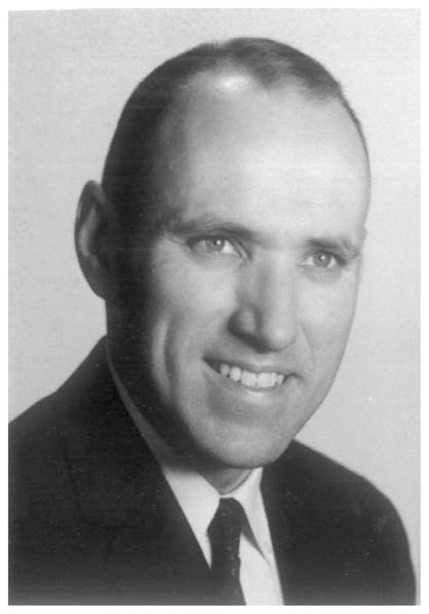
James Nora. Reprinted with permission from Michael Nihill, MD, Lillie Frank Abercrombie Section of Pediatric Cardiology, Texas Children’s Hospital.
Nora’s meaning about this model changed over time. When initially proposed,33 he proffered polygenic inheritance, which had a long history in human genetics. The first ideas about this form of complex genetics were developed by Francis Galton in the 1800s, before Mendel’s work or the discovery of genes.34 Spurred by the work of his first cousin, Charles Darwin, on variation in domesticated animals, Galton applied mathematics to the study of certain human traits with continuous values, such as intelligence and height, developing the field of biometry. He described the normal distribution for complex genetic traits and pointed out the phenomenon of reversion to the mean. Galton’s successors came to interpret his work in light of the discovery of genes, resulting in the notion that the Gaussian distribution in the population for continuous traits, such as height, resulted from small effects of a large number of genes, polygenic inheritance. For binary traits, such as CHD, they developed the concept of threshold effects—a large number of genes contribute to susceptibility for binary traits; those exhibiting it are on the tail of the Gaussian distribution. These thinkers also recognized the role of environmental factors in complex genetic traits. With height, for instance, it was clear that overall nutrition set the position of the mean and SD for the population normal curve. For binary traits, such as CHD, environmental exposures were suggested to shift the curve adversely, rendering a larger proportion of the population at risk.
For the polygenic model that Nora first suggested as relevant for CHD, certain predictions devolve from the underlying mathematics. Among these is the fact that recurrence risks should be equal among different classes of first-degree relatives, such as siblings and offspring. As noted before, risks for recurrence of CHD among siblings were well established but those for offspring were uncertain because of small numbers. In 1987, Ruth Whittemore at Yale published the results of her study of 373 infants born to 233 mothers with CHD.35 Unlike the 2% to 3% recurrence risk for siblings, she found a 16% offspring recurrence risk. Moreover, Whittemore observed ≈60% lesion concordance risk, a strong genetic signal. Finally, she found variability depending on the mother’s heart lesion with some forms (eg, left ventricular outflow tract obstructive defects) having high recurrence risk, whereas others (eg, muscular ventricular septal defects) appearing not to be associated with CHD recurrence. Subsequent studies examined offspring recurrence risk, some studying fathers with CHD, as well as affected mothers.36–38 Although the estimates of the magnitude of those risks have varied, it seems clear that offspring recurrence risks significantly exceed sibling recurrence risks. Thus, polygenic inheritance was ultimately rejected as the primary model for CHD.
In response, Nora altered his multifactorial model.39,40 His central tenet remained unchanged: CHD arose from gene–environmental interactions with threshold effects. For his revised model, Nora tapped into an intellectual struggle that had arisen between the biometricians and the Mendelians about how to interpret variation in human traits.34 Starting with Udney Yule and later through the work of R.A. Fisher, Sewell Wright and the aforementioned J.B.S. Haldane, reconciliation between the 2 schools had been accomplished. Thus, it had been demonstrated that one could generate Gaussian distributions for phenotypes without resorting to polygenic inheritance; a few alleles for a limited number of genes, oligogenetic inheritance, or even a single gene can produce the same phenotype distribution pattern in the population. In addition to including Mendelian effects, Nora broadened the range of genetic mechanisms to include nontraditional inheritance (mitochondrial, genomic imprinting, germline mosaicism, and uniparental disomy). Others have written about CHD cause in the ensuing 25 years, but Nora’s work stands as the last attempt at a holistic model.
Molecular Genetics Era
Gene Discovery for Mendelian CHD
In 1986, 1 year before Whittemore published her landmark study, the era of discovery of genes for Mendelian traits in humans was born when Stuart Orkin discovered the gene for chronic granulomatous disease using only molecular genetic methods.41 Initially termed reverse genetics and later positional cloning, this approach allowed investigators to find mutations for Mendelian disorders without previous knowledge of the pathogenesis. As this approach became increasingly robust, particularly after the human genome was sequenced, numerous genes were found for Mendelian disorders with CHD. Although some of those traits were isolated CHD, most were syndromes with CHD. Of note, elucidation of the genetic programs driving cardiovascular development, generally through animal studies, strongly informed the human genetic research, identifying candidate genes and illuminating disease pathogenesis. Overall, CHD genes uncovered through human genetic studies predominantly encode transcription factors and signal transduction proteins. The proportion of isolated CHD attributable to these Mendelian factors is very low, an unsurprising result given the more-than-one-hundred-year observation that CHD is uncommonly inherited in families.
Copy Number Variants
A major breakthrough in elucidating the genetic causes of CHD was the discovery of copy number variants (CNVs). As Nora had anticipated,33 there were genomic lesions at the submicroscopic level. The first important observation relevant for CHD genetics was the discovery of 22q11 deletions in DiGeorge and velocardiofacial syndromes. Loss of that chromosomal region, first implicated from a translocation and then observed in a modest percentage of affected individuals using high-resolution karyotyping,42,43 became robustly detectable with molecular testing (fluorescence in situ hybridization).44 Subsequent genetic epidemiological studies established that this genomic lesion accounts for ≈2% of CHD and 20% of conotruncal forms of CHD.45 Moreover, the genetic findings exposed limitations in our classification of CHD as 22q11 deletions were prevalent among patients with certain conotruncal defects (eg, truncus arteriosus) but rare among those with others (eg, double-outlet right ventricle). The mechanism for these recurrent deletions was established46 and proved relevant for the submicroscopic deletions at the elastin locus on chromosome 7 found to underlie Williams syndrome, another trait with CHD.47
After the human genome had been sequenced, CNVs, defined as gains or losses of DNA sequences >1 kb, were discovered as prevalent genomic variability.48,49 In aggregate, roughly 12% of the human genome is altered by CNVs in the general population. The average person harbors >1000 CNVs, covering ≈0.8% of his/her genome.50 Rare CNVs, often large and de novo, have been associated with disease, first neurodevelopmental and psychiatric disorders, such as autism and schizophrenia and, more recently, CHD.51 Overall, ≈10% of individuals with CHD harbor such CNVs; such CNVs are more prevalent among those who also have extracardiac abnormalities, such as intellectual and developmental disabilities, and children harboring them have poorer outcomes in somatic growth and neurocognition.52,53 Although certain submicroscopic defects, such as 22q11 deletions, which we would now classify as pathological CNVs, are associated with readily recognizable syndromes with facial dysmorphia, many of the recurrent pathological CNVs underlying CHD cannot be suspected from clinical examination and are associated with rather variable phenotypes including none at all. To summarize, pathological CNVs in aggregate account for >10% of CHD, making them the largest pathogenic class for CHD identified to date.
De Novo Mutations
Campbell’s work about the paternal age effect implied that de novo point mutations and small insertions or deletions (indels) were likely to explain some proportion of CHD. Until recently, technical limitations (ie, reliance on polymerase chain reaction and Sanger sequencing) dictated that this class of genetic cause could only be investigated for a limited number of genes, introducing a selection bias. With the development of massively parallel DNA sequencing (next-generation sequencing) as well methods for physically selecting portions of the genomes with which to generate sublibraries, sequencing of all coding portions of the human genome (whole-exome sequencing) or even the entire genome (whole-genome sequencing) became technically and economically possible. The Pediatric Cardiac Genomics Consortium used whole-exome sequencing to investigate the hypothesis that de novo mutations underlie severe forms of CHD.54 By sequencing a cohort of 362 individuals with critical forms of CHD, as well as their unaffected parents, the rate of de novo point mutations and small insertions/deletions (indels) was shown to be no different from that in the general population. Although not burdened by de novo mutations quantitatively, individuals in this CHD cohort were more likely to harbor de novo mutations altering genes that are more highly expressed in the embryonic heart, used as a surrogate for importance for cardiac development. Examining biological functions of the mutated genes, a burden of de novo mutations altering genes encoding histone-modifying enzymes, particularly those relevant for methylation of the histone 3 lysine 4, was observed. As the authors of this study noted, “evidence of dosage sensitivity of many chromatin-modifying genes raises the possibility that environmental perturbations of these pathways in critical developmental windows might phenocopy the effects of these mutations.”
Looking Forward
Discovery
At present, specific genetic factors contributing importantly to CHD pathogenesis can be identified in roughly 1 of 3 of affected individuals. Although certain environmental factors causing CHD are known (eg, fetal exposure to rubella or substantial alcohol levels, and maternal insulin-dependent diabetes mellitus) and others are at least implicated epidemiologically (eg, maternal obesity), the percentage of cases of CHD for which an environmental toxicant is identified definitively remains small. To identify the cause of CHD for the remaining 2 in 3 of cases, genetic/genomic studies are likely to be the drivers, at least for the near term.
Nora was right in thinking that many types of genetic mechanisms are relevant for CHD, and not all of them have been investigated robustly. To date, studies have focused on de novo single-nucleotide variants and indels in the exome and on CNVs altering at least 3 exons.52 Although there will be many more mutations belonging to those categories, which will be elaborated through additional studies of the full range of CHD, whole-genome sequencing will enable the study of de novo single-nucleotide variants/indels altering noncoding sequences and a broader array of structural variation, including smaller CNVs. Challenges remain in interpreting mutations of noncoding sequences in particular, but these are likely to be overcome as whole-genome sequencing data from increasing numbers of individuals become available.
Because CHD is inherited in Mendelian fashion infrequently, dominantly inherited variants of large effect are not likely to contribute importantly to CHD causation. The consistent evidence that consanguinity increases CHD risk suggests that autosomal recessive inheritance may account for some fraction of cases55; robust studies of probands with parental consanguinity, perhaps recruited from nations where that practice is customary, would seem worthwhile. Examinations of the role of common variants, assessed to date with cohorts of limited size with genome-wide association studies, have provided little evidence that there are variants of relatively large effect sizes for CHD in general, although there may be ones of modest size for atrial septal defects.56,57 This result, if sustained with further study, would align with the view that the poor reproductive fitness of CHD evolutionarily would have swept away most CHD variants. Unaddressed so far is the possibility intermediate between monogenic and polygenic inheritance: oligogenetic inheritance of genetic factors of modest size.
Nora suggested that other types of genetic and epigenetic alterations might be relevant for CHD.39 Somatic mosaicism can be detected in an unbiased manner using whole-exome sequencing or whole-genome sequencing with paired samples (eg, DNAs from heart and peripheral blood leukocytes). Because heart tissue is readily procured during surgery from patients with CHD, this issue could readily be examined. Abnormalities in DNA methylation, either inherited or de novo, can also be detected throughout the genome using bisulfite conversion-based methods. At present, the case for searching for mutations in the mitochondrial genome seems less compelling.
Understanding the role of specific environmental factors causing CHD will continue to be a challenge. Given that the relevant exposure window is during fetal life and precedes detection of the CHD by weeks or months, large, prospective studies of pregnant women would seem necessary. Given the relative infrequency of CHD and the variety of forms, which probably do not all arise from exposures to the same toxicants, execution of robust studies is challenging. Other epidemiological approaches, particularly if geographical hotspots are identified, might be more plausible. If the ongoing genetic work continues to emphasize the role of epigenetics, either through histone modifications or DNA methylation, the need for identifying environmental factors phenocopying the former or engendering the latter will become even more compelling. Finally, the elaboration of gene by environment interactions, as Nora envisioned, is highly desired but will require large cohorts, well beyond those being assembled currently, unless methodologic advances occur.
Clinical Effects
As the genetic basis for CHD can be identified in a growing percentage of affected individuals, the opportunities to use that information to improve clinical care and outcomes are also increasing. Genotype–phenotype associations are enabling care providers to more accurately prognosticate for individual patients. This can inform certain clinical decisions (eg, early intervention for infants at the greatest risk for intellectual and developmental disabilities) and counseling around reproductive issues. Eventually, genetic information may be able to drive certain aspects of cardiac care, for example, knowing when it is likely that pulmonary valve stenosis will progress or a subaortic membrane will develop. The identification of mutations with higher risks for poorer outcomes (eg, certain CNVs) will enable more robust designs of some CHD clinical trials, depending on the end points. Although not economically relevant, elucidation of the cause of an offspring’s CHD is greatly valued by parents, providing comfort that the defect was because of genetic randomness beyond their control and that certain associated problems arose from the same underlying genetic issue, not from preventable errors.
Finally, pathogenic information about CHD, being inherently mechanistic, will provide rich opportunities to develop therapies. If important environmental toxicants are identified as causal, eliminating or at least reducing exposure to developing fetuses for the relevant developmental window could reduce CHD incidence. Fetal interventions, such as with small molecules, to ameliorate the effect of genetic lesions is not inconceivable but will be extremely challenging, both logistically (ie, discovering mutations and intervening in a timely fashion) and from a safety perspective, most biological pathways having importance in the development of several organ systems. Interventions postnatally seem more feasible. Preventing the progression of obstructive lesions, enhancing the closure of certain septal defects, and improving myocardial performance based on genetic cause are imaginable. Genotype-specific small molecule therapies to improve neurocognition are being pursued for certain syndromes. Similar approaches could be contemplated for the genetic-based intellectual and developmental disabilities in CHD. Gene therapies designed to correct mutations or replace missing protein products could theoretically be curative, but the hurdles for implementing them for CHD remain substantial.
In summary, elucidation of the genetic causes of CHD has the potential to drive a paradigm shift in care for affected individuals. The time may finally be close at hand for the vision that Taussig offered 50 years ago, “Our next great step forward will come in the field of cause and the prevention of malformations.”15
Acknowledgments
This article is based, in part, on the Alexander S. Nadas Memorial Lecture delivered by B.D. Gelb at the American Heart Association’s 2014 Scientific Sessions in Chicago, IL.
Sources of Funding
This work was supported in part from a grant the National Heart, Lung and Blood Institute (HL098123).
Footnotes
Disclosures
None.
References
- 1.Newburger JW, Sleeper LA, Bellinger DC, Goldberg CS, Tabbutt S, Lu, et al. Pediatric Heart Network Investigators. Early developmental outcome in children with hypoplastic left heart syndrome and related anomalies: the single ventricle reconstruction trial. Circulation. 2012;125:2081–2091. doi: 10.1161/CIRCULATIONAHA.111.064113. [DOI] [PMC free article] [PubMed] [Google Scholar]
- 2.Garg V, Muth AN, Ransom JF, Schluterman MK, Barnes R, King IN, et al. Mutations in NOTCH1 cause aortic valve disease. Nature. 2005;437:270–274. doi: 10.1038/nature03940. [DOI] [PubMed] [Google Scholar]
- 3.Porter IH. The nineteenth-century physician and cardiologist Thomas Bevill PEACOCK (1812–82) Med Hist. 1962;6:240–254. doi: 10.1017/s0025727300027393. [DOI] [PMC free article] [PubMed] [Google Scholar]
- 4.Fye WB. Thomas Bevill Peacock. Clin Cardiol. 1990;13:447–448. doi: 10.1002/clc.4960130615. [DOI] [PubMed] [Google Scholar]
- 5.Peacock T. On Malformations of the Human Heart. London: John Churchill; 1858. [Google Scholar]
- 6.Abbott ME. Congenital cardiac disease. In: Osler W, editor. Modern Medicine: Its Theory and Practice. Philadelphia: Lea & Febiger; 1908. pp. 323–425. [Google Scholar]
- 7.Mendel JG. Versuche über Pfanzenhybriden. Verhandlungen des naturforschenden Vereines in Brünn, Bd. IV fur das Jahr 1865. Leipzig: Abhandlungen; 1866. pp. 3–47. [Google Scholar]
- 8.Evans WN, Béland MJ. The paediatric cardiology Hall of Fame: Maude Elizabeth Abbott. Cardiol Young. 2010;20:124–132. doi: 10.1017/s1047951110000119. [DOI] [PubMed] [Google Scholar]
- 9.Abbott ME. Unique case of malformation of the heart? Defect of the interventricular septum; rudimentary right ventricles patent foramen ovales great dilation of the right auricle and right auricular appendix. Montreal Med J. 1901;30:522–532. [Google Scholar]
- 10.de Maupertuis PLM. Letter XIV. Oeuvres de Mr De Maupertuis. Nouvelle edition, corrigee et augmentee. Lyon: J-M Bruyset; 1756. [Google Scholar]
- 11.Abbott ME. Congenital cardiac disease. In: Osler W, editor. Modern medicine: Its Theory and Practice. Philadelphia: Lea & Febiger; 1927. [Google Scholar]
- 12.Taussig HB. Congenital Malformations of the Heart. New York: Commonwealth Fund; 1947. [Google Scholar]
- 13.Gross RE, Hubbard JP. Surgical ligation of a patent ductus arteriosus. JAMA. 1939;112:729–731. doi: 10.1001/jama.251.9.1201. [DOI] [PubMed] [Google Scholar]
- 14.Blalock A, Taussig HB. The surgical treatment of malformations of the heart in which there is pulmonary stenosis or pulmonary atresia. JAMA. 1945;128:189–202. doi: 10.1001/jama.251.16.2123. [DOI] [PubMed] [Google Scholar]
- 15.Taussig HB. On the evolution of our knowledge of congenital malformations of the heart; the T. Duckett Jones memorial lecture. Circulation. 1965;31:768–777. doi: 10.1161/01.cir.31.5.768. [DOI] [PubMed] [Google Scholar]
- 16.Taussig HB. World survey of the common cardiac malformations: developmental error or genetic variant? Am J Cardiol. 1982;50:544–559. doi: 10.1016/0002-9149(82)90324-1. [DOI] [PubMed] [Google Scholar]
- 17.Meckel JF. Halle. 1802. De cordis conditionibus abnormibus. [Google Scholar]
- 18.Von Rokitansky K. Die Defecte der Scheidewand des Herzens. Pathologish-Anatomisch. Vienna: W. Braumüller; 1875. [Google Scholar]
- 19.Spitzer A. Über den Bauplan des normalen und mißbildeten Herzens. Versuch Einer phylogenetischen Theorie. Virchows Arch Path Anat. 1923;243:6–272. [Google Scholar]
- 20.Taussig HB. Evolutionary origin of cardiac malformations. J Am Coll Cardiol. 1988;12:1079–1086. doi: 10.1016/0735-1097(88)90479-2. [DOI] [PubMed] [Google Scholar]
- 21.Silverman ME. Maurice Campbell: first editor of heart. Heart. 2003;89:1379–1381. doi: 10.1136/heart.89.12.1379. [DOI] [PMC free article] [PubMed] [Google Scholar]
- 22.Brock RC, Campbell M. Valvulotomy for pulmonary valvular stenosis. Br Heart J. 1950;12:377–402. doi: 10.1136/hrt.12.4.377. [DOI] [PMC free article] [PubMed] [Google Scholar]
- 23.Campbell JMH. Heredity in acholuric jaundice. Q J Med. 1926;19:333–355. [Google Scholar]
- 24.Campbell JM, Douglas CG, Haldane JS, Hobson FG. The response of the respiratory centre to carbonic acid, oxygen, and hydrogen ion concentration. J Physiol. 1913;46:301–318. doi: 10.1113/jphysiol.1913.sp001593. [DOI] [PMC free article] [PubMed] [Google Scholar]
- 25.Douglas CG, Haldane JS, Haldane JB. The laws of combination of haemoglobin with carbon monoxide and oxygen. J Physiol. 1912;44:275–304. doi: 10.1113/jphysiol.1912.sp001517. [DOI] [PMC free article] [PubMed] [Google Scholar]
- 26.Haldane JS, Pembrey MS. An Improved Method of Determining Moisture and Carbonic Acid in Air. Vol. 29. London, Edinburgh: Dublin Philosophical Magazine; 1890. pp. 303–316. [Google Scholar]
- 27.Haldane JBS, Sprunt AD, Haldane NM. Reduplication in mice (preliminary communication) J Genet. 1915;5:133–135. [Google Scholar]
- 28.Campbell M. Genetic and environmental factors in congenital heart disease. Q J Med. 1949;18:379–391. [PubMed] [Google Scholar]
- 29.Campbell M. Causes of malformations of the heart. Br Med J. 1965;2:895–904. doi: 10.1136/bmj.2.5467.895. [DOI] [PMC free article] [PubMed] [Google Scholar]
- 30.Penrose LS. Parental age and mutation. Lancet. 1955;269:312–313. doi: 10.1016/s0140-6736(55)92305-9. [DOI] [PubMed] [Google Scholar]
- 31.Lamy M, De Grouchy J, Schweisguth O. Genetic and non-genetic factors in the etiology of congenital heart disease: a study of 1188 cases. Am J Hum Genet. 1957;9:17–41. [PMC free article] [PubMed] [Google Scholar]
- 32.Fuhrmann W. Research on the etiology of congenital angiocardiopathy. Acta Genet Stat Med. 1961;11:289–316. [PubMed] [Google Scholar]
- 33.Nora JJ. Multifactorial inheritance hypothesis for the etiology of congenital heart diseases. The genetic-environmental interaction. Circulation M. 1968;38:604–617. doi: 10.1161/01.cir.38.3.604. [DOI] [PubMed] [Google Scholar]
- 34.Bulmer M. Francis Galton: Pioneer of Heredity and Biometry. Baltimore: The Johns Hopkins University Press; 2003. [Google Scholar]
- 35.Whittemore R, Hobbins JC, Engle MA. Pregnancy and its outcome in women with and without surgical treatment of congenital heart disease. Am J Cardiol. 1982;50:641–651. doi: 10.1016/0002-9149(82)90334-4. [DOI] [PubMed] [Google Scholar]
- 36.Czeizel A, Pornoi A, Péterffy E, Tarcal E. Study of children of parents operated on for congenital cardiovascular malformations. Br Heart J. 1982;47:290–293. doi: 10.1136/hrt.47.3.290. [DOI] [PMC free article] [PubMed] [Google Scholar]
- 37.Dennis NR, Warren J. Risks to the offspring of patients with some common congenital heart defects. J Med Genet. 1981;18:8–16. doi: 10.1136/jmg.18.1.8. [DOI] [PMC free article] [PubMed] [Google Scholar]
- 38.Rose V, Gold RJ, Lindsay G, Allen M. A possible increase in the incidence of congenital heart defects among the offspring of affected parents. J Am Coll Cardiol. 1985;6:376–382. doi: 10.1016/s0735-1097(85)80175-3. [DOI] [PubMed] [Google Scholar]
- 39.Nora JJ. Causes of congenital heart diseases: old and new modes, mechanisms, and models. Am Heart J. 1993;125(5 Pt 1):1409–1419. doi: 10.1016/0002-8703(93)91014-6. [DOI] [PubMed] [Google Scholar]
- 40.Nora JJ. From generational studies to a multilevel genetic-environmental interaction. J Am Coll Cardiol. 1994;23:1468–1471. doi: 10.1016/0735-1097(94)90393-x. [DOI] [PubMed] [Google Scholar]
- 41.Royer-Pokora B, Kunkel LM, Monaco AP, Goff SC, Newburger PE, Baehner RL, et al. Cloning the gene for an inherited human disorder–chronic granulomatous disease–on the basis of its chromosomal location. Nature. 1986;322:32–38. doi: 10.1038/322032a0. [DOI] [PubMed] [Google Scholar]
- 42.de la Chapelle A, Herva R, Koivisto M, Aula P. A deletion in chromosome 22 can cause DiGeorge syndrome. Hum Genet. 1981;57:253–256. doi: 10.1007/BF00278938. [DOI] [PubMed] [Google Scholar]
- 43.Greenberg F, Elder FF, Haffner P, Northrup H, Ledbetter DH. Cytogenetic findings in a prospective series of patients with DiGeorge anomaly. Am J Hum Genet. 1988;43:605–611. [PMC free article] [PubMed] [Google Scholar]
- 44.Scambler PJ, Kelly D, Lindsay E, Williamson R, Goldberg R, Shprintzen R, et al. Velo-cardio-facial syndrome associated with chromosome 22 deletions encompassing the DiGeorge locus. Lancet. 1992;339:1138–1139. doi: 10.1016/0140-6736(92)90734-k. [DOI] [PubMed] [Google Scholar]
- 45.Goldmuntz E, Clark BJ, Mitchell LE, Jawad AF, Cuneo BF, Reed L, et al. Frequency of 22q11 deletions in patients with conotruncal defects. J Am Coll Cardiol. 1998;32:492–498. doi: 10.1016/s0735-1097(98)00259-9. [DOI] [PubMed] [Google Scholar]
- 46.Edelmann L, Pandita RK, Spiteri E, Funke B, Goldberg R, Palanisamy N, et al. A common molecular basis for rearrangement disorders on chromosome 22q11. Hum Mol Genet. 1999;8:1157–1167. doi: 10.1093/hmg/8.7.1157. [DOI] [PubMed] [Google Scholar]
- 47.Ewart AK, Morris CA, Atkinson D, Jin W, Sternes K, Spallone P, et al. Hemizygosity at the elastin locus in a developmental disorder, Williams syndrome. Nat Genet. 1993;5:11–16. doi: 10.1038/ng0993-11. [DOI] [PubMed] [Google Scholar]
- 48.Iafrate AJ, Feuk L, Rivera MN, Listewnik ML, Donahoe PK, Qi Y, et al. Detection of large-scale variation in the human genome. Nat Genet. 2004;36:949–951. doi: 10.1038/ng1416. [DOI] [PubMed] [Google Scholar]
- 49.Sebat J, Lakshmi B, Troge J, Alexander J, Young J, Lundin P, et al. Large-scale copy number polymorphism in the human genome. Science. 2004;305:525–528. doi: 10.1126/science.1098918. [DOI] [PubMed] [Google Scholar]
- 50.Conrad DF, Pinto D, Redon R, Feuk L, Gokcumen O, Zhang Y, et al. Wellcome Trust Case Control Consortium. Origins and functional impact of copy number variation in the human genome. Nature. 2010;464:704–712. doi: 10.1038/nature08516. [DOI] [PMC free article] [PubMed] [Google Scholar]
- 51.Thienpont B, Mertens L, de Ravel T, Eyskens B, Boshoff D, Maas N, et al. Submicroscopic chromosomal imbalances detected by array-CGH are a frequent cause of congenital heart defects in selected patients. Eur Heart J. 2007;28:2778–2784. doi: 10.1093/eurheartj/ehl560. [DOI] [PubMed] [Google Scholar]
- 52.Glessner JT, Bick AG, Ito K, Homsy JG, Rodriguez-Murillo L, Fromer M, et al. Increased frequency of de novo copy number variants in congenital heart disease by integrative analysis of single nucleotide polymorphism array and exome sequence data. Circ Res. 2014;115:884–896. doi: 10.1161/CIRCRESAHA.115.304458. [DOI] [PMC free article] [PubMed] [Google Scholar]
- 53.Carey AS, Liang L, Edwards J, Brandt T, Mei H, Sharp AJ, et al. Effect of copy number variants on outcomes for infants with single ventricle heart defects. Circ Cardiovasc Genet. 2013;6:444–451. doi: 10.1161/CIRCGENETICS.113.000189. [DOI] [PMC free article] [PubMed] [Google Scholar]
- 54.Zaidi S, Choi M, Wakimoto H, Ma L, Jiang J, Overton JD, et al. De novo mutations in histone-modifying genes in congenital heart disease. Nature. 2013;498:220–223. doi: 10.1038/nature12141. [DOI] [PMC free article] [PubMed] [Google Scholar]
- 55.Fahed AC, Gelb BD, Seidman JG, Seidman CE. Genetics of congenital heart disease: the glass half empty. Circ Res. 2013;112:707–720. doi: 10.1161/CIRCRESAHA.112.300853. [DOI] [PMC free article] [PubMed] [Google Scholar]
- 56.Cordell HJ, Töpf A, Mamasoula C, Postma AV, Bentham J, Zelenika D, et al. Genome-wide association study identifies loci on 12q24 and 13q32 associated with tetralogy of Fallot. Hum Mol Genet. 2013;22:1473–1481. doi: 10.1093/hmg/dds552. [DOI] [PMC free article] [PubMed] [Google Scholar]
- 57.Hu Z, Shi Y, Mo X, Xu J, Zhao B, Lin Y, et al. A genome-wide association study identifies two risk loci for congenital heart malformations in Han Chinese populations. Nat Genet. 2013;45:818–821. doi: 10.1038/ng.2636. [DOI] [PubMed] [Google Scholar]