Abstract
Invasive evaluations play important roles in identifying epileptogenic zones and functional areas in patients with intractable focal epilepsy. This article reviews the usefulness, methods, and limitations of invasive evaluations for epilepsy surgery. Invasive evaluations include various types of intracranial electrodes such as stereotactically implanted intracranial depth electrodes (stereo-EEG), chronic subdural electrodes, and intraoperative electrocorticography. Scalp EEG is distorted by the skull, meninges, and skin. On the other hand, intracranial electrodes provide spatial information with higher resolution than scalp electrodes, thereby enabling further delineation of epileptogenic zones and mapping of functional areas with electrical stimulation. In addition, intracranial electrodes record a wide frequency range of electrical activity, which is not possible with scalp electrodes. The very slow potentials in ictal recordings, known as ictal direct current (DC) shifts and ictal/interictal high frequency oscillations, such as ripples (100–200 Hz) and fast ripples (200–500 Hz), have been correlated with the ictal onset zone and are a sensitive and specific marker for epileptogenicity. Furthermore, several studies reported that the electrical stimulation of epileptogenic zones elicited enhanced cortical evoked potentials, abnormal delayed or repetitive responses, and fast ripples. These responses may assist in the delineation of the epileptogenic cortex as a potential new marker. There are definite risks of complications associated with the use of intracranial electrodes. However, when an invasive evaluation is selected based on careful consideration of the risks and benefits, it provides useful information for establishing a strategy for epilepsy surgery.
Keywords: intracranial electrodes, functional mapping, cortical stimulation, electrocorticography, focal epilepsy
Introduction
The electrical current recorded by scalp electroencephalography (EEG) is distorted by high-resistance tissues such as the skull, meninges, and skin. In addition, epileptiform discharges must involve the synchronized activity of a significant extension of the cortex (approximately 6 cm2) in order to be detected by scalp EEG.1) Therefore, scalp EEG is limited in its detection, precise localization, and determination of the extent of epileptogenic zones. Direct intracranial recordings from the brain are used to further delineate the regions of epileptogenicity in cases in which scalp EEG has failed to localize the epileptogenic zone. Intracranial electrodes are advantageous because they provide a markedly higher resolution than scalp electrodes and a clearer view of small loci of activity that are difficult to detect on the scalp.2) In addition to the localization of epileptogenic zones, intracranial electrodes aid in the mapping of eloquent brain areas in order to preserve brain function. The invasive electrodes used by institutions vary greatly in indications, techniques, types of electrodes, and number of implanted electrodes. These variabilities in the techniques used for invasive recording have led to different approaches to the treatment of epilepsy.3) This article will review invasive evaluations for epilepsy surgery, including types of invasive electrodes, surgical procedures, monitoring of epileptic activity, methodology for electrical stimulation, complications, and new approaches to identify epileptogenic zones.
Types of Intracranial Electrodes
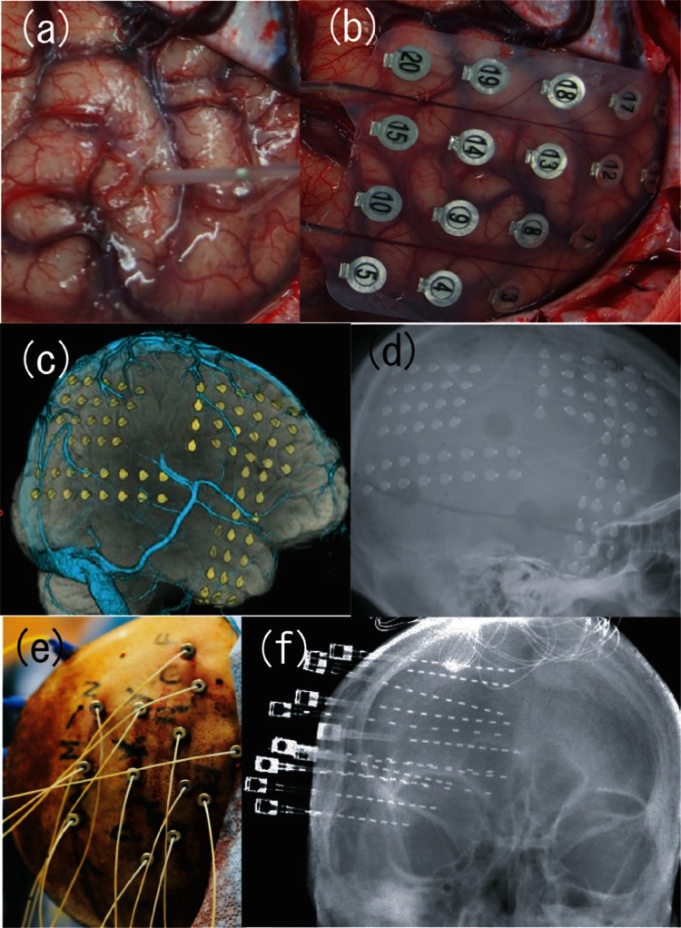
The implantation of an intracranial electrode is indicated when the precise localization of an epileptogenic zone and functional cortex is required in order to plan surgical resection with the preservation of brain function. Intracranial electrodes include two types of electrodes, i.e., depth electrodes and subdural grids/strip electrodes (Fig. 1a, b).3) The techniques and types of recordings used by institutions vary widely. Most major epilepsy centers in North America and Japan employ subdural electrodes or a combination of subdural and depth electrodes (Fig. 1c, d). European epilepsy centers rely primarily on stereotactically-inserted depth electrodes (stereoelectroencephalography, SEEG), which were introduced by Bancaud and Talairach in the 1950s (Fig. 1e, f).4,5) Furthermore, the choice of electrodes depends on the areas that need to be explored. Invasive recordings with subdural electrodes are helpful in the precise description of seizure onset and spread in the cortical surface, and provide a more precise definition of the functional cortex and delineation of the interface between epileptogenic zones and functional cortex. However, subdural electrodes cannot explore deeper generators such as the insula and amygdalo-hippocampal complex.3,6) On the other hand, depth electrodes penetrate the brain and are indicated when recording is needed from these deep brain structures.3) SEEG has a number of advantages including its ability to sample deep areas of the brain and anatomically distant regions and both hemispheres.7) However, the main constraint of depth electrodes is limited spatial sampling and difficulties in precise anatomical delineation between contiguous cortical regions of the epileptogenic zone and functional cortex.3,6) Careful consideration of their advantages and disadvantages needs to be coupled with a hypothesis about the epileptogenic zone derived from a non-invasive evaluation in order to decide the electrode, technique, and location for electrode implantation.
Fig. 1.
Various types of intracranial electrodes. Intraoperative view of (a) the depth electrode and (b) the subdural grid electrode. Three-dimensional reconstructed computed tomography (c) and radiography of implanted subdural grid electrodes (d). Intraoperative view (e) and radiography of stereotactically inserted depth electrodes (f) (stereoelectroencephalography, SEEG).
Surgical Procedure of Electrode Placement
Intracranial electrodes are implanted under general anesthesia. The placement of subdural electrodes is performed through burr holes for subdural strip electrodes and via craniotomy for grid electrodes. Most subdural grids are placed under direct visualization in order to avoid bridging veins or bony deformities and additional smaller grids or strips sliding over or under the brain without direct visualization, especially at orbito-frontal, inferior-temporal, and interhemispheric locations.2) When intraoperative electrocorticography is recorded, the effects of anesthesia need to be considered.8,9) A specific study on the effects of anesthesia reported that high-dose (1.5 MAC) sevoflurane anesthesia and isoflurane anesthesia provoked interictal spike frequency, whereas low-dose (0.3 MAC) isoflurane anesthesia suppressed interictal spikes.8)
The planning of SEEG electrode implantation requires a specific hypothesis of epileptic lesions based on non-invasive evaluation tests.7) After the localizing hypothesis is formulated, the targets are reached using depth electrodes of various lengths and number of contacts, depending on the specific brain region to be explored. Electrodes are implanted using a conventional stereotactic technique through drill holes. These intracranial electrodes are typically left implanted for a period of 5–14 days.2)
Localization of Epileptogenic Zones
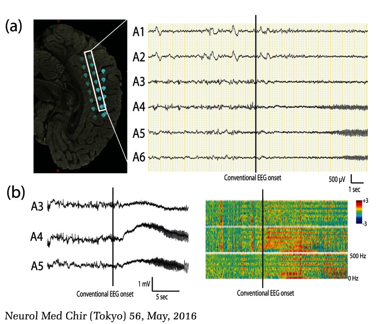
An epileptogenic zone is defined as the area of the brain that is necessary and sufficient for initiating seizures and whose removal or disconnection is needed for the abolition of seizures.10,11) This area is a theoretical concept and cannot be measured directly. Therefore, the location of the epileptogenic zone is considered to reside within an area defined by a concordance of findings from several investigations, including clinical seizure semiology, the scalp/intracranial EEG localization of epileptiform abnormalities, neuroimaging, and nuclear imaging. The identification of irritative zones and seizure onset zones detected with intracranial electrodes is the most concordant finding with the epileptogenic zone.10,11) Early ictal patterns recorded by intracranial electrodes may take various morphologies, including rhythmic sinusoidal waves, irregular spike discharges, spike and wave activity, and low voltage fast activity (Fig. 2a).12) Familiarity with these patterns is crucial for identifying the epileptogenic zone. The interpretation of ictal patterns requires knowledge of not only pathological, but also physiological and artefactual patterns. Moreover, invasive recordings provide new information on wide frequency-band EEG data such as ictal direct current (DC) shifts and high-frequency oscillations (HFOs), which play important roles in the definition of the epileptogenic zone (Fig. 2b). Ictal DC shifts are slow negative shifts at the beginning of seizures, and were initially recorded in animal epilepsy experiments.13,14) In order to record these very slow potentials, electrocorticogram recordings must employ a very low setting for the high-pass filter; no higher than 0.016 Hz. It has been speculated that ictal DC shifts reflect passively depolarized glial activity following the highly synchronized paroxysmal depolarization shifts (PDSs) of pyramidal cells, which occur in association with ictal activity. Therefore, ictal DC recordings may provide very important information on core epileptogenicity in patients with intractable partial epilepsy as surgical candidates. DC recordings are especially useful in cases of neocortical epilepsy in which conventionally filtered EEG shows only ill-defined broad electrodecremental patterns.2) The recent development of technology has enabled us to record focal HFOs with intracranial electrodes over regions of primary epileptogenicity in patients with intractable focal epilepsy. (HFOs) between 100 Hz and 500 Hz (such as ripples for 100–200 Hz and fast ripples for 200–500 Hz) were identified in the epileptogenic area as interictal activity in animal experiments and human epilepsy patients.15,16) Previous studies reported that ictal HFOs were well-defined in the primary seizure focus of patients with mesial temporal lobe epilepsy and neocortical epilepsy, and thus, delineate core epileptogenicity and the common properties of epileptogenicity regardless of etiology.17) Kanazawa et al. reported that ictal DC shifts and ictal HFOs were observed in 75.0% and 50.0% of patients, and 71.3% and 46.3% of analyzed seizures, respectively.18) The onset of ictal DC shifts preceded that of ictal HFOs, and the spatial extent of ictal DC shifts or HFOs was smaller than that of the conventionally defined seizure onset zone. Therefore, ictal DC shifts and HFOs may both represent the core of tissue generating seizures. The opening of an analysis window to extremely slow and fast activities may be very important for the addition of clinically useful information to invasive presurgical recordings.
Fig. 2.
Data from the implanted subdural grid electrodes. a: The ictal recordings, with conventional recording setting (time constant: 0.1 sec, low-pass filter: 600 Hz) showed electrographic seizure onset, consisting of low voltage fast activities on the several electrodes (A3–6). b: The same ictal recording with the setting for very slow potential (time constant: 10 sec) and the time-frequency analysis revealed the ictal direct current (DC) shift (left column) and the ictal high-frequency oscillations (HFOs) (right column), limited to A4 electrode, respectively. Both ictal DC shifts and HFOs might represent the core of tissue generating seizures. EEG: electroencephalography.
Function Brain Mapping
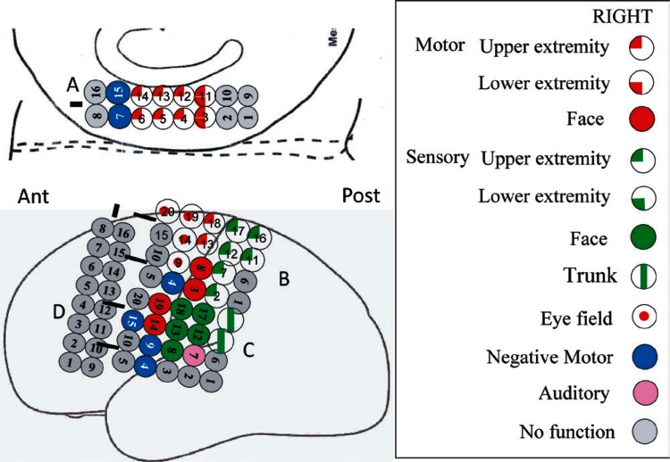
Electrical stimulation of the cerebral cortex has been used to localize the eloquent cortex, and mapping of these functional brain areas plays an important role in an invasive evaluation as well as the identification of the epileptogenic zone (Fig. 3).2) Electrical cortical stimulation produces two effects: (a) a positive phenomenon, i.e., the activation of some function, such as tonic or clonic movements or (b) a negative phenomenon, i.e., interference with a function, such as speech arrest, the cessation of motor activity, or loss of muscle tone. The stimulation parameters used at our institution are 50 Hz, biphasic, constant current stimuli, a duration of 300 μs, with intensity ranging from 1 mA to 15 mA. Stimulus intensity is increased stepwise at 1–2 mA while monitoring for neurological symptoms and afterdischarges. Since cortical stimulation may also induce seizures, continuous electrocorticographic monitoring is essential for checking afterdischarges induced by the electrical stimulation. Although patients are typically weaned from their anticonvulsants in order to better assure that seizures are captured, they need to be restarted on adequate doses of their anticonvulsants prior to the beginning of cortical stimulation. Surgical resection of eloquent cortical areas identified by cortical stimulation will generally lead to neurological deficits. However, there are exceptions and surgical removal of some cortical areas showing neurological symptoms during cortical stimulation (such as the anterior portion of the basal temporal language area, primary negative motor areas, and supplementary sensorimotor areas) leads only to temporary neurological deficits, with complete or almost complete neurological recovery.
Fig. 3.
Diagram of the functional brain areas defined by electrical stimulation in a patient with intractable left frontal lobe epilepsy. Red color indicates positive motor responses and green for sensory symptoms. Negative motor response is defined as the inability to continue voluntary movements or sustained muscle contraction without disturbance of consciousness.
Complications
There are definite medical risks associated with the use of intracranial electrodes. The complication rate of subdural electrodes has been reported to range between 6% and 26%.19–23) Relatively common adverse events associated with subdural electrodes are fever, headache, and nausea.24) Another group reported transient cerebrospinal fluid (CSF) leakage (13–31%), infection (6–8%), intracranial bleeding (8%), and cerebral edema in addition to an intracranial mass effect.25) Nair et al. reported that complications included (in the order of their frequency) infection, transient neurological deficit, epidural hematoma, increased intracranial pressure, and infarction.2) An increase in the complication rate was associated with (a) a greater number of grids/electrodes, (b) longer duration of monitoring, (c) older age of the patient, (d) left-sided grid insertion, (e) the use of burr holes in addition to craniotomy, and (f) an earlier year of monitoring (most likely a reflection of the aforementioned surgeon’s experience).
On the other hand, the complication rate of SEEG has been reported to be 3–5.6%.7,26) Previously reported complications of SEEG were intracranial bleeding and infection.4,20,26–28) Subdural electrodes have been perceived to have lower permanent morbidity (0–3%) than that of depth electrodes (3–6%) because there is no intraparenchymal penetration.19) Careful patient selection and consideration of the risk-benefit ratio of this modality are critically important, as is the case with all invasive techniques.
Cortical Stimulation for Epilepsy Evaluation
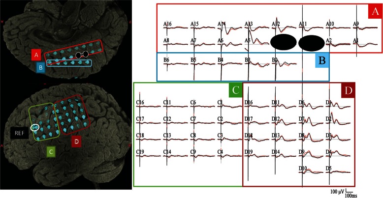
Adrian was the first to record the “spread of activity” of evoked cortical neuronal activity using a single-pulse electrical stimulation in animals.29) Several studies showed that direct electrical stimulation of the human cortex produced similar activities, forming an initial negative response (15–30 ms) followed by a slow positive response and then a slower negative response (lasting 250–300 ms).30,31) These potentials were called direct cortical responses (DCRs).30,32) Matsumoto et al. introduced an electrical stimulation technique termed cortical-cortical evoked potentials (CCEPs) in order to investigate the language network of the human brain.33) CCEPs have been used to evaluate various brain networks.34) CCEPs may provide information on pathological connectivity in the epileptic cortex as well as functional networks. Previous CCEP studies revealed that the stimulation of ictal onset zones elicited larger CCEP responses at the surrounding cortices than a normal cortex stimulation.35) Other CCEP studies attempted to track seizure propagation pathways (Fig. 4).36,37) Furthermore, Lega et al. reported that CCEPs and post-stimulation changes in gamma band activity at early spread sites (ictal spread within 3 seconds) were enhanced more than late spread sites (spread later than 3 seconds).37) These findings suggest that the size of CCEP responses reflect cortical excitability or dysfunctional inhibition.
Fig. 4.
Cortico-cortical evoked potential (CCEP) with ictal onset stimulation. CCEP distribution is correlated with seizure spread areas. Black circle indicates the stimulation sites.
Valentin et al. used a single-pulse electrical stimulation to study the epileptogenic cortex.38,39) This group also revealed that a favorable surgical outcome was associated with the removal of areas underlying electrodes showing a delayed and/or repetitive response to electrical stimulation.39)
A recent study revealed that fast ripples (250–520 Hz) induced by a single pulse electrical stimulation assisted in the delineation of the epileptogenic cortex.40) The median sensitivity for the seizure onset zone was 100% for single pulse-evoked spikes and 67% for single pulse-evoked fast ripples, whereas the median specificity was 17% for single pulse-evoked spikes to 79% for single pulse-evoked fast ripples. A median positive predictive value for the evoked responses in the seizure onset zone of 17% was found for spikes, 26% for ripples, and 37% for fast ripples. Single pulse-evoked fast ripples have potential as a new marker of epileptogenicity.
Conclusion
Invasive evaluations play important roles in the localization of epileptogenic zones in candidates indicated for epilepsy surgery. Subdural electrodes are useful for recordings in neocortical epilepsy in order to localize superficial epileptic foci and for mapping adjacent to the eloquent cortex. Depth electrodes are advantageous for recording epileptic activity arising from deep sources in the brain. Improvements in electrodes will allow the coverage of more extensive regions of the brain. Further advancements in intracranial electrodes, technology, and neurophysiological findings will lead to invasive evaluations that provide more precise information about not only the epileptogenic focus, but also brain functions and networks.
Acknowledgments
The authors wish to thank Dr. Jorge Gonzalez-Martinez at Cleveland Clinic for providing the images of SEEG.
References
- 1). Cooper R, Winter AL, Crow HJ, Walter WG: Comparison of subcortical, cortical and scalp activity using chronically indwelling electrodes in man. Electroencephalogr Clin Neurophysiol 18: 217– 228, 1965. [DOI] [PubMed] [Google Scholar]
- 2). Nair DR, Burgess R, McIntyre CC, Lüders H: Chronic subdural electrodes in the management of epilepsy. Clin Neurophysiol 119: 11– 28, 2008. [DOI] [PubMed] [Google Scholar]
- 3). Zumsteg D, Wieser HG: Presurgical evaluation: current role of invasive EEG. Epilepsia 41 (Suppl 3): S55– S60, 2000. [DOI] [PubMed] [Google Scholar]
- 4). Bancaud J, Angelergues R, Bernouilli C, Bonis A, Bordas-Ferrer M, Bresson M, Buser P, Covello L, Morel P, Szikla G, Takeda A, Talairach J: Functional stereotaxic exploration (SEEG) of epilepsy. Electroencephalogr Clin Neurophysiol 28: 85– 86, 1970. [PubMed] [Google Scholar]
- 5). Kahane P, Francione S: Stereoelectroencephalography, in Lüders HO. (ed): Textbook of Epilepsy Surgery. London, Informa Healthcare, Taylor & Francis distributor, 2008, pp 649– 658 [Google Scholar]
- 6). Enatsu R, Bulacio J, Najm I, Wyllie E, So NK, Nair DR, Foldvary-Schaefer N, Bingaman W, Gonzalez-Martinez J: Combining stereo-electroencephalography and subdural electrodes in the diagnosis and treatment of medically intractable epilepsy. J Clin Neurosci 21: 1441– 1445, 2014. [DOI] [PubMed] [Google Scholar]
- 7). Gonzalez-Martinez J, Bulacio J, Alexopoulos A, Jehi L, Bingaman W, Najm I: Stereoelectroencephalography in the “difficult to localize” refractory focal epilepsy: early experience from a North American epilepsy center. Epilepsia 54: 323– 330, 2013. [DOI] [PubMed] [Google Scholar]
- 8). Watts AD, Herrick IA, McLachlan RS, Craen RA, Gelb AW: The effect of sevoflurane and isoflurane anesthesia on interictal spike activity among patients with refractory epilepsy. Anesth Analg 89: 1275– 1281, 1999. [PubMed] [Google Scholar]
- 9). Fernández IS, Loddenkemper T: Electrocorticography for seizure foci mapping in epilepsy surgery. J Clin Neurophysiol 30: 554– 570, 2013. [DOI] [PubMed] [Google Scholar]
- 10). Rosenow F, Lüders H: Presurgical evaluation of epilepsy. Brain 124: 1683– 1700, 2001. [DOI] [PubMed] [Google Scholar]
- 11). Siegel AM: Presurgical evaluation and surgical treatment of medically refractory epilepsy. Neurosurg Rev 27: 1– 18; discussion 19–21, 2004. [DOI] [PubMed] [Google Scholar]
- 12). Koubeissi M: Subdural electrodes, in Lüders HO. (ed): Textbook of Epilepsy Surgery. London, FL: Informa Healthcare, Taylor & Francis distributor, 2008, pp 641– 648 [Google Scholar]
- 13). Ikeda A, Yazawa S, Kunieda T, Araki K, Aoki T, Hattori H, Taki W, Shibasaki H: Scalp-recorded, ictal focal DC shift in a patient with tonic seizure. Epilepsia 38: 1350– 1354, 1997. [DOI] [PubMed] [Google Scholar]
- 14). Ikeda A: DC recordings to localize the ictal onset zone, in Lüders HO. (ed): Textbook of Epilepsy Surgery. London, Informa Healthcare, Taylor & Francis distributor, 2008, pp 659– 666 [Google Scholar]
- 15). Bragin A, Engel J, Jr, Wilson CL, Fried I, Mathern GW: Hippocampal and entorhinal cortex high-frequency oscillations (100–500 Hz) in human epileptic brain and in kainic acid—treated rats with chronic seizures. Epilepsia 40: 127– 137, 1999. [DOI] [PubMed] [Google Scholar]
- 16). Bragin A, Staba R, Engel J, Jr: The significance of interictal fast ripples in the evaluation of the epileptogenic zone, in Lüders HO. (ed): Textbook of Epilepsy Surgery. London, Informa Healthcare, Taylor & Francis distributor, 2008, pp 530– 536 [Google Scholar]
- 17). Jirsch JD, Urrestarazu E, LeVan P, Olivier A, Dubeau F, Gotman J: High-frequency oscillations during human focal seizures. Brain 129: 1593– 1608, 2006. [DOI] [PubMed] [Google Scholar]
- 18). Kanazawa K, Matsumoto R, Imamura H, Matsuhashi M, Kikuchi T, Kunieda T, Mikuni N, Miyamoto S, Takahashi R, Ikeda A: Intracranially recorded ictal direct current shifts may precede high frequency oscillations in human epilepsy. Clin Neurophysiol 126: 47– 59, 2015. [DOI] [PubMed] [Google Scholar]
- 19). Rydenhag B, Silander HC: Complications of epilepsy surgery after 654 procedures in Sweden, September 1990–1995: a multicenter study based on the Swedish National Epilepsy Surgery Register. Neurosurgery 49: 51– 56; discussion 56–57, 2001. [DOI] [PubMed] [Google Scholar]
- 20). Hamer HM, Morris HH, Mascha EJ, Karafa MT, Bingaman WE, Bej MD, Burgess RC, Dinner DS, Foldvary NR, Hahn JF, Kotagal P, Najm I, Wyllie E, Lüders HO: Complications of invasive video-EEG monitoring with subdural grid electrodes. Neurology 58: 97– 103, 2002. [DOI] [PubMed] [Google Scholar]
- 21). Burneo JG, Steven DA, McLachlan RS, Parrent AG: Morbidity associated with the use of intracranial electrodes for epilepsy surgery. Can J Neurol Sci 33: 223– 227, 2006. [DOI] [PubMed] [Google Scholar]
- 22). Widdess-Walsh P, Jeha L, Nair D, Kotagal P, Bingaman W, Najm I: Subdural electrode analysis in focal cortical dysplasia: predictors of surgical outcome. Neurology 69: 660– 667, 2007. [DOI] [PubMed] [Google Scholar]
- 23). Wong CH, Birkett J, Byth K, Dexter M, Somerville E, Gill D, Chaseling R, Fearnside M, Bleasel A: Risk factors for complications during intracranial electrode recording in presurgical evaluation of drug resistant partial epilepsy. Acta Neurochir (Wien) 151: 37– 50, 2009. [DOI] [PubMed] [Google Scholar]
- 24). Swartz BE, Rich JR, Dwan PS, DeSalles A, Kaufman MH, Walsh GO, Delgado-Escueta AV: The safety and efficacy of chronically implanted subdural electrodes: a prospective study. Surg Neurol 46: 87– 93, 1996. [DOI] [PubMed] [Google Scholar]
- 25). Salazar F, Bingaman W: Placement of subdural grids, in Lüders HO. (ed): Textbook of Epilepsy Surgery. London, Informa Healthcare, Taylor & Francis distributor, 2008, pp 931– 937 [Google Scholar]
- 26). Cossu M, Cardinale F, Castana L, Citterio A, Francione S, Tassi L, Benabid AL, Lo Russo G: Stereoelectroencephalography in the presurgical evaluation of focal epilepsy: a retrospective analysis of 215 procedures. Neurosurgery 57: 706– 718; discussion 706–718, 2005. [PubMed] [Google Scholar]
- 27). Guenot M, Isnard J, Ryvlin P, Fischer C, Ostrowsky K, Mauguiere F, Sindou M: Neurophysiological monitoring for epilepsy surgery: the Talairach SEEG method. StereoElectroEncephaloGraphy. Indications, results, complications and therapeutic applications in a series of 100 consecutive cases. Stereotact Funct Neurosurg 77: 29– 32, 2001. [DOI] [PubMed] [Google Scholar]
- 28). De Almeida AN, Olivier A, Quesney F, Dubeau F, Savard G, Andermann F: Efficacy of and morbidity associated with stereoelectroencephalography using computerized tomography—or magnetic resonance imaging-guided electrode implantation. J Neurosurg 104: 483– 487, 2006. [DOI] [PubMed] [Google Scholar]
- 29). Adrian ED: The spread of activity in the cerebral cortex. J Physiol (Lond) 88: 127– 161, 1936. [DOI] [PMC free article] [PubMed] [Google Scholar]
- 30). Goldring S, Jerva MJ, Holmes TG, O’Leary JL, Shields JR: Direct response of human cerebral cortex. Arch Neurol 4: 590– 598, 1961. [DOI] [PubMed] [Google Scholar]
- 31). Purpura DP, Pool JL, Ransohoff J, Frumin MJ, Housepian EM: Observations on evoked dendritic potentials of human cortex. Electroencephalogr Clin Neurophysiol 9: 453– 459, 1957. [DOI] [PubMed] [Google Scholar]
- 32). Barth DS, Sutherling W: Current source-density and neuromagnetic analysis of the direct cortical response in rat cortex. Brain Res 450: 280– 294, 1988. [DOI] [PubMed] [Google Scholar]
- 33). Matsumoto R, Nair DR, LaPresto E, Najm I, Bingaman W, Shibasaki H, Lüders HO: Functional connectivity in the human language system: a cortico-cortical evoked potential study. Brain 127: 2316– 2330, 2004. [DOI] [PubMed] [Google Scholar]
- 34). Kunieda T, Yamao Y, Kikuchi T, Matsumoto R: New approach for exploring cerebral functional connectivity: review of cortico-cortical evoked potential. Neurol Med Chir (Tokyo) 55: 374– 382, 2015. [DOI] [PMC free article] [PubMed] [Google Scholar]
- 35). Iwasaki M, Enatsu R, Matsumoto R, Novak E, Thankappen B, Piao Z, O’Connor T, Horning K, Bingaman W, Nair D: Accentuated cortico-cortical evoked potentials in neocortical epilepsy in areas of ictal onset. Epileptic Disord 12: 292– 302, 2010. [DOI] [PubMed] [Google Scholar]
- 36). Enatsu R, Jin K, Elwan S, Kubota Y, Piao Z, O’Connor T, Horning K, Burgess RC, Bingaman W, Nair DR: Correlations between ictal propagation and response to electrical cortical stimulation: a cortico-cortical evoked potential study. Epilepsy Res 101: 76– 87, 2012. [DOI] [PubMed] [Google Scholar]
- 37). Lega B, Dionisio S, Flanigan P, Bingaman W, Najm I, Nair D, Gonzalez-Martinez J: Cortico-cortical evoked potentials for sites of early versus late seizure spread in stereoelectroencephalography. Epilepsy Res 115: 17– 29, 2015. [DOI] [PubMed] [Google Scholar]
- 38). Valentín A, Anderson M, Alarcón G, Seoane JJ, Selway R, Binnie CD, Polkey CE: Responses to single pulse electrical stimulation identify epileptogenesis in the human brain in vivo. Brain 125: 1709– 1718, 2002. [DOI] [PubMed] [Google Scholar]
- 39). Valentín A, Alarcón G, Honavar M, García Seoane JJ, Selway RP, Polkey CE, Binnie CD: Single pulse electrical stimulation for identification of structural abnormalities and prediction of seizure outcome after epilepsy surgery: a prospective study. Lancet Neurol 4: 718– 726, 2005. [DOI] [PubMed] [Google Scholar]
- 40). van ’t Klooster MA, Zijlmans M, Leijten FS, Ferrier CH, van Putten MJ, Huiskamp GJ: Time-frequency analysis of single pulse electrical stimulation to assist delineation of epileptogenic cortex. Brain 134: 2855– 2866, 2011. [DOI] [PubMed] [Google Scholar]


