Abstract
Purpose
This study was undertaken in order to establish a new reference value for the total antioxidant capacity (TAC) in seminal plasma as a predictor of fertility. This study also aims to propose a detailed protocol for the TAC assay including calculation of assay results and assessment of sensitivity and specificity over possible cutoff values in infertile men and controls with proven and unproven fertility.
Methods
Seminal plasma from 279 infertile patients and 46 normal healthy men referred to a male infertility testing laboratory were tested to measure TAC by a colorimetric assay kit. Receiver operating characteristics (ROC) curves were generated to establish cutoff values, sensitivity, and specificity, and the distribution of cutoff values in controls and infertile patients was calculated.
Results
Infertile patients showed significantly lower levels (mean ± SEM) of total antioxidants (micromolar Trolox equivalents) in their seminal plasma (1863.84 ± 27.16 μM) compared to those from fertile men (2013 ± 56.04 μM, P = 0.019). A preferred cutoff TAC value of 1947 μM could facilitate better diagnosis of oxidative stress (OS) in men with male factor infertility. At this threshold, the specificity of TAC assay was 63.0 % and the sensitivity 59.5 % with a positive predictive value of 90.7 % and a negative predictive value of 20.4 %.
Conclusions
Our results establish a new diagnostic cutoff TAC value of 1947 μM in seminal plasma to distinguish prevalence of OS in infertile patients compared to healthy men. This study provides a robust reference value of seminal plasma TAC that may provide an important diagnostic tool to the physicians for managing OS and male factor infertility in such patients.
Keywords: Male infertility, Oxidative stress, Seminal plasma, Total antioxidant capacity, Diagnostic value, Specificity and sensitivity
Introduction
Male infertility factors are present in about 20 % of infertile couples and contributory in about 30 to 40 %. An infertility evaluation should be performed if a couple has not achieved conception after 1 year of unprotected intercourse [1]. Oxidative stress (OS) is a common pathology in approximately half of all infertile men and is associated with pathogenesis of sperm DNA damage and dysfunction [2–6]. It results in negative changes in semen parameters such as sperm concentration, motility and morphology, and eventually male factor infertility [7].
Spermatozoa rely on oxidative phosphorylation in the mitochondria and glycolysis in the head and principal piece of the flagellum for energy. Oxidative phosphorylation is coupled with respiratory (electron transport) chain—a process accompanied by reactive oxygen species (ROS) generation [8]. ROS are natural products of cellular metabolism which, in physiological amounts, are essential for sperm processes such as capacitation, hyperactivation, and acrosome reaction leading to successful fertilization [9, 10]. Immature spermatozoa with excessive cytoplasm around the mid piece is also a concerning source of ROS resulting in impaired motility and abnormal morphology that impacts negatively on its fertilization potential [11]. In addition, seminal leukocytes are capable of producing up to 1000 times more ROS than immature spermatozoa causing high OS [12, 13]. Spermatozoa have limited antioxidant defense mechanisms and low concentration of ROS scavenging enzymes [8]. Also, spermatozoa are rich in polyunsaturated fatty acids and therefore susceptible to ROS attack [14]. However, seminal plasma has a very effective antioxidant defense system that provides spermatozoa with a protective environment against OS [15]. Antioxidants can protect cells against OS via three mechanisms: prevention, interception, and repair [16, 17]. OS occurs when there is excessive ROS production and limited antioxidant scavenging capacity resulting in an imbalance [18–21]. OS has been implicated in several male infertility-associated pathologies, including leukocytospermia, varicocele, as well as idiopathic infertility [22–24]. Our group has recently proposed a reference value and range of seminal ROS [25, 26]. Still there is a lack of complete clarity concerning the guidelines for determination of what is excessive ROS production or limited antioxidant capacity. It is therefore clinically important to establish the reference values for measuring the total antioxidant capacity (TAC) in the seminal plasma.
In this study, our goal was to establish a reliable and robust cutoff value of TAC in seminal plasma by investigating a large sample size. We established the cutoff value, including assay sensitivity, specificity, and negative and positive predictive values, for better diagnosis and management of OS in male factor infertility patients.
Materials and methods
Subjects
The study was approved by the Institutional Review Board of the Cleveland Clinic, and all the patient as well as donor samples used in the study were obtained with informed consent. Semen samples were used from 279 infertile patients referred to our andrology lab and 46 healthy controls with proven and unproven fertility screened from June 2014 through October 2015. The infertile men attending our male infertility clinic had confirmed male factor infertility after at least 1 year of unprotected intercourse. All patients were evaluated by a male infertility specialist. Patients with azoospermia, severe oligozoospermia, retrograde ejaculation, and collection problems (partial ejaculate) were excluded. The female partners of these patients underwent a complete gynecological investigation and were found to be healthy.
The controls consisted of healthy male volunteers selected on the basis of normal semen analysis (n = 21) according to the 2010 World Health Organization guidelines [27] or were of proven fertility (n = 25). All the male volunteers were tested for leukocytospermia and those men with >1.0 × 10⁶/mL white blood cells in semen as detected by myeloperoxidase or Endtz test were excluded from the study. Only 20.07 % (56/279) of infertile patients were identified with leukocytospermia and hence not grouped separately.
Semen collection and preparation
-
(i)
Semen specimens were collected by masturbation after 48 to 72 h of sexual abstinence. After complete liquefaction at 37 °C for 20 min, a 5-μL aliquot of each specimen was loaded on a 20-μM MicroCell chamber (Vitrolife, San Diego, CA) where it was analyzed for sperm concentration and motility.
-
(ii)
All samples were centrifuged at 300×g for 10 min at room temperature. Clear seminal plasma was aliquoted and frozen at −80 °C until the time of TAC assay.
TAC assay
Seminal plasma TAC measurement was done using the antioxidant assay kit and reagents (Cat #709001; Cayman Chemical, Ann Arbor, MI). The principle of the assay is based upon the ability of all antioxidants in the seminal plasma to inhibit the oxidation of the 2,2′-azino-di-[3-ethylbenzthiazoline sulfonate] (ABTS) to ABTS+ resulting in change of the absorbance at 750 nm to a degree that is proportional to their concentration. The capacity of the antioxidants present in the sample to prevent ABTS oxidation was compared with that of standard Trolox (6-hydroxy-2,5,7,8-tetramethylchroman-2-carboxylic acid), a water-soluble tocopherol analog. Results are reported as micromoles of Trolox equivalent.
The steps followed in the assay are given below.
-
(i)
All reagents and samples were equilibrated to room temperature at least 30 min prior to the beginning of the assay. Trolox standards and reagents were prepared as per the manufacturer’s instructions at the time of the assay.
-
(ii)
The antioxidant assay buffer concentrate was diluted 1:9 in a 15-mL conical tube. The reconstituted vial is stable for 6 months when stored at 4 °C.
-
(iii)
The lyophilized metmyoglobin powder was reconstituted with 600 μL of assay buffer. It is adequate for 60 wells. The reconstituted reagent is stable for 1 month when stored at −20 °C.
-
(iv)
One milliliter of reconstituted lyophilized Trolox was used to prepare the standard curve. It is stable for 24 h at 4 °C.
-
(v)
The working solution of 441 μM was prepared by two serial dilution, first by adding 10 μL of 8.82 M hydrogen peroxide to 990 μL of ultrapure water and then further by removing 20 μL and diluting with 3.98 mL of ultrapure water. It is stable for 4 h at room temperature.
-
(vi)
Chromogen (containing ABTS) was reconstituted with 6 mL of ultrapure water which was sufficient for 40 wells. The reconstituted vial is stable for 24 h at 4 °C. It is light sensitive and was prepared in indirect light.
-
(vii)
All the frozen samples of seminal plasma were thawed and centrifuged at 300×g for 7 min to ensure precipitation of any cellular component such as spermatozoa or cell debris, and the supernatant was used. Each sample was diluted 1:9 with assay buffer in clear microfuge tubes to avoid variability because of interference by the plasma proteins or sample dilution. All the samples were analyzed in duplicate.
-
(viii)
Samples as well as Trolox standards that were added to each well in duplicate were noted down using a plate template.
-
(ix)
Ten microliters of Trolox standard and test samples were loaded into the corresponding wells of a 96-well plate and assayed in duplicate (Cayman Chemical, Ann Arbor, MI).
-
(x)
Ten microliters of metmyoglobin and 150 μL of chromogen were added to all standard/sample wells. The reaction was initiated by adding 40 μL of hydrogen peroxide as quickly as possible.
-
(xi)
The plate was covered and incubated for 5 min on a horizontal plate shaker at room temperature (Eppendorf MixMate, Hamburg, Germany).
-
(xii)
Absorbance was monitored at 750 nm using a microplate reader (BioTek Instruments, Inc., Winooski, VT).
Calculation of assay results
-
(i)
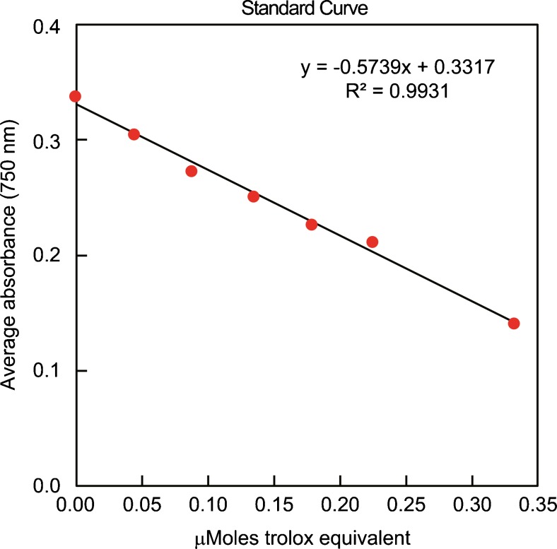
Determination of the reaction rate was done by calculating the average absorbance of each standard and sample. The average absorbance of the standards as a function of the final Trolox concentration (μM of Trolox equivalent) was plotted for the standard curve in each run, from which the unknown samples were determined (Fig. 1).
-
(ii)The total antioxidant concentration of each sample was calculated using the equation obtained from the linear regression of the standard curve by substituting the average absorbance values for each sample into the equation:
Fig. 1.
Example of a standard curve for TAC measurement: standard Trolox concentrations are represented on the X-axis and the absorbance on the Y-axis
Statistical analysis
The difference in distributions of TAC levels between infertile patients and healthy donors was assessed using the Wilcoxon rank sum test or chi-square test. Summaries of the distributions include frequency (%) and mean ± standard error of the mean (SEM). A receiver operating characteristics (ROC) curve was plotted to display estimated sensitivity, specificity, positive predictive value, negative predictive value, and accuracy over a range of possible cutoff points for TAC as a predictor of fertility. A cutoff value was chosen to maximize the sum of sensitivity and specificity.
Results
The background information on semen parameters in healthy donors (n = 46) and infertile patients (n = 279) used in the study is presented in Table 1. In the group of healthy men, the age was 37.57 ± 11.37 years, the ejaculatory abstinence was 3.54 ± 1.72 days, and the sperm concentration was 54.50 ± 33.53 × 10⁶/mL expressed as mean ± standard deviation. In the group of infertile patients, the age was 35.61 ± 6.06 years, the ejaculatory abstinence was 3.98 ± 2.75 days, and the sperm concentration was 47.27 ± 53.58 × 10⁶/mL. Among the infertile patients, the level of leukocytes was 0.27 ± 0.89 × 10⁶/mL and 34.05 % (95/279) men presented with a varicocele.
Table 1.
Background information on semen parameters in healthy donors and infertile men used in the study
| Parameter | Healthy donors (n = 46) | Infertile patients (n = 279) |
|---|---|---|
| Age (years) | 37.57 ± 11.37 | 35.61 ± 6.06 |
| Ejaculatory abstinence (days) | 3.54 ± 1.72 | 3.98 ± 2.75 |
| Sperm concentration (×106/mL) | 54.50 ± 33.53 | 47.27 ± 53.58 |
| Leukocytes (×106/mL) | 0 | 0.27 ± 0.89 |
| Diagnosis of varicocele (%) | 0 | 34.05 |
Data are expressed as mean ± standard deviation
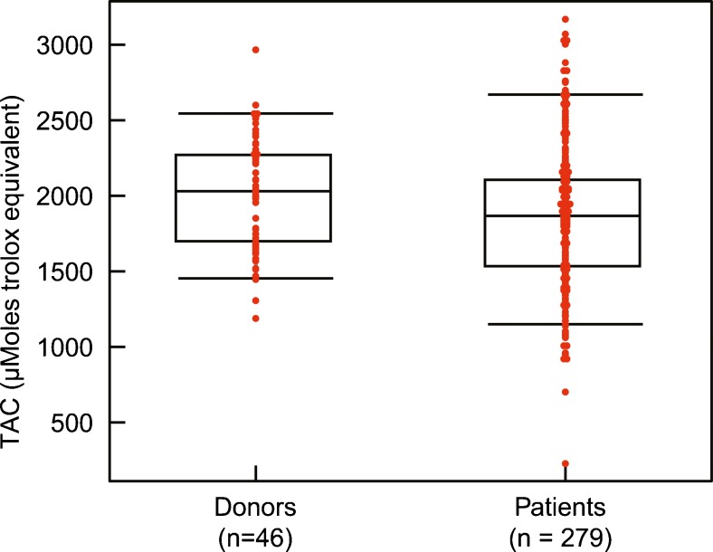
Distribution of TAC levels between healthy donors and infertile patients is shown in Fig. 2. A total of 325 TAC measurements were conducted in order to associate TAC levels with status of a subject, as either a healthy donor (control) or an infertile patient. The mean ± SEM TAC values were 2013 ± 56.04 μM in healthy donors as compared to 1863.84 ± 27.16 μM in infertile patients (P = 0.019).
Fig. 2.
Distribution of TAC levels between healthy donors (n = 46) and infertile patients (n = 279)
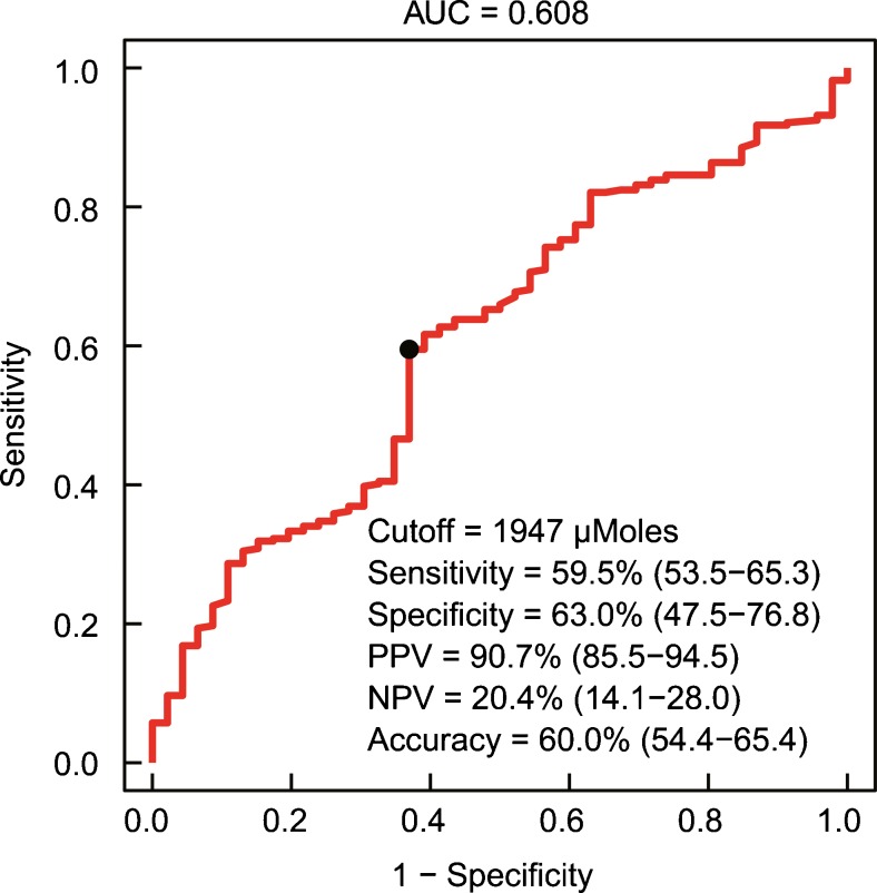
In our study, an ROC curve was plotted to determine cutoff value, sensitivity, and specificity of the TAC assay. A preferred cutoff value of 1947 μM could facilitate better diagnosis of OS in men with male factor infertility. At this cutoff point, the sensitivity was 59.5 %, specificity 63.0 %, positive predictive value 90.7 %, and negative predictive value 20.4 %. The accuracy of the test was 60.0 % (AUC = 0.608) (Fig. 3).
Fig. 3.
Receiver operating characteristics (ROC) curve showing the area under curve (AUC), cutoff, sensitivity, specificity, positive predictive value, negative predictive value, and accuracy of the assay
The status of a subject was also determined either as a healthy donor or an infertile patient depending upon two potential cutoff settings for antioxidants: (a) TAC levels higher than 1790 μM and (b) TAC levels higher than 1947 μM. For the potential cutoff level higher than 1790 μM, healthy donors as well as infertile patients did not show any significant difference (P = 0.46); 65.2 % donors (n = 30) and 59.5 % patients (n = 166) presented with more than this TAC level. On the other hand, a significantly higher proportion (P < 0.05) of healthy donors (63 %; n = 29) presented the second potential cutoff value for TAC levels higher than 1947 μM in comparison to that of infertile patients (40.50 %; n = 113). These findings led to the establishment of seminal plasma TAC levels higher than 1947 μM as the preferred cutoff value.
Discussion
The variability in TAC levels among infertile patients as shown in Fig. 2 can be explained in terms of varying degrees of OS [28] or any other clinical diagnosis such as varicocele. The background information on semen parameters in the infertile patient population as presented in Table 1 revealed that 34.05 % (95/279) men presented with a varicocele. Wang et al. reported that a significant portion of TAC has a genetic component [29]. How this relates to OS and/or TAC and their role in infertility is not yet understood. However, such information will be very useful to the clinicians in their decision-making for treating these infertility cases.
The impact of factors such as paternal age as well as the rate and time of centrifugation on seminal oxidative stress has been investigated. In the present study, the average age of healthy men was 37.57 ± 11.37 years, while it was 35.61 ± 6.06 years in the case of infertile patients. Cocuzza et al. demonstrated that ROS levels are significantly higher in seminal ejaculates for fertile older men (≥40 years) compared to younger ones (<40 years). In the category of fertile older men, the average age was 43.5 years, whereas the average age of fertile younger men was 33.5 years. The average age of infertile men who served as positive control was 42.65 years in the case of older men, whereas it was 27.95 years in the case of younger men [30]. On the other hand, a previous study by our group did not report any significant difference in seminal ejaculate ROS levels between older and younger infertile men. In this study, the average age of older infertile men (>40 years) was 46.6 years, followed by 35.3 years (31–40 years category) and 28.2 years (≤30 years) in the groups of younger infertile men [31]. The effect of centrifugation rate and time (200×g for 2 or 10 min and 500×g for 2 or 10 min) on the formation of ROS in semen of healthy and infertile men was investigated by Shekarriz and coworkers [32]. They concluded that the time of centrifugation was more important than g-force for inducing ROS formation in semen as the increase in ROS was less when semen was centrifuged for 2 as compared to 10 min. Bani Hani suggested that the rate of centrifugation can also influence seminal TAC levels, and noted a decrease in TAC upon raising the centrifugation force from 220 to 400×g [33].
The impact of ROS has widespread implications in the genitourinary system, including sperm physiology, fertility evaluation, reproductive outcomes, and antioxidant therapy [34]. Although ROS is formed during normal enzymatic reactions, cellular damage is prevented by the antioxidant scavenging system via enzymatic and nonenzymatic antioxidant pathways [35, 36]. This finely balanced oxidant-antioxidant system allows the formation of beneficial oxidants for normal cellular functions and concurrently prevents the damaging effects of excess OS [34]. TAC assesses the cumulative effect of all antioxidants present within the semen, based on their ability to scavenge free radicals with any specific or nonspecific mechanism(s) available [37–39].
Although multiple tests are available to measure TAC [39], the Trolox equivalent antioxidant capacity assay and the ferric reducing ability of the seminal plasma are the two most widely used in recent times. The first method, also called as ABTS assay, is based on the inhibition of oxidation of the ABTS to ABTS+. The second, ferric reducing antioxidant power (FRAP) assay, is based on the formation of [Fe2+] tripyridyltriazine from its oxidized [Fe3+] form. These assays have used colorimetric techniques to measure the seminal TAC. Our laboratory standardized a colorimetric TAC assay in a kit form that was used successfully with seminal plasma [40].
The evaluation of seminal plasma TAC levels has been emphasized to decide on the prognosis and the diagnosis of male infertility, especially in idiopathic male infertility [41, 42]. These authors reported average TAC levels of 3239 ± 562.25 μM (expressed as mean ± standard deviation) in healthy men (control) compared to 1916.4 ± 575.39 μM in asthenoteratospermic and 1896.7 ± 650.86 μM in oligoasthenoteratozoospermic infertility patients, respectively. However, in our study, infertile patients showed significantly lower (1863.84 ± 27.16 μM expressed as mean ± SEM) seminal plasma TAC levels compared with healthy men (2013 ± 56.04 μM; P = 0.019). For seminal plasma TAC levels higher than 1947 μM, the proportion of healthy donors was significantly more in comparison to infertile patients (P < 0.05). The seminal plasma ROC curve suggested 60 % accuracy of the test with good sensitivity and specificity over a cutoff TAC value of 1947 μM. It also indicated a high positive predictive value (90.7 %) and a low negative predictive value (20.4 %). The assay is reported to have an inter-assay coefficient of variation of 3 % and intra-assay coefficient of variation of 3.4 %. Such minimal variability suggests our TAC assay to be more robust and reliable and the cutoff value to be more precise that may help the clinicians in their decision-making to offer appropriate therapy to these patients.
ABTS assay has been used to measure the seminal plasma TAC for a variety of diagnostic purposes. In an earlier study, significantly higher TAC was reported in varicocele patients in comparison to patients with genitourinary inflammation or normal fertile controls, suggesting possible involvement of systemic hormones prolactin and tetraiodothyronine in the regulation of seminal TAC [43]. Further analysis between these hormones and seminal parameters showed an inverse correlation between prolactin and sperm motility, while a direct correlation of TAC with prolactin and tetraiodothyronine was observed [43]. The effect of spermatic vein ligation on seminal TAC in patients with varicocele has also been assessed by the same method. The evaluation of seminal TAC levels in these patients before and after surgery revealed that spermatic vein ligation can improve the seminal TAC in patients with middle- and high-grade varicocele [44]. They noted seminal plasma levels of 2580 ± 250 μM in preoperative grade I varicocele patients as against 2610 ± 160 μM after surgery. In varicocele grade II and III patients, the TAC levels were 2360 ± 230 μM in preoperative and 2660 ± 150 μM in postoperative cases. Further studies are needed to establish associations between such surgical procedures or hormones with TAC scores and implications of such association in the management of infertility patients.
It is interesting to find that by using the same assay, higher seminal TAC levels were obtained after a shorter period of ejaculatory abstinence (1 vs. 4 days) before oocyte insemination for IVF, suggesting that it may protect sperm from OS-induced damage by a mechanism independent of lipid peroxidation of sperm membranes [45]. The same authors hypothesized that higher seminal TAC would reduce exposure of sperm to harmful ROS and may be a protective mechanism for improving sperm quality. They argued that purging sperm from the cauda of the epididymis or vas deferens by shortening the period of ejaculatory abstinence could diminish the population of senescent sperm, thus reducing ROS and perhaps lessening the protective effect of TAC [45]. Another study reported a positive correlation of the magnitude of decrease in the seminal plasma TAC levels with the abnormal semen parameters such as concentration, motility, and morphology in men with idiopathic infertility [46]. They suggested TAC levels of 1098 ± 120 μM in oligozoospermic patients compared to 1267 ± 75 μM in normozoospermic men.
Many environmental factors are known to affect fertility. How these are linked to OS and TAC in the semen samples is not clear. Kumar and colleagues measured TAC values in the seminal plasma of health workers occupationally exposed to ionizing radiation using the ABTS assay and reported a positive correlation of TAC with sperm chromatin integrity [47]. Recently, this technique was used to measure seminal TAC in males environmentally exposed to lead (1470 ± 240 μM in low-exposure group vs. 1320 ± 170 μM in high-exposure group). It was reported that lead exposure induces OS in seminal plasma and modulates antioxidant defense systems [48]. Similarly, many other environmental issues linked to male factor infertility and their prevention/treatment could benefit from such TAC evaluation of semen samples collected in a clinical lab or at the exposure sites.
Any correlation of TAC with antioxidant therapy may also play a useful role in such patient care. The supplementation of antioxidant alpha-lipoic acid (also called thioctic acid) in infertile men has been shown to result in a significant increase in seminal TAC levels in comparison to placebo, suggesting this as an oral antioxidant therapy of asthenoteratospermia [49]. Raigani and colleagues studied the impact of zinc sulfate and folic acid supplementation on seminal plasma TAC levels and sperm functional parameters in oligoasthenoteratozoospermic men [50]. However, no statistically significant differences in seminal TAC could be detected before 16 weeks of randomized double-blind treatment.
A recent prospective, randomized, controlled clinical tamoxifen treatment in idiopathic oligoasthenospermic patients resulted in increased serum and seminal plasma TAC and, significantly, spermatozoa intracellular ROS, indicating the antioxidant role of tamoxifen [51]. From the abovementioned studies, it is evident that the seminal plasma TAC assay has assumed importance as a component of clinical trials in the prevention of OS-related male factor infertility as seminal TAC can provide useful information [49–51]. As such, results of such trials are expected to have wide clinical importance in fertility clinics and laboratories, thereby establishing the practical utility of the TAC assay.
The FRAP assay has also been used widely for measurement of seminal plasma TAC levels. FRAP is a ferric-reducing agent with antioxidant potential that measures change in absorbance at 593 nm due to the formation of a blue-colored [Fe2+] tripyridyltriazine compound from colorless oxidized [Fe3+] form, by the action of electron-donating antioxidants [42]. Using this agent in a simple colorimetric method, they reported significantly lower seminal plasma TAC levels in oligoasthenozoospermic (1389.72 ± 242.11 μM), asthenozoospermic (1777.88 ± 239.87 μM), and azoospermic (1702.67 ± 485.95 μM) patients than in seminal plasma of normozoospermic (1980.82 ± 160.58 μM) men.
Recently, Layali and colleagues have reported lower TAC levels (1230.25 ± 352 μM) in hyperviscous semen samples as compared to nonhyperviscous semen samples (1710.31 ± 242.11 μM) [52]. They attributed this to a severe impairment of seminal antioxidant systems and an increased sperm membrane lipid peroxidation in patients with hyperviscous semen. Increased ROS in the seminal plasma of patients with hyperviscous semen, originating from leukocytes and abnormal sperm, may decrease the effective TAC and increase the harmful effects of ROS [52]. A severe impairment of the high and low molecular weight antioxidants in semen of patients with hyperviscous semen has also been reported [53]. However, in the present study, we did not include hyperviscous semen samples as a separate group. Significantly higher TAC levels have been reported in azoospermia (2913 ± 900 μM) than in teratozoospermia (2269 ± 570 μM) or the normal controls (2120 ± 900 μM) [54]. This relationship needs to be investigated further.
Another study by the same group suggested significantly higher TAC levels in normozoospermic (1672 ± 631 μM) and oligozoospermic (1763 ± 561 μM) men compared to fertile men (1065 ± 377 μM) [55]. The seminal TAC levels even in normal fertile men in these two studies by the same group were different. It could be due to the lack of standardization in the TAC assay unlike in our present study. Those authors supported their findings by suggesting that TAC reflects the effect of low molecular weight antioxidants but not the activity of endogenous antioxidant enzymes (e.g., catalase, SOD, etc.) or metal-binding proteins. Kratz et al. further suggested that such inconsistency in the TAC values of infertile patients might be caused by different study conditions and that any increase in the antioxidant capacity of male semen may not necessarily lead to improved sperm parameters or the subsequent fertilization process [55]. Recently, Yousefniapasha and colleagues also used this assay to measure the seminal plasma TAC of infertile men with history of smoking and showed higher TAC levels in their semen (1785.45 ± 718.18 μM) [56]. They suggested that the increased nitric oxide levels associated with smoking might exceed the capacity of the antioxidant defense system leading to increased oxidative damage and decreased fertility and thus associated with higher TAC levels. This needs further investigation.
Our group previously measured ROS production and TAC levels by chemiluminescence assay to create a ROS-TAC score to provide to the clinicians with a means to suggest early varicocele repair for patients and prevent further sperm damage by OS [57]. Infertile patients with varicocele had lower semen quality scores, higher ROS, and lower TAC and ROS-TAC scores compared to healthy men. TAC levels of 1186 ± 96.9 μM were noted in infertile varicocele patients, while the levels were 1443 ± 105 μM in healthy donors (control) and 939 ± 107 μM in patients with varicocele [57]. Semen quality scores were derived from concentration, motility, curvilinear velocity, straight-line velocity, average path velocity, amplitude of lateral head displacement, linearity, and morphology of spermatozoa. Semen and ROS-TAC scores serve as good indicators about fertilizing potential because an imbalance between ROS production and TAC in seminal plasma leads to OS-induced male infertility [58]. Pasqualotto and coworkers also suggested that the ROS-TAC score may be more predictive of subfertility than TAC or ROS levels alone as it gives the benefit of measuring two reliable parameters [57]. In the present study, we established a new cutoff value of seminal plasma TAC by investigating a large number of healthy donors as well as infertile patients. By plotting the ROC curve over a range of possible cutoff points, the sensitivity, specificity, and negative and positive predictive values have been estimated with minimal variability to distinguish between healthy men and male factor infertility patients. A cutoff value of 1947 μM seminal plasma TAC was chosen to maximize the sum of sensitivity and specificity.
One limitation of our study was that the subjects were grouped either as healthy donors (based on normal semen analysis of proven fertile as well as normozoospermic men according to WHO 2010 guidelines) or as infertile patients (attending a tertiary care hospital). Only some of the donors were of proven fertility, and hence, we did not include proven fertile men as a separate group in our study. Secondly, although the leukocytospermic men were excluded in the group of healthy donors, only a limited number of infertile patients were leukocytospermic and hence not included as a separate group. Furthermore, healthy donors (control group) had a smaller sample size in comparison to infertile patients. Another limitation was that except for varicocele, we did not examine any other clinical diagnosis of our infertile population regarding the exact etiology of infertility or reproductive potential in these patients.
Conclusions
In conclusion, we have provided a detailed protocol to measure seminal antioxidant levels and established the reference values below which the TAC levels are abnormal and may result in OS in the presence of high ROS. The results of the present study establish a new diagnostic cutoff value of 1947 μM seminal plasma TAC to distinguish between healthy donors and male factor infertility patients. Each laboratory must establish their own reference values using the protocol described. This study provides a reference value taking into account a large number of infertile patients, which will help in better diagnosis and management of male factor infertility.
Acknowledgments
The study was supported by funds from the American Center for Reproductive Medicine. Dr. Shubhadeep Roychoudhury was supported by a fellowship from the Department of Biotechnology, Government of India. The authors are grateful to the Andrology Center technologists for scheduling the study subjects and Jeff Hammel, senior biostatistician, for his contribution to data analysis.
Footnotes
Capsule Our results establish a new diagnostic cutoff TAC value of 1947 μM in seminal plasma to distinguish prevalence of OS in infertile patients compared to healthy men.
References
- 1.Gangel EK. AUA and ASRM produce recommendations for male infertility. Am Fam Physician. 2002;65(12):2589–90. [PubMed] [Google Scholar]
- 2.Tremellen K. Oxidative stress and male infertility—a clinical perspective. Hum Reprod Update. 2008;14(3):243–58. doi: 10.1093/humupd/dmn004. [DOI] [PubMed] [Google Scholar]
- 3.Chen H, Zhao HX, Huang XF, Chen GW, Yang ZX, Sun WJ, et al. Does high load of oxidants in human semen contribute to male factor infertility? Antioxid Redox Signal. 2012;16(8):754–59. doi: 10.1089/ars.2011.4461. [DOI] [PubMed] [Google Scholar]
- 4.Aitken RJ, De Iuliis GN, Finnie JM, Hedges A, McLachlan RI. Analysis of the relationships between oxidative stress, DNA damage and sperm vitality in a patient population: development of diagnostic criteria. Hum Reprod. 2010;25(10):2415–26. doi: 10.1093/humrep/deq214. [DOI] [PubMed] [Google Scholar]
- 5.Aitken RJ, Roman SD. Antioxidant systems and oxidative stress in the testes. Adv Exp Med Biol. 2008;636:154–71. doi: 10.1007/978-0-387-09597-4_9. [DOI] [PubMed] [Google Scholar]
- 6.Iwasaki A, Gagnon C. Formation of reactive oxygen species in spermatozoa of infertile patients. Fertil Steril. 1992;57(2):409–16. doi: 10.1016/S0015-0282(16)54855-9. [DOI] [PubMed] [Google Scholar]
- 7.Khosrowbeygi A, Zarghami N. Levels of oxidative stress biomarkers in seminal plasma and their relationship with seminal parameters. BMC Clin Pathol. 2007;7:6. doi: 10.1186/1472-6890-7-6. [DOI] [PMC free article] [PubMed] [Google Scholar]
- 8.du Plessis SS, Makker K, Desai NR, Agarwal A. Impact of oxidative stress on IVF. Expet Rev Obstet Gynecol. 2008;3(4):539–54. doi: 10.1586/17474108.3.4.539. [DOI] [Google Scholar]
- 9.Agarwal A, Allamaneni SS, Said TM. Chemiluminescence technique for measuring reactive oxygen species. Reprod Biomed Online. 2004;9(4):466–68. doi: 10.1016/S1472-6483(10)61284-9. [DOI] [PubMed] [Google Scholar]
- 10.de Lamirande E, Gagnon C. Human sperm hyperactivation and capacitation as parts of an oxidative process. Free Radic Biol Med. 1993;14(2):157–166. doi: 10.1016/0891-5849(93)90006-G. [DOI] [PubMed] [Google Scholar]
- 11.Rengan AK, Agarwal A, van der Linde M, du Plessis SS. An investigation of excess residual cytoplasm in human spermatozoa and its distinction from the cytoplasmic droplet. Reprod Biol Endocrinol. 2012;10:92. doi: 10.1186/1477-7827-10-92. [DOI] [PMC free article] [PubMed] [Google Scholar]
- 12.Kessopoulou E, Tomlinson MJ, Barratt CL, Bolton AE, Cooke ID. Origin of reactive oxygen species in human semen: spermatozoa or leucocytes? J Reprod Fertil. 1992;94(2):463–70. doi: 10.1530/jrf.0.0940463. [DOI] [PubMed] [Google Scholar]
- 13.Whittington K, Ford WC. Relative contribution of leukocytes and of spermatozoa to reactive oxygen species production in human sperm suspensions. Int J Androl. 1999;22(4):229–35. doi: 10.1046/j.1365-2605.1999.00173.x. [DOI] [PubMed] [Google Scholar]
- 14.Aitken RJ, Clarkson JS, Fishel S. Generation of reactive oxygen species, lipid peroxidation, and human sperm function. Biol Reprod. 1989;41(1):183–97. doi: 10.1095/biolreprod41.1.183. [DOI] [PubMed] [Google Scholar]
- 15.Aitken RJ. The Amoroso Lecture. The human spermatozoon—a cell in crisis? J Reprod Fertil. 1999;115(1):1–7. doi: 10.1530/jrf.0.1150001. [DOI] [PubMed] [Google Scholar]
- 16.Guerin P, El Mouatassim S, Menezo Y. Oxidative stress and protection against reactive oxygen species in the pre-implantation embryo and its surroundings. Hum Reprod Update. 2001;7(2):175–89. doi: 10.1093/humupd/7.2.175. [DOI] [PubMed] [Google Scholar]
- 17.Agarwal A, Nallella KP, Allamaneni SS, Said TM. Role of antioxidants in treatment of male infertility: an overview of the literature. Reprod Biomed Online. 2004;8(6):616–27. doi: 10.1016/S1472-6483(10)61641-0. [DOI] [PubMed] [Google Scholar]
- 18.Sikka SC, Rajasekaran M, Hellstrom WJ. Role of oxidative stress and antioxidants in male infertility. J Androl. 1995;16(6):464–68. [PubMed] [Google Scholar]
- 19.Sharma RK, Agarwal A. Role of reactive oxygen species in male infertility. Urology. 1996;48(6):835–50. doi: 10.1016/S0090-4295(96)00313-5. [DOI] [PubMed] [Google Scholar]
- 20.Sikka SC. Relative impact of oxidative stress on male reproductive function. Curr Med Chem. 2001;8(7):851–62. doi: 10.2174/0929867013373039. [DOI] [PubMed] [Google Scholar]
- 21.Lewis SE, Aitken RJ. DNA damage to spermatozoa has impacts on fertilization and pregnancy. Cell Tissue Res. 2005;322(1):33–41. doi: 10.1007/s00441-005-1097-5. [DOI] [PubMed] [Google Scholar]
- 22.Pasqualotto FF, Sharma RK, Potts JM, Nelson DR, Thomas AJ, Agarwal A. Seminal oxidative stress in patients with chronic prostatitis. Urology. 2000;55(6):881–85. doi: 10.1016/S0090-4295(99)00613-5. [DOI] [PubMed] [Google Scholar]
- 23.Shiraishi K, Matsuyama H, Takihara H. Pathophysiology of varicocele in male infertility in the era of assisted reproductive technology. Int J Urol. 2012;19(6):538–50. doi: 10.1111/j.1442-2042.2012.02982.x. [DOI] [PubMed] [Google Scholar]
- 24.Doshi SB, Khullar K, Sharma RK, Agarwal A. Role of reactive nitrogen species in male infertility. Reprod Biol Endocrinol. 2012;10:109. doi: 10.1186/1477-7827-10-109. [DOI] [PMC free article] [PubMed] [Google Scholar]
- 25.Agarwal A, Ahmad G, Sharma R. Reference values of reactive oxygen species in seminal ejaculates using chemiluminescence assay. J Assist Reprod Genet. 2015;32(12):1721–29. doi: 10.1007/s10815-015-0584-1. [DOI] [PMC free article] [PubMed] [Google Scholar]
- 26.Homa ST, Vessey W, Perez-Miranda A, Riyait T, Agarwal A. Reactive oxygen species (ROS) in human semen: determination of a reference range. J Assist Reprod Genet. 2015;32(5):757–64. doi: 10.1007/s10815-015-0454-x. [DOI] [PMC free article] [PubMed] [Google Scholar]
- 27.WHO: World Health Organization laboratory manual for the examination and processing of human semen. 5th edition. Geneva, Switzerland, 2010.
- 28.Alvarez JG, Touchstone JC, Blasco L, Storey BT. Spontaneous lipid peroxidation and production of hydrogen peroxide and superoxide in human spermatozoa. Superoxide dismutase as major enzyme protectant against oxygen toxicity. J Androl. 1987;8(5):338–48. doi: 10.1002/j.1939-4640.1987.tb00973.x. [DOI] [PubMed] [Google Scholar]
- 29.Wang XL, Rainwater DL, VandeBerg JF, Mitchell BD, Mahaney MC. Genetic contributions to plasma total antioxidant activity. Arterioscler Thromb Vasc Biol. 2001;21(7):1190–95. doi: 10.1161/hq0701.092146. [DOI] [PubMed] [Google Scholar]
- 30.Cocuzza M, Athayde KS, Agarwal A, Sharma R, Pagani R, Lucon AM, et al. Age-related increase of reactive oxygen species in neat semen in healthy fertile men. Urology. 2008;71(3):490–94. doi: 10.1016/j.urology.2007.11.041. [DOI] [PubMed] [Google Scholar]
- 31.Alshahrani S, Agarwal A, Assidi M, Abuzenadah AM, Durairajanayagam D, Ayaz A, et al. Infertile men older than 40 years are at higher risk of sperm DNA damage. Reprod Biol Endocrinol. 2014;12:103. doi: 10.1186/1477-7827-12-103. [DOI] [PMC free article] [PubMed] [Google Scholar]
- 32.Shekarriz M, DeWire DM, Thomas AJ, Jr, Agarwal A. A method of human semen centrifugation to minimize the iatrogenic sperm injuries caused by reactive oxygen species. Eur Urol. 1995;28(1):31–35. doi: 10.1159/000475016. [DOI] [PubMed] [Google Scholar]
- 33.Bani Hani SA. Semen quality and chemical oxidative stress: quantification and remediation. PhD thesis in Analytical Biochemistry from Cleveland State University, Cleveland, Ohio, USA 2011, 153 p.
- 34.Ko EY, Sabanegh ES, Agarwal A. Male infertility testing: reactive oxygen species and antioxidant capacity. Fertil Steril. 2014;102(6):1518–27. doi: 10.1016/j.fertnstert.2014.10.020. [DOI] [PubMed] [Google Scholar]
- 35.Agarwal A, Prabakaran S, Allamaneni SS. Relationship between oxidative stress, varicocele and infertility: a meta-analysis. Reprod Biomed Online. 2006;12(5):630–33. doi: 10.1016/S1472-6483(10)61190-X. [DOI] [PubMed] [Google Scholar]
- 36.Agarwal A, Durairajanayagam D, Halabi J, Peng J, Vazquez-Levin M. Proteomics, oxidative stress and male infertility. Reprod Biomed Online. 2014;29(1):32–58. doi: 10.1016/j.rbmo.2014.02.013. [DOI] [PubMed] [Google Scholar]
- 37.Agarwal A, Said TM, Bedaiwy MA, Banerjee J, Alvarez JG. Oxidative stress in an assisted reproductive techniques setting. Fertil Steril. 2006;86(3):503–12. doi: 10.1016/j.fertnstert.2006.02.088. [DOI] [PubMed] [Google Scholar]
- 38.Agarwal A, Virk G, Ong C, du Plessis SS. Effect of oxidative stress on male reproduction. World J Mens Health. 2014;32(1):1–17. doi: 10.5534/wjmh.2014.32.1.1. [DOI] [PMC free article] [PubMed] [Google Scholar]
- 39.Muller CH, Lee TK, Montano MA. Improved chemiluminescence assay for measuring antioxidant capacity of seminal plasma. Methods Mol Biol. 2013;927:363–76. doi: 10.1007/978-1-62703-038-0_31. [DOI] [PubMed] [Google Scholar]
- 40.Mahfouz R, Sharma R, Sharma D, Sabanegh E, Agarwal A. Diagnostic value of the total antioxidant capacity (TAC) in human seminal plasma. Fertil Steril. 2009;91(3):805–11. doi: 10.1016/j.fertnstert.2008.01.022. [DOI] [PubMed] [Google Scholar]
- 41.Hosseinzadeh Colagar A, Karimi F, Jorsaraei SG. Correlation of sperm parameters with semen lipid peroxidation and total antioxidants levels in astheno- and oligoasheno-teratospermic men. Iran Red Crescent Med J. 2013;15(9):780–85. doi: 10.5812/ircmj.6409. [DOI] [PMC free article] [PubMed] [Google Scholar]
- 42.Pahune PP, Choudhari AR, Muley PA. The total antioxidant power of semen and its correlation with the fertility potential of human male subjects. J Clin Diagn Res. 2013;7(6):991–95. doi: 10.7860/JCDR/2013/4974.3040. [DOI] [PMC free article] [PubMed] [Google Scholar]
- 43.Mancini A, Festa R, Silvestrini A, Nicolotti N, Di Donna V, La Torre G, et al. Hormonal regulation of total antioxidant capacity in seminal plasma. J Androl. 2009;30(5):534–40. doi: 10.2164/jandrol.108.006148. [DOI] [PubMed] [Google Scholar]
- 44.Ozturk U, Ozdemir E, Buyukkagnici U, Dede O, Sucak A, Celen S, et al. Effect of spermatic vein ligation on seminal total antioxidant capacity in terms of varicocele grading. Andrologia. 2012;44(Suppl 1):199–204. doi: 10.1111/j.1439-0272.2011.01164.x. [DOI] [PubMed] [Google Scholar]
- 45.Marshburn PB, Giddings A, Causby S, Matthews ML, Usadi RS, Steuerwald N, et al. Influence of ejaculatory abstinence on seminal total antioxidant capacity and sperm membrane lipid peroxidation. Fertil Steril. 2014;102(3):705–10. doi: 10.1016/j.fertnstert.2014.05.039. [DOI] [PubMed] [Google Scholar]
- 46.Eroglu M, Sahin S, Durukan B, Ozakpinar OB, Erdinc N, Turkgeldi L, et al. Blood serum and seminal plasma selenium, total antioxidant capacity and coenzyme q10 levels in relation to semen parameters in men with idiopathic infertility. Biol Trace Elem Res. 2014;159(1–3):46–51. doi: 10.1007/s12011-014-9978-7. [DOI] [PubMed] [Google Scholar]
- 47.Kumar D, Salian SR, Kalthur G, Uppangala S, Kumari S, Challapalli S, et al. Association between sperm DNA integrity and seminal plasma antioxidant levels in health workers occupationally exposed to ionizing radiation. Environ Res. 2014;132:297–304. doi: 10.1016/j.envres.2014.04.023. [DOI] [PubMed] [Google Scholar]
- 48.Kasperczyk A, Dobrakowski M, Czuba ZP, Horak S, Kasperczyk S. Environmental exposure to lead induces oxidative stress and modulates the function of the antioxidant defense system and the immune system in the semen of males with normal semen profile. Toxicol Appl Pharmacol. 2015;284(3):339–44. doi: 10.1016/j.taap.2015.03.001. [DOI] [PubMed] [Google Scholar]
- 49.Haghighian HK, Haidari F, Mohammadi-Asl J, Dadfar M. Randomized, triple-blind, placebo-controlled clinical trial examining the effects of alpha-lipoic acid supplement on the spermatogram and seminal oxidative stress in infertile men. Fertil Steril. 2015;104(2):318–24. doi: 10.1016/j.fertnstert.2015.05.014. [DOI] [PubMed] [Google Scholar]
- 50.Raigani M, Yaghmaei B, Amirjannti N, Lakpour N, Akhondi MM, Zeraati H, et al. The micronutrient supplements, zinc sulphate and folic acid, did not ameliorate sperm functional parameters in oligoasthenoteratozoospermic men. Andrologia. 2014;46(9):956–62. doi: 10.1111/and.12180. [DOI] [PubMed] [Google Scholar]
- 51.Guo L, Jing J, Feng YM, Yao B. Tamoxifen is a potent antioxidant modulator for sperm quality in patients with idiopathic oligoasthenospermia. Int Urol Nephrol. 2015;47(9):1463–69. doi: 10.1007/s11255-015-1065-2. [DOI] [PubMed] [Google Scholar]
- 52.Layali I, Tahmasbpour E, Joulaei M, Jorsaraei SG, Farzanegi P. Total antioxidant capacity and lipid peroxidation in semen of patient with hyperviscosity. Cell J. 2015;16(4):554–59. doi: 10.22074/cellj.2015.500. [DOI] [PMC free article] [PubMed] [Google Scholar]
- 53.Siciliano L, Tarantino P, Longobardi F, Rago V, De Stefano C, Carpino A. Impaired seminal antioxidant capacity in human semen with hyperviscosity or oligoasthenozoospermia. J Androl. 2001;22(5):798–803. [PubMed] [Google Scholar]
- 54.Kratz EM, Piwowar A, Zeman M, Stebelova K, Thalhammer T. Decreased melatonin levels and increased levels of advanced oxidation protein products in the seminal plasma are related to male infertility. Reprod Fertil Dev. 2016. doi:10.1071/RD14165. [DOI] [PubMed]
- 55.Kratz EM, Kaluza A, Ferens-Sieczkowska M, Olejnik B, Fiutek R, Zimmer M. et al. Gelatinases and their tissue inhibitors are associated with oxidative stress: a potential set of markers connected with male infertility. Reprod Fertil Dev. 2016. doi:10.1071/RD14268. [DOI] [PubMed]
- 56.Yousefniapasha Y, Jorsaraei G, Gholinezhadchari M, Mahjoub S, Hajiahmadi M, Farsi M. Nitric oxide levels and total antioxidant capacity in the seminal plasma of infertile smoking men. Cell J. 2015;17(1):129–36. doi: 10.22074/cellj.2015.519. [DOI] [PMC free article] [PubMed] [Google Scholar]
- 57.Pasqualotto FF, Sundaram A, Sharma RK, Borges E, Jr, Pasqualotto EB, Agarwal A. Semen quality and oxidative stress scores in fertile and infertile patients with varicocele. Fertil Steril. 2008;89(3):602–7. doi: 10.1016/j.fertnstert.2007.03.057. [DOI] [PubMed] [Google Scholar]
- 58.Sharma RK, Pasqualotto FF, Nelson DR, Thomas AJ, Jr, Agarwal A. The reactive oxygen species-total antioxidant capacity score is a new measure of oxidative stress to predict male infertility. Hum Reprod. 1999;14(11):2801–07. doi: 10.1093/humrep/14.11.2801. [DOI] [PubMed] [Google Scholar]


