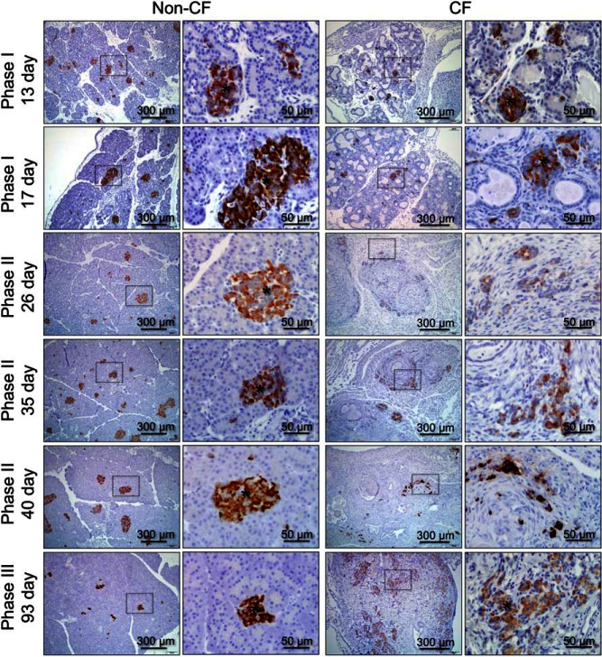
Figure 2.
Age-dependent changes in islet structure and insulin protein expression in the pancreas. Pancreatic paraffin sections of non-CF (left) and CF (right) animals were immunostained for insulin and counterstained with hematoxylin. Ages of the animals evaluated are given on the left. The right column of photomicrographs for each genotype is an enlargement of the boxes region in the left set of photomicrographs. Supplemental Figure 1 shows H&E-stained sections for the same age groups. Asterisks mark discernable islets.

