Abstract
Detection and intervention at an early stage is a critical factor to impede arthritis progress. Here we present a non-invasive method to detect inflammatory changes in joints of arthritic mice. Inflammation was monitored by dual fluorescence optical imaging for near-infrared fluorescent (750F) matrix-metalloproteinase activatable agent and allophycocyanin-conjugated anti-mouse CD11b. Increased intensity of allophycocyanin (indication of macrophage accumulation) and 750F (indication of matrix-metalloproteinase activity) showed a biological relationship with the arthritis severity score and the histopathology score of arthritic joints. Our results demonstrate that this method can be used to detect early stages of arthritis with minimum intervention in small animal models.
OCIS codes: (170.0170) Medical optics and biotechnology, (170.2655) Functional monitoring and imaging
1. Introduction
Arthritis is a chronic progressive inflammatory disease of joints with a substantial morbidity in approximately 22.7% of the US adult population [1]. Arthritis not only results in damage to joints but also increases the risk of systemic complications [2]. Early diagnosis and treatment can delay development or progression of painful joint erosions, and affect outcomes of the disease [3,4]. Currently, clinical diagnosis of arthritis is in very late stages of the disease, and only 20% of patients can be diagnosed for arthritis clinically [5]. To date, various techniques are available for the detection of arthritis, ranging from enzymatic assays of blood and joint fluid to radiographic analysis using X-rays, magnetic resonance imaging (MRI) and positron emission tomography (PET). Apart from being time-consuming and expensive, these techniques have limitations in detecting the early stages of joint destruction. Thus, there is a need to develop new techniques for non-invasive and sensitive imaging of arthritic joints in early phases of arthritis, as early intervention has been shown to prevent joint disability and damage [6]. Localization, progression and therapeutic intervention have been successfully monitored in preclinical research by using a non-invasive in vivo optical fluorescence imaging technique [7,8]. In addition, techniques such as enzyme-activatable fluorescent probes and fluorochrome-conjugated target-specific antibody probes can be utilized for in vivo imaging [9,10]. Previously, we developed a method for early diagnosis of arthritis in vivo and serial measurement of total disease in individual joints in a small animal model using fluorescent-labeled antibodies that detect type II collagen (which is the major collagen in articular cartilage) and an imaging system [8]. This technique can be easily transferred to other target materials such as specific types of cells, antibodies, peptides, proteins, or enzymes. Furthermore, recently we described a dual fluorescence imaging technique to detect joint destruction in a mouse model of collagen-induced arthritis [7].
The pathogenesis of arthritis involves infiltration and activation of various populations of immune cells along with the release of inflammatory cytokines and mediators into the synovium. Inflammatory cytokines, including IL-1β and TNFα, induce the production of matrix metalloproteinases (MMPs), enzymes that degrade extracellular matrix components of the cells of synovium and chondrocytes [11]. In addition to secreting multiple potent inflammatory mediators, activated macrophages are known to participate in antigen presentation, and contribute in the activation and proliferation of antigen-specific T-cells and their consequent destructive activities [12–14]. Thus, the biological activity of macrophages greatly contributes to both acute and chronic stages of arthritis [14]. The number and activity level of macrophages in the synovial lining and sub-lining area directly correlate with disease activity, including joint inflammation, pain and bone erosion [15–17]. Therefore, a sensitive technique that can monitor accumulation of macrophages and/or enzyme activity in joints would be a useful tool for early detection of arthritis. In the present study, we investigate whether infiltration of inflammatory immune cells and enzyme activity at the site of inflammation (arthritic joints) can be simultaneously monitored in vivo by a small animal imaging system and whether fluorescence intensity in the joints is related to arthritis severity using the interleukin-1 receptor antagonist-deficient (IL-1rn−/−) mouse, which spontaneously develops arthritis [18].
2. Methods and materials
2.1 Animals
IL-1rn−/− mice (arthritic mice) and IL-1rn−/−/MyD88−/− mice (non-arthritic mice) on a BALB/c background were bred at the VA Medical Center at Memphis. All experimental animals were age- and gender-matched. All animal care and housing requirements set forth by the National Institutes of Health Committee on Care and Use of Laboratory Animals of the Institute of Laboratory Animal Resources were followed, and animal protocols were reviewed and approved by the Institutional Animal Care and Use Committee at the VA Medical Center in Memphis.
2.2 Dual fluorescent imaging and quantification
Mice were injected retro-orbitally with 100 μl of a solution containing a mixture of MMPSense 750 FAST Fluorescent Imaging Agent (750F) (Perkin Elmer, Hopkinton, MA) and APC-conjugated monoclonal anti-mouse CD11b antibody (αCD11b-APC, eBioscience, San Diego, CA). Twenty four (24) hours later, mice were anesthetized and then scanned using an in vivo imaging system (IVIS Lumina XR System, Perkin Elmer, Hopkinton, MA) with a high range filter set. The excitation/emission wavelengths used for 750F and αCD11b-APC were 745 nm/800 nm and 640 nm/720 nm, respectively. Living Image 4.0 software was used to calculate the flux radiating omni-directionally from the region of interest (ROI) in each joint. ROI was defined as the area where the foot and leg meet. This area consists of the following three joints: true ankle joint, subtalar joint, and inferior tibiofibular joint. Fluorescence intensities were quantified within ROI by using Living Image 4.0 software. Each sample was quantified thrice and the average was taken. The average of each sample per group represented the mean value of each group. Calculations are represented graphically as radiant efficiency (photons/s/cm2/str)/(μW/cm2). Standardized ROI of the joint fluorescence was measured by capturing the same area for each mouse. Background fluorescence was removed by subtracting the fluorescence of the null or background capture area (consisting of muscle and skin tissue) from each articular reading.
2.3 Macroscopic arthritis score and histology
Macroscopic examination was done based on a scoring system that ranges from 0 to 4 as follows: 0 = No sign of arthritis; 1 = Swelling/redness observed in 1-2 interphalangeal (IP) joints; 2 = Additional signs of arthritis in one larger joint or 3-4 IP joints; 3 = More than 4 joints have swelling/redness; 4 = Severe arthritis in the whole paw. The maximum arthritis severity score for each paw is 4 and for each mouse is 16. To perform histological evaluation mouse legs were fixed in 10% (w/v) neutral-buffered formalin solution (Sigma, St. Louis, MO) and decalcified with decalcifying solution (Thermo Fisher Scientific, Waltham, MA) for 48 hr followed by embedding in paraffin. Twenty (n = 20) serial sections from each knee joint (~5-7 μm) were analyzed. The sections were stained with hematoxylin and eosin (H&E) and Safranin-O/Fast Green staining (S&F) [8]. The sections were scored by two (2) independent investigators blinded to the experimental groups. The maximum H&E score for each paw is 12 and thus for each mouse is 48. Each paw was scored as follows: Inflammatory reactions in the synovial tissue (enlargement of the lining layers and cellular density of the synovial stroma), 0–3 points; pannus formation, 0–3; cartilage damage 0–3 points; subchondral bone erosion, 0–3 points [17,19].
2.4 Statistical analysis
All experiments were performed independently in triplicate. Student’s t-test was performed to determine statistical significance. A P value of ≤ 0.05 was considered statistically significant.
3. Results
3.1 Detection of APC and 750F by IVIS
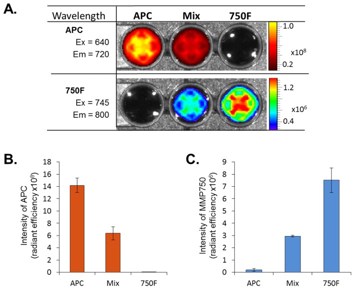
We selected two fluorophores, APC and 750F, to be conjugated to antibodies specific for the selected cell surface marker and enzyme substrate. To evaluate if the fluorescence signals from APC and 750F interfere with each other, the two dyes were measured by IVIS scan with different filter sets in vitro. As demonstrated in Fig. 1, APC was only detected at excitation wavelength of 640 nm and emission wavelength of 720 nm. On the other hand, 750F was only detected at excitation wavelength of 745 nm and emission wavelength of 800 nm. The signal also decreased according to the reduced amount of fluorescence for both dyes. Further, no overlapping signal was confirmed by mixing the two dyes in equal proportion and measuring with IVIS scan at the respective wavelengths.
Fig. 1.
In vitro detection of APC and 750F. (A) Representative image showing detection of APC and 750F. Left: Solution containing APC only; Middle: Solution containing mixture of APC and 750F in equal proportions; Right: Solution containing 750F only. (B) Graphical representation of APC and 750F. Data are expressed as mean ± SD.
3.2 Comparison of dual fluorescence to clinical assessment
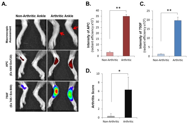
Number and biological activity level of macrophages in the synovial lining and sub-lining area directly correlate with disease activity of arthritis, including joint inflammation, articular pain, and bone erosion [15–17]. Based on this, we hypothesized that levels of the infiltrated macrophages and local activity of destructive enzymes, such as MMPs, produced by infiltrated leukocytes and inflamed synovial cells in joint cavities might be related to disease severity. Thus, simultaneous detection of infiltrated macrophages and MMP enzyme activity in joints can be a sensitive way to detect arthritis. It is known that between the ages of 5-12 weeks, IL-1rn−/− mice in BALB/c background spontaneously develop an autoimmune T cell-mediated arthritis that exhibits several characteristics of rheumatoid arthritis due to excessive IL-1 signaling [18,19]. We have observed that deletion of MyD88, the major signaling adaptor molecule in the IL-1R1 signaling pathway, completely suppresses development of arthritis in IL-1rn−/− mice (unpublished data). We performed clinical evaluation of arthritis in MyD88-sufficient IL-1rn−/−(MyD88+/+/IL-1rn−/−, develop arthritis spontaneously) mice and MyD88-deficient IL-1rn−/− (MyD88−/−/IL-1rn−/−, do not develop arthritis, used as a control strain) twice a week for the designated time period. We selected mice with different clinical arthritis severity as determined by microscopic examination and evaluated accumulation of CD11b+ cells and MMP activity in the joints of these mice by administration of αCD11b-APC and 750F followed by in vivo dual fluorescence imaging using IVIS. As shown in Fig. 2(A), very low levels of fluorescent signal was detected in joints of non-arthritic mice injected with a combination of αCD11b-APC and 750F. In contrast, fluorescent signals for both dyes were drastically increased in the joints of arthritic mice, indicating increased accumulation of CD11b+ cells and MMP enzyme activity in arthritic joints. The levels of inflammatory activities (CD11b+ cell accumulation and increased MMP activity) in joints detected by imaging and quantification of fluorescent signals showed a relationship with the clinical disease severity score determined by conventional macroscopic examination of joints (Fig. 2 and data not shown). These results demonstrate that CD11b+ cell accumulation, MMP activity, and hence clinical arthritis severity score may be related. Taken together, our results demonstrated that leukocyte infiltration and MMP activation in the inflamed joints can be quantified and visualized, using a non-invasive dual fluorescence in vivo imaging system. Our results also suggest that simultaneous detection of leukocyte infiltration and MMP activity using this imaging system can be an efficient and sensitive way to monitor the inflammatory status of joints and a useful clinical tool to detect arthritis.
Fig. 2.
Assessment of ankle joints of arthritic MyD88+/+/IL-1rn−/− mice and non-arthritic MyD88−/−/IL-1rn−/− mice. (A) Macroscopic analysis and IVIS images. Macroscopic score approximately equal to 3 is obvious in arthritic ankle (red arrow) (B) Intensity of APC signal and (C) 750F signal in ankle joints. (D) Arthritis score of ankle joints. Data are expressed as mean ± SD. (*p < 0.05; **p < 0.01)
3.3. Comparison of dual fluorescence to joint histopathology
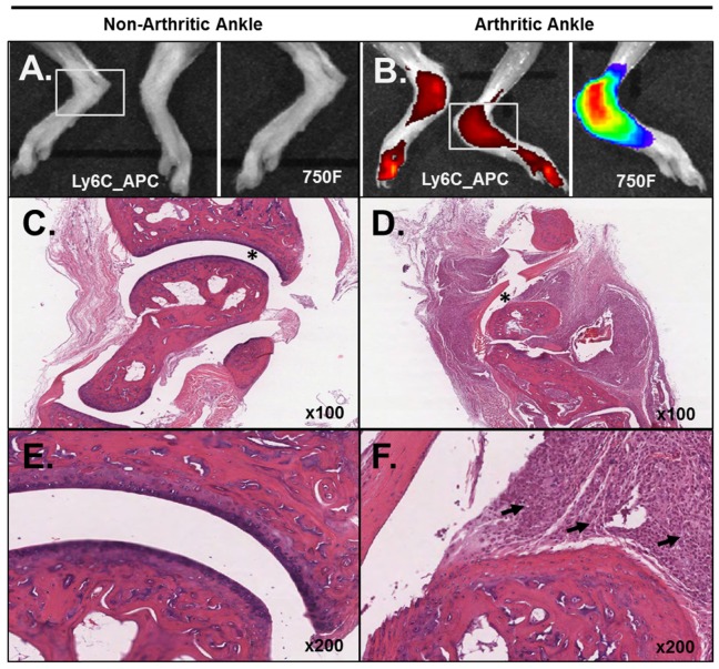
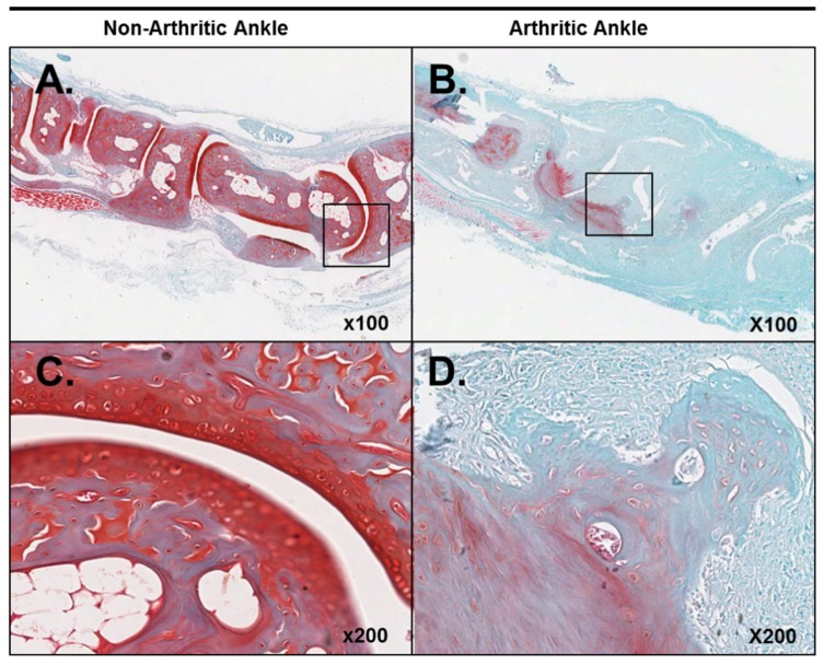
We further investigated whether an increase in signal for CD11b+ cell accumulation and MMP activity in joints also reflects histopathologic changes of the joints. Histopathologic features of arthritic joints are marked elevation of inflammatory cell infiltration in a joint cavity, bone and cartilage erosion, pannus formation, and fibrin deposition [20]. H&E staining was performed to determine inflammatory cell influx and chondrocyte death. S&F staining was performed to assess proteoglycan depletion and cartilage and bone destruction. The joints that showed strong fluorescent signals for αCD11b-APC and 750F also vividly showed massive infiltration of inflammatory cells, severe pannus formation, cartilage destruction, fibrillation, delamination, bone erosion and proteoglycan loss (Figs. 3(B), 3(D), 3(F), 4(B) and 4(D)).
Fig. 3.
Relationship between IVIS and histopathology. (A, B) IVIS images of non-arthritic and arthritic ankle joints. (C, D) H&E section showing loss of integrity of structure in arthritic joint (*). (E, F) Comparison between infiltrations of leukocytes in arthritic joint as compared with non-arthritic joint. Black arrows indicate infiltrating leukocytes.
Fig. 4.
Proteoglycan content of ankle joints. (A, B) Loss of proteoglycans is apparent in arthritic joint along with the loss of joint integrity. (C, D) Magnified representation of selected regions shown in images A and B.
In contrast, no histopathologic alteration of the joint was observed in non-arthritic joints that showed no detectable fluorescent signals for αCD11b-APC and 750F (Figs. 3(A), 3(C), 3(E), 4(A) and 4(C)).
Histopathological scoring also showed significant differences between non-arthritic and arthritic ankles (Table 1).
Table 1. Histopathology scores of non-arthritic and arthritic joints.
| Animal | Inflammation | Pannus | Cartilage Damage | Bone Damage | Total |
|---|---|---|---|---|---|
| Non- Arthritic Ankles | 0.1 ± 0.32 | 0.1 ± 0.32 | 0.4 ± 0.52 | 0 ± 0.00 | 0.6 ± 0.70 |
| Arthritic Ankles | 2.6 ± 0.70 | 2.3 ± 0.67 | 2.7 ± 0.48 | 2.1 ± 0.57 | 9.7 ± 2.06 |
| p | <0.01 | <0.01 | <0.01 | <0.01 | <0.01 |
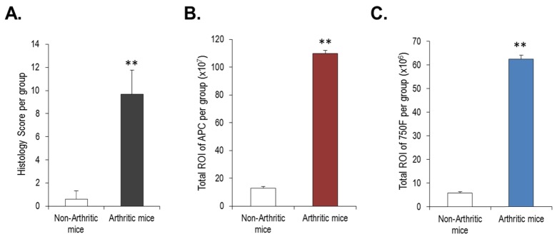
Arthritic ankles showed severe signs of inflammation, pannus formation and damage to cartilage and bone. Furthermore, we found that as the histology score increases so does the ROI of APC in arthritic ankles (Table 1 and Fig. 5). There was highly significant difference between non-arthritic and arthritic ankles (Fig. 5(A)). Total ROI, calculated as the sum of fluorescent signals for APC and 750F per group, also showed a highly significant difference between non-arthritic and arthritic ankles (Fig. 5(B) and 5(C)). Thus, both dual fluorescence imaging and histopathological analysis confirmed inflammatory changes in arthritic joints distinctively from non-arthritic joints.
Fig. 5.
Relationship between histological score and ROI. (A) Comparison of histology score between non-arthritic and arthritic mice. (B) Sum of APC signal from non-arthritic and arthritic mice (C) Sum of 750F signal from non-arthritic and arthritic mice. Total ROI represents fluorescence intensities of APC and 750 per group. Data are expressed as mean ± SD. (**p < 0.01).
4. Discussion and conclusions
In recent times fluorophores have been widely used in pre-clinical studies owing to their specificity [21]. This is an important parameter in multiplexed imaging where different fluorophores are detected simultaneously. In our study we used APC and a near-infrared emitting dye, 750F, with non-overlapping excitation and emission properties, to detect two different indicators of tissue inflammation simultaneously in vivo. We showed that a combination of αCD11b-APC and MMP750 can be used to monitor infiltration of immune cells and MMP activity in inflamed arthritic joints in the same animal. We used MMPSense 750 instead of MMPSense 680 in order to prevent overlapping signal with APC, which has an excitation range of 680 nm. Further, before administration to animals we confirmed in vitro through IVIS that these two fluorochromes do not interfere each other. It was found that both dyes can be detected at their respective wavelengths with no overlapping signal. Our results also verify that these two fluorochromes can be utilized to detect two different properties both in vitro and in vivo.
Arthritis is a complex musculoskeletal disorder characterized by inflammation that damages the joint architecture, including cartilage, bone and connective tissues. Furthermore, arthritis involves many catabolic cascades. For instance, the inflammatory response has several phases such as inflammatory inducers (infection or tissue damage), inflammatory sensors (immune cells such as monocytes and macrophages), and inflammatory mediators (cytokines, chemokines). All of these factor play roles in disease progression. Increases in leukocyte infiltration, proinflammatory mediators and proteolytic enzymes in the joint synovium contribute to the painful and progressive destruction of the joints. Based on characteristics of arthritis, we exploited αCD11b antibody conjugated with APC and 750F, as in vivo fluorescence imaging agents, to detect infiltrated leukocytes and enzyme activity of MMPs, respectively, as indicators of active joint inflammation in an arthritic mouse model.
Inflammation in joints generally occurs when natural mediators are replaced by pro-inflammatory mediators that recruit lymphocytes to the synovium. CD11b is expressed on the surface of various leukocytes, including monocytes, macrophages, granulocytes, and natural killer cells, and used to identify macrophages [22]. Our histology results showed significant number of infiltrated leukocytes in arthritic joints as compared to non-arthritic joints. It is evident that the αCD11b-APC fluorescence intensity in the affected joints of arthritic mice was substantially higher as compared to joint regions of the non-arthritic mouse group. Fluorescent imaging of murine ankle sagittal sections clearly demonstrated the localization of a higher concentration of αCD11b-APC within the inflamed joints, while the soft tissues showed relatively lower fluorescent intensity. Anti-CD11b antibodies detect infiltration of monocytes and macrophages in the inflamed joint. Thus αCD11b-APC can be used as a probe for non-invasive early detection of arthritis.
MMP activity is involved in the pathogenesis of many diseases, including cancer, pulmonary diseases, cardiovascular diseases, and rheumatoid arthritis. Thus, detection of MMP activity can be used to monitor the progression of these disease activities. MMP-activatable fluorescent probes, such as MMPSense (750F), emits near infra-red fluorescence upon cleavage by a broad range of MMPs, including MMP-2, 3, 7, 9, 12, and 13 [20,23]. A study using a mouse model of arthritis showed that increased fluorescent intensity of MMPSense (750F) in joints is due to the local activation of proteases in the inflamed synovium [20]. The fluorescent activity steadily increases as joint damage progresses, beginning in the early stages of arthritis. It could be said that MMP-mediated monitoring of early arthritis may enable early monitoring of destructive inflammatory events. This is also supported by our results that showed drastic increase in the fluorescent intensity of 750F in arthritic joints of IL-1rn−/− mice.
We observed that both αCD11b-APC and 750F were detected in the same ROI present in the hind paw. Therefore, we investigated further to reveal the histological parameters of the arthritic joints. Histological analysis showed infiltration of immune cells in the arthritic joints as compared to non-arthritic joints. This confirmed the inflammatory signal yielded earlier by fluorescence imaging. Furthermore, there was increased cartilage damage in the arthritic joints as revealed by the loss of proteoglycans, massive fibrillation and lamination.
Further, we compared the ROI with the histology score. We found that the ROI of APC and 750F increased with an increase in the histological score. This observation clearly shows that our dual fluorescence imaging technique can clearly detect arthritis-related changes in joints as compared with histology. Inflammation in the joints of IL-1rn−/− mice resulted in cartilage damage and hence arthritis. We emphasize on the basis of histopathology that our dual fluorescence methodology was able to detect the inflammation of the arthritic joints with minimum intervention in the living animal. Further advantages of this non-invasive dual fluorescence in vivo imaging are detection of active inflammation in areas that are difficult to monitor, such as knee and hip joints, and detection of types of infiltrating cells in vivo by using fluorochrome-conjugated specific cell surface markers. This is important if animals have to be kept long-term to study arthritis. This technique will allow analysis of the arthritic joints at different time points during the course of the study.
In summary, we present here a non-invasive technique that can be used to detect early stages of inflammation in arthritis. We were able to detect MMP activity and infiltrated CD11b+ leukocytes simultaneously in the same joint by using two dyes at their respective filter settings. We also presented the biological relationship between fluorescence intensity and the degree of disease manifestation. This is a reliable, inexpensive, time-effective and non-invasive method for detecting early stages of arthritis. This method can be used to monitor the progress of arthritis and is a useful tool for studying the mechanism of inflammatory cascades in arthritis in vivo.
Acknowledgments
H.C. was supported by grants from William and Ella Owens Medical Research Foundation. K.A.H was supported by grants from the Department of Veterans Affairs (VA Merit Review award), NIH (AR060408) and the UTHSC (CTSI award). J.M.S. was supported by grants from the Department of Veterans Affairs (Program Project Grant). A.K.Y. was supported by grants from NIH/NIAMS (AR064723) and the Arthritis Foundation (IRG 5942). The contents of this publication are solely the responsibility of the authors and do not necessarily represent the official views of the NIH, NIAMS, Arthritis Foundation, William and Ella Owens Medical Research Foundation, Department of Veterans Affairs, or University of Tennessee Health Science Center. We thank Drs. S. Akira (Osaka Univ., Osaka, Japan) and Yoichiro Iwakura (The University of Tokyo, Tokyo, Japan) for kindly providing MyD88−/− mice and IL-1rn−/− mice, respectively. We thank Dr. Anand Kulkarni (Director, Tissue Services Core, UTHSC, Memphis) for scanning histology slides using Scanscope®XT and Mrs. Andrea Patters for her excellent assistance with preparation of the manuscript.
References and links
- 1.Centers for Disease Control and Prevention (CDC) , “Prevalence of doctor-diagnosed arthritis and arthritis-attributable activity limitation--United States, 2010-2012,” MMWR Morb. Mortal. Wkly. Rep. 62(44), 869–873 (2013). [PMC free article] [PubMed] [Google Scholar]
- 2.Picerno V., Ferro F., Adinolfi A., Valentini E., Tani C., Alunno A., “One year in review: the pathogenesis of rheumatoid arthritis,” Clin. Exp. Rheumatol. 33(4), 551–558 (2015). [PubMed] [Google Scholar]
- 3.Finckh A., Liang M. H., van Herckenrode C. M., de Pablo P., “Long-term impact of early treatment on radiographic progression in rheumatoid arthritis: A meta-analysis,” Arthritis Rheum. 55(6), 864–872 (2006). 10.1002/art.22353 [DOI] [PubMed] [Google Scholar]
- 4.Goekoop-Ruiterman Y. P., de Vries-Bouwstra J. K., Allaart C. F., van Zeben D., Kerstens P. J., Hazes J. M., Zwinderman A. H., Peeters A. J., de Jonge-Bok J. M., Mallée C., de Beus W. M., de Sonnaville P. B., Ewals J. A., Breedveld F. C., Dijkmans B. A., “Comparison of treatment strategies in early rheumatoid arthritis: a randomized trial,” Ann. Intern. Med. 146(6), 406–415 (2007). 10.7326/0003-4819-146-6-200703200-00005 [DOI] [PubMed] [Google Scholar]
- 5.Lawrence R. C., Helmick C. G., Arnett F. C., Deyo R. A., Felson D. T., Giannini E. H., Heyse S. P., Hirsch R., Hochberg M. C., Hunder G. G., Liang M. H., Pillemer S. R., Steen V. D., Wolfe F., “Estimates of the prevalence of arthritis and selected musculoskeletal disorders in the United States,” Arthritis Rheum. 41(5), 778–799 (1998). [DOI] [PubMed] [Google Scholar]
- 6.Quinn M. A., Conaghan P. G., Emery P., “The therapeutic approach of early intervention for rheumatoid arthritis: what is the evidence?” Rheumatology (Oxford) 40(11), 1211–1220 (2001). 10.1093/rheumatology/40.11.1211 [DOI] [PubMed] [Google Scholar]
- 7.Cho H., Bhatti F., Lee S., Brand D., Yi A., Hasty K. A., “In vivo dual fluorescence imaging to detect joint destruction,” Artif. Organs (2015). [DOI] [PMC free article] [PubMed]
- 8.Cho H., Pinkhassik E., David V., Stuart J. M., Hasty K. A., “Detection of early cartilage damage using targeted nanosomes in a post-traumatic osteoarthritis mouse model,” Nanomedicine (Lond.) 11(4), 939–946 (2015). [DOI] [PubMed] [Google Scholar]
- 9.Chan M. M., Gray B. D., Pak K. Y., Fong D., “Non-invasive in vivo imaging of arthritis in a collagen-induced murine model with phosphatidylserine-binding near-infrared (NIR) dye,” Arthritis Res. Ther. 17(1), 50 (2015). 10.1186/s13075-015-0565-x [DOI] [PMC free article] [PubMed] [Google Scholar]
- 10.Bender A. T., Spyvee M., Satoh T., Gershman B., Teceno T., Burgess L., Kumar V., Wu Y., Yang H., Ding Y., Akare S., Chen Q., “Evaluation of a candidate anti-arthritic drug using the mouse collagen antibody induced arthritis model and clinically relevant biomarkers,” Am. J. Transl. Res. 5(1), 92–102 (2013). [PMC free article] [PubMed] [Google Scholar]
- 11.Burrage P. S., Mix K. S., Brinckerhoff C. E., “Matrix metalloproteinases: role in arthritis,” Front. Biosci. 11(1), 529–543 (2006). 10.2741/1817 [DOI] [PubMed] [Google Scholar]
- 12.Bresnihan B., “Pathogenesis of joint damage in rheumatoid arthritis,” J. Rheumatol. 26(3), 717–719 (1999). [PubMed] [Google Scholar]
- 13.Burmester G. R., Stuhlmüller B., Keyszer G., Kinne R. W., “Mononuclear phagocytes and rheumatoid synovitis. Mastermind or workhorse in arthritis?” Arthritis Rheum. 40(1), 5–18 (1997). 10.1002/art.1780400104 [DOI] [PubMed] [Google Scholar]
- 14.Kinne R. W., Bräuer R., Stuhlmüller B., Palombo-Kinne E., Burmester G. R., “Macrophages in rheumatoid arthritis,” Arthritis Res. 2(3), 189–202 (2000). 10.1186/ar86 [DOI] [PMC free article] [PubMed] [Google Scholar]
- 15.Mulherin D., Fitzgerald O., Bresnihan B., “Synovial tissue macrophage populations and articular damage in rheumatoid arthritis,” Arthritis Rheum. 39(1), 115–124 (1996). 10.1002/art.1780390116 [DOI] [PubMed] [Google Scholar]
- 16.Tak P. P., Smeets T. J., Daha M. R., Kluin P. M., Meijers K. A., Brand R., Meinders A. E., Breedveld F. C., “Analysis of the synovial cell infiltrate in early rheumatoid synovial tissue in relation to local disease activity,” Arthritis Rheum. 40(2), 217–225 (1997). 10.1002/art.1780400206 [DOI] [PubMed] [Google Scholar]
- 17.Yanni G., Whelan A., Feighery C., Bresnihan B., “Synovial tissue macrophages and joint erosion in rheumatoid arthritis,” Ann. Rheum. Dis. 53(1), 39–44 (1994). 10.1136/ard.53.1.39 [DOI] [PMC free article] [PubMed] [Google Scholar]
- 18.Zhou F., He X., Iwakura Y., Horai R., Stuart J. M., “Arthritis in mice that are deficient in interleukin-1 receptor antagonist is dependent on genetic background,” Arthritis Rheum. 52(12), 3731–3738 (2005). 10.1002/art.21481 [DOI] [PubMed] [Google Scholar]
- 19.Horai R., Saijo S., Tanioka H., Nakae S., Sudo K., Okahara A., Ikuse T., Asano M., Iwakura Y., “Development of chronic inflammatory arthropathy resembling rheumatoid arthritis in interleukin 1 receptor antagonist-deficient mice,” J. Exp. Med. 191(2), 313–320 (2000). 10.1084/jem.191.2.313 [DOI] [PMC free article] [PubMed] [Google Scholar]
- 20.Ibarra J. M., Jimenez F., Martinez H. G., Clark K., Ahuja S. S., “MMP-Activated Fluorescence Imaging Detects Early Joint Inflammation in Collagen-Antibody-Induced Arthritis in CC-Chemokine Receptor-2-Null Mice, In-Vivo,” Int. J. Inflamm. 2011, 691587 (2011). 10.4061/2011/691587 [DOI] [PMC free article] [PubMed] [Google Scholar]
- 21.Krohn M., Ohrndorf S., Werner S. G., Schicke B., Burmester G. R., Hamm B., Backhaus M., Hermann K. G., “Near-infrared Fluorescence Optical Imaging in Early Rheumatoid Arthritis: A Comparison to Magnetic Resonance Imaging and Ultrasonography,” J. Rheumatol. 42(7), 1112–1118 (2015). 10.3899/jrheum.141244 [DOI] [PubMed] [Google Scholar]
- 22.Solovjov D. A., Pluskota E., Plow E. F., “Distinct roles for the alpha and beta subunits in the functions of integrin alphaMbeta2,” J. Biol. Chem. 280(2), 1336–1345 (2005). 10.1074/jbc.M406968200 [DOI] [PubMed] [Google Scholar]
- 23.Leahy A. A., Esfahani S. A., Foote A. T., Hui C. K., Rainbow R. S., Nakamura D. S., Tracey B. H., Mahmood U., Zeng L., “Analysis of the trajectory of osteoarthritis development in a mouse model by serial near-infrared fluorescence imaging of matrix metalloproteinase activities,” Arthritis Rheumatol. 67(2), 442–453 (2015). 10.1002/art.38957 [DOI] [PMC free article] [PubMed] [Google Scholar]


