Abstract
Cysteine peptidases play a central role in the biology of Leishmania. In this work, we sought to further elucidate the mechanism(s) by which the cysteine peptidase CPB contributes to L. mexicana virulence and whether CPB participates in the formation of large communal parasitophorous vacuoles induced by these parasites. We initially examined the impact of L. mexicana infection on the trafficking of VAMP3 and VAMP8, two endocytic SNARE proteins associated with phagolysosome biogenesis and function. Using a CPB-deficient mutant, we found that both VAMP3 and VAMP8 were down-modulated in a CPB-dependent manner. We also discovered that expression of the virulence-associated GPI-anchored metalloprotease GP63 was inhibited in the absence of CPB. Expression of GP63 in the CPB-deficient mutant was sufficient to down-modulate VAMP3 and VAMP8. Similarly, episomal expression of GP63 enabled the CPB-deficient mutant to establish infection in macrophages, induce the formation of large communal parasitophorous vacuoles, and cause lesions in mice. These findings implicate CPB in the regulation of GP63 expression and provide evidence that both GP63 and CPB are key virulence factors in L. mexicana.
Author Summary
The parasite Leishmania mexicana expresses several cysteine peptidases of the papain family that are involved in processes such as virulence and evasion of host immune responses. The cysteine peptidase CPB is required for survival within macrophages and for lesion formation in susceptible mice. Upon their internalization by macrophages, parasites of the L. mexicana complex induce the formation of large communal parasitophorous vacuoles in which they replicate, and expansion of those large vacuoles correlates with the ability of the parasites to survive inside macrophages. Here, we found that CPB contributes to L. mexicana virulence (macrophage survival, formation and expansion of communal parasitophorous vacuoles, lesion formation in mice) through the regulation of the virulence factor GP63, a Leishmania zinc-metalloprotease that acts by cleaving key host cell proteins. This work thus elucidates a novel Leishmania virulence regulatory mechanism whereby CPB controls the expression of GP63.
Introduction
The protozoan Leishmania parasitizes macrophages and causes a spectrum of human diseases ranging from self-healing cutaneous lesions to a progressive visceral infection that can be fatal if left untreated. Infection is initiated when promastigote forms of the parasite are inoculated into the mammalian host by infected sand flies and are internalized by phagocytes. Inside macrophages, promastigotes differentiate into amastigotes to replicate within phagolysosomal compartments also known as parasitophorous vacuoles (PVs). Upon their internalization, L. donovani and L. major promastigotes arrest phagolysosomal biogenesis and create an intracellular niche favorable to the establishment of infection and to the evasion of the immune system [1, 2]. Disruption of the macrophage membrane fusion machinery through the action of virulence factors plays an critical role in this PV remodeling. Hence, insertion of the promastigote surface glycolipid lipophosphoglycan (LPG) into the PV membrane destabilizes lipid microdomains and causes exclusion of the membrane fusion regulator synaptotagmin V from the PV [2–4]. Similarly, the parasite GPI-anchored metalloprotease GP63 [5, 6] redistributes within the infected cells and cleaves key Soluble NSF Attachment Protein Receptors (SNAREs) and synaptotagmins to impair phagosome functions [1, 7].
Whereas L. major and L. donovani multiply in tight individual PVs, parasites of the L. mexicana complex (L. mexicana, L. amazonensis) replicate within large communal PVs. Relatively little is known about the host and parasite factors involved in the biogenesis and expansion of those communal PVs. Studies with L. amazonensis revealed that phagosomes containing promastigotes fuse extensively with late endosomes/lysosomes within 30 minutes post-infection [8]. At that stage, parasites are located within small individual compartments and by 18 to 24 hours large PVs containing several parasites are observed. The rapid increase in the size of those PVs requires extensive fusion with secondary lysosomes and correlates with the depletion of those organelles in infected cells [9–11]. Homotypic fusion between L. amazonensis-containing PVs also occurs, but its contribution to PV enlargement remains to be further investigated [12]. These studies highlighted the contribution of the host cell membrane fusion machinery in the biogenesis and expansion of large communal PVs and are consistent with a role for endocytic SNAREs in this process [13]. Interestingly, communal PVs interact with the host cell’s endoplasmic reticulum (ER) and disruption of the fusion machinery associated with the ER and Golgi inhibits parasite replication and PV enlargement [14–16].
The Leishmania-derived molecules involved in the expansion of the communal PVs remains to be identified. LPG and other phosphoglycans do not play a significant role in L. mexicana promastigote virulence and PV formation [17], in contrast to L. major and L. donovani [2]. Cysteine peptidases (CP) are a large family of papain-like enzymes that play important roles in the biology of Leishmania [18]. Three members of these papain-like proteases are expressed by L. mexicana and the generation of CP-deficient mutants revealed that CPB contributes to the ability to infect macrophages and to induce lesions in BALB/c mice [19–21]. The precise mechanism(s) by which CPB participates in the virulence of L. mexicana is poorly understood. Previous studies revealed that CPB traffics within and outside infected macrophages [18]. In infected macrophages, CPB alters signal transduction and gene expression through the activation of the protein tyrosine phosphatase PTP-1B and the cleavage of transcription factors responsible for the expression of genes involved in host defense and immunity [20, 22]. The observation that CPs interfere with the host immune response through the degradation of MHC class II molecules and invariant chains present in PVs housing L. amazonensis [23], raises the possibility that CPB participates in the modulation of PV composition and function.
In this study, we sought to gain insight into the mechanism by which CPB contributes to L. mexicana virulence, with a focus on the PV. We provide evidence that CPB participates in PV biogenesis and virulence through the regulation of GP63 expression.
Results
CPB enables L. mexicana to down-modulate VAMP3 and VAMP8
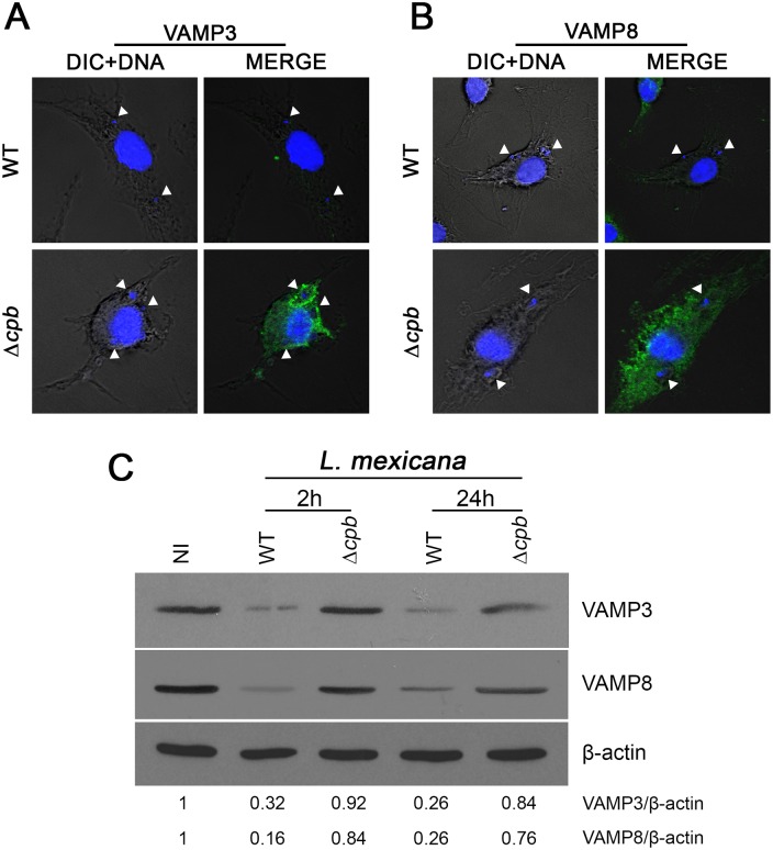
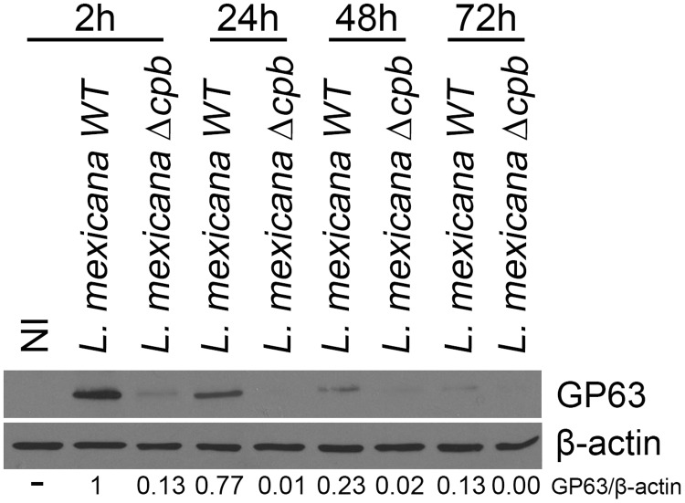
Formation and expansion of communal PVs hosting L. mexicana involve fusion between PVs and endocytic organelles, as well as homotypic fusion among PVs [10–12]. To identify the host and parasite factors involved in this process, we embarked on a study to elucidate the fate of endosomal SNAREs during infection of macrophages with L. mexicana. Given the requirement of CPB for L. mexicana to replicate normally inside macrophages [19], we included a L. mexicana CPB-deficient mutant (Δcpb) in our investigation. We infected BMM with either WT or Δcpb L. mexicana promastigotes for 2 h and we assessed the distribution of the endosomal SNAREs VAMP3 and VAMP8 by confocal immunofluorescence microscopy. As previously observed during infection with L. major promastigotes [1], we found a notable reduction in the staining intensity for both VAMP3 (Fig 1A) and VAMP8 (Fig 1B) in BMM infected with WT L. mexicana, but this was not observed with Δcpb. This reduction in staining intensity correlated with a down-modulation of VAMP3 and VAMP8 proteins in BMM infected with WT L. mexicana, compared to cells infected with Δcpb (Fig 1C). These results suggested that L. mexicana causes the reduction of VAMP3 and VAMP8 levels in infected BMM through the action of CPB. However, we considered the possibility that CPB acted indirectly on VAMP3 and VAMP8 because we previously found that GP63 targets those SNAREs in L. major-infected BMM [1]. We therefore ensured that similar levels of GP63 were present in lysates of BMM infected with WT and Δcpb L. mexicana promastigotes. As shown in Fig 2, GP63 was detected in lysates of BMM infected with WT L. mexicana up to 72 h post-infection, when the parasites replicate as amastigotes. The important reduction in GP63 levels at this time point is consistent with previously published data showing a 90% reduction in the amount of GP63 detected in amastigotes with respect to promastigotes [24, 25]. Surprisingly, we found that GP63 was barely detectable in BMM infected with Δcpb at all time points tested. This observation raised the possibility that the lack of VAMP3 and VAMP8 down-regulation in Δcpb-infected BMM was due to defective expression of GP63.
Fig 1. Down-modulation of VAMP3 and VAMP8 by L. mexicana.
BMM were infected with serum-opsonized stationary phase L. mexicana (WT and Δcpb) promastigotes for 2 h. VAMP3 (A) and VAMP8 (B) levels (green) were then visualized by confocal microscopy. Macrophage and parasite nuclei are shown in blue (DAPI). Internalized parasites are denoted by white arrowheads. In (C), VAMP3 and VAMP8 levels in total cell extracts were assessed by Western blot analysis. Each immunofluorescence assay was done on 300 phagosomes on triplicate coverslips in two independent experiments and Western blot analyses were performed twice in three independent experiments. VAMP3 and VAMP8/β-actin ratios were determined by densitometry. Original magnification X63.
Fig 2. GP63 is down-modulated in the L. mexicana Δcpb mutant.
BMM were infected with serum-opsonized stationary phase L. mexicana (WT and Δcpb) promastigotes for 2 h, 24 h, 48 h and 72 h. Total cell extracts were assayed for GP63 levels by Western blot analysis. GP63/ β-actin ratios were determined by densitometry. Similar results were obtained in three independent experiments.
CPB is required for GP63 expression
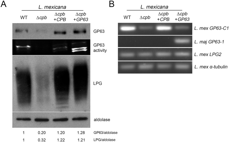
To address the issue of GP63 down-regulation in L. mexicana Δcpb, we first determined whether complementation of Δcpb with the CPB gene array (Δcpb+CPB) restores wild type GP63 levels. As shown in Fig 3A, GP63 levels and activity are down-modulated in the Δcpb mutant, and complementation with the CPB array restored GP63 levels and activity similar to those observed in WT parasites. It was previously reported that expression of the cell surface glycolipid LPG and of GP63 may share common biosynthetic steps [26–29]. We therefore evaluated the levels of LPG in lysates of WT, Δcpb, Δcpb+CPB, and Δcpb+GP63 parasites by Western blot analysis. Strikingly, similar to GP63, LPG levels were also down-modulated in the Δcpb mutant and complementation with either the CPB array or GP63 restored wild type LPG levels. To further investigate the possible role of CPB in the regulation of GP63 expression, we determined the levels of GP63 mRNA in WT, Δcpb, Δcpb+CPB, and Δcpb+GP63 parasites by RT-PCR. As shown in Fig 3B, GP63 mRNA levels were highly down-regulated in Δcpb and complementation with the CPB array restored wild type levels of GP63 mRNA. Interestingly, complementation of Δcpb with L. major GP63 did not increase endogenous GP63 mRNAs. RT-PCR using L. major GP63-specific primers showed that this gene is expressed only in Δcpb+GP63. Together, these results suggest that CPB controls GP63 mRNA levels at the post-transcriptional level. Clearly, additional studies will be required to elucidate the underlying mechanism(s). Our results also raised the possibility that down-modulation of GP63 in the Δcpb mutant may have accounted for the inability of Δcpb to down-regulate VAMP3 and VAMP8. The finding that expression of GP63 in Δcpb restored LPG levels was unexpected and suggested a role for GP63 in the expression of LPG in L. mexicana. As it is estimated that at least 25 genes are required for the synthesis, assembly, and transport of the various components of LPG [30], it may be difficult to determine whether GP63 acts on the expression of a LPG biosynthetic gene or on a biosynthetic step. Assessment of LPG2 gene expression revealed that it was equally expressed WT, Δcpb, Δcpb+CPB, and Δcpb+GP63 parasites. Further studies will be necessary to understand how GP63 expression restores LPG synthesis in Δcpb. Since LPG does not play a major role in the virulence of L. mexicana [17], the Δcpb mutant expressing exogenous GP63 provides a unique opportunity to address the impact of GP63 on SNARE cleavage, as well as on the in vitro and in vivo virulence of L. mexicana.
Fig 3. Expression of GP63 and LPG is impaired in the absence of CPB.
(A) Stationary phase promastigotes were lysed and total cell extracts were analysed by Western blotting and zymography for GP63 levels and activity and for LPG levels. Aldolase was used as a loading control. GP63 and LPG/aldolase ratios were determined by densitometry. (B) Promastigote total RNA was extracted and reverse transcription followed by PCR was performed to assess mRNA levels for L. mexicana GP63-C1, LPG2, and α-tubulin, and L. major GP63-1. Similar results were obtained in three independent experiments.
GP63 is responsible for the cleavage of VAMP3 and VAMP8 by L. mexicana
We next assessed the impact of GP63 on VAMP3 and VAMP8 during L. mexicana infection. To this end, we infected BMM with either WT, Δcpb, Δcpb+CPB, or Δcpb+GP63 L. mexicana promastigotes for various time points, and we assessed VAMP3 and VAMP8 levels and intracellular distribution. Fig 4A shows that GP63 is present at high levels in lysates of BMM infected for 2 h with WT, Δcpb+CPB, and Δcpb+GP63 promastigotes (compared to lysates of BMM infected with Δcpb). At 72 h post-infection, GP63 levels are strongly reduced in BMM infected with WT and Δcpb+CPB, whereas they remain elevated in BMM infected with the Δcpb+GP63 (Fig 4A) [25]. The high levels of GP63 present in BMM infected with Δcpb+GP63 for 72 h may be related to the fact that expression of the L. major GP63 gene from the pLEXNeo episomal vector [31] is not under the control of endogenous GP63 3' untranslated regions. Western blot analyses revealed that down-regulation of VAMP3 and VAMP8 correlated with GP63 levels expressed by the parasites. Consistently, the staining intensity of VAMP3 and VAMP8 was reduced in BMM infected with GP63-expressing parasites, as assessed by confocal immunofluorescence microscopy (Fig 4D and 4E). These results suggest that GP63 is responsible for the down-modulation of the endosomal SNAREs VAMP3 and VAMP8 in L. mexicana-infected BMM. Interestingly, we observed recruitment of VAMP3 to PVs containing L. mexicana parasites at later time points, when promastigotes have differentiated into amastigotes, with the exception of Δcpb+GP63 L. mexicana promastigotes (Fig 4B). In contrast, we found that VAMP8 is excluded from L. mexicana-containing PVs both at early and later time points post-infection, independently of GP63 levels, suggesting that additional mechanisms regulate VAMP8 recruitment to L. mexicana PVs.
Fig 4. GP63 is responsible for the down-modulation of VAMP3 and VAMP8 in L. mexicana-infected macrophages.
BMM were infected with serum-opsonized stationary phase L. mexicana (WT, Δcpb, Δcpb+CPB and Δcpb+GP63) promastigotes for 2 h and 72 h. Total cell extracts were analysed by Western blot (A). Similar results were obtained in three independent experiments. VAMP3 and VAMP8 recruitment to the phagosome was visualized by immunofluorescence microscopy and quantified for 300 phagosomes on triplicate coverslips (B and C). Representative pictures from each condition are shown (D and E). Immunofluorescence assays were performed on 300 phagosomes on triplicate coverslips for three independent experiments. *p<0.0001. Original magnification X63.
GP63 expression restores virulence of Δcpb
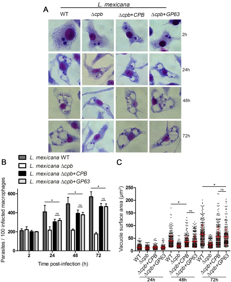
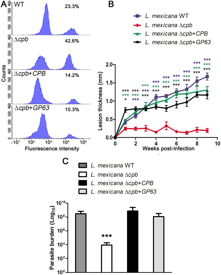
Since GP63 was shown to contribute to L. major virulence [32], we next sought to determine whether expression of GP63 is sufficient to restore the ability of Δcpb to replicate inside macrophages and to cause lesions in mice [19]. To this end, we first infected BMM with either WT, Δcpb, Δcpb+CPB, or Δcpb+GP63 stationary phase promastigotes and we assessed parasite burden and PV surface area at various time points post-infection. We found that Δcpb had an impaired capacity to replicate inside macrophages and to induce the formation of large communal PVs compared to WT and Δcpb+CPB parasites (Fig 5A, 5B and 5C). Strikingly, expression of GP63 in Δcpb restored its ability to replicate in macrophages and to induce large communal PVs up to 72 h post-infection. These results underline the role of GP63 in the ability of L. mexicana to infect and replicate in macrophages, even in the absence of CPB. Following inoculation inside the mammalian host, promastigotes are exposed to complement and both GP63 and LPG confer resistance to complement-mediated lysis [32, 33]. L. mexicana promastigotes were therefore analyzed for their sensitivity to complement-mediated lysis in the presence of fresh human serum. As shown in Fig 6A, over 40% of Δcpb was killed after 30 min in the presence of 20% serum, whereas Δcpb+CPB, and Δcpb+GP63 were more resistant to serum lysis at 14% and 10%, respectively. Absence of both GP63 and LPG may be responsible for the serum sensitivity of Δcpb. Finally, to assess the impact of GP63 on the ability of Δcpb to cause lesions, we used a mouse model of cutaneous leishmaniasis. Susceptible BALB/c mice were infected in the hind footpad with either WT, Δcpb, Δcpb+CPB, or Δcpb+GP63 promastigotes and disease progression was monitored for 9 weeks. Consistent with its reduced capacity to replicate inside macrophages, Δcpb failed to cause significant lesions compared to WT parasites [19] and Δcpb complemented with CPB (Fig 6B). Remarkably, expression of GP63 in Δcpb restored its capacity to cause lesions, albeit to a lower level than Δcpb complemented with CPB. Lesion size correlated with parasite burden, as measured at 9 weeks post-infection (Fig 6C). Collectively, these results indicate that expression of GP63 is sufficient to restore virulence of Δcpb.
Fig 5. GP63 enables L. mexicana Δcpb to infect macrophages and induce large PVs.
BMM were infected with stationary phase serum-opsonized L. mexicana (WT, Δcpb, Δcpb+cpb and Δcpb+GP63) promastigotes for 2 h, 24 h, 48 h and 72 h. Macrophages were stained with the HEMA 3 kit. Representative pictures from each condition are shown (A) Parasites were counted in 300 macrophages on triplicate coverslips (B). Macrophages were stained with the LAMP1 antibody and vacuole sizes were measured with the ZEN 2012 software (C). Parasitemia and vacuole size was determined on 300 phagosomes in triplicate in three independent experiments. *p<0.0001.
Fig 6. GP63 confers virulence to L. mexicana Δcpb.
Stationary phase L. mexicana (WT, Δcpb, Δcpb+cpb and Δcpb+GP63) promastigotes were incubated in the presence of 20% human serum for 30 min, stained with a fixable viability dye, and then subjected to flow cytometry (A). Mice were challenged with 5x10⁶ late-stationary phase L. mexicana (WT, Δcpb, Δcpb+cpb and Δcpb+GP63) promastigotes that were injected subcutaneously into the hind footpad. Disease progression was monitored at weekly intervals, by measuring the thickness of the infected footpad and the contralateral uninfected footpad. (B). Parasite burden was obtained by limiting dilution assay from infected homogenized footpads 9 weeks after inoculation (C). Human serum lyses were performed in two independent experiments and six mice per group were used for the determination of lesion formation and parasite burden. Each data point represents the average ± SEM of 6 mice per group, and statistical significance was denoted by * p≤0.01, and *** p≤0.0001.
Discussion
This study aimed at investigating the mechanism(s) by which CBP contributes to L. mexicana virulence. To this end, we initially examined PV biogenesis by assessing the impact of L. mexicana infection on the trafficking of VAMP3 and VAMP8, two endocytic SNAREs associated with phagosome biogenesis and function [1, 34]. We found that both SNAREs were down-modulated in a CPB-dependent manner, which hampered VAMP3 recruitment to PVs. We also discovered that expression of GP63, which we previously showed to be responsible for cleaving SNAREs in L. major-infected macrophages [1], was down-modulated in the L. mexicana Δcpb. Strikingly, restoration of GP63 expression in Δcpb bypassed the need for CPB for SNARE cleavage. Similarly, episomal expression of GP63 enabled the Δcpb mutant to establish infection in macrophages, induce larger PVs and cause lesions in mice. These findings imply that CPB contributes to L. mexicana virulence in part through the regulation of GP63 expression, and provide evidence that GP63 is a key virulence factor for L. mexicana.
The observation that CPB regulates GP63 expression at the mRNA levels was both unexpected and intriguing. Insight into the possible mechanism(s) may be deduced from a recent study on the role of cathepsin B in L. donovani, which is homologous to the L. mexicana CPC [35]. Similar to L. mexicana Δcpb, L. donovani ΔcatB displays reduced virulence in macrophages. To investigate the role of cathepsin B in virulence, the authors performed quantitative proteome profiling of WT and ΔcatB parasites and identified 83 proteins whose expression is altered in the absence of cathepsin B, with the majority being down-modulated [35]. Among those were a group of proteins involved in post-transcriptional regulation of gene expression (RNA stability, processing, translation) [35]. Whether this is the case in Δcpb deserves further investigation. Clearly, a detailed analysis of wild-type and Δcpb parasites may provide the information required to understand the extent of the impact of CBP on the expression and synthesis of virulence factors and the exact role of CPB in L. mexicana virulence. The observation that episomal expression of GP63 in Δcpb restored LPG synthesis is an intriguing issue, as it suggests that GP63 acts on a LPG biosynthetic step. This role for GP63 is likely redundant, since L. major Δgp63 promastigotes express LPG levels similar to that of wild type parasites (S1 Fig).
It has been proposed that expansion of the PVs hosting parasites of the L. mexicana complex leads to the dilution of the microbicidal effectors to which the parasites are exposed, thereby contributing to parasite survival [36]. Both host and parasite factors may be involved in the control of PV enlargement. On the host side, it has been previously reported that L. amazonensis cannot survive in cells overexpressing LYST, a host gene that restricts Leishmania growth by counteracting PV expansion [37]. Similarly, disrupting the fusion between PVs housing L. amazonensis and the endoplasmic reticulum resulted in limited PV expansion and inhibition of parasite replication [15, 16]. On the parasite side, virulence of L. amazonensis isolates was shown to correlate with the ability to induce larger PVs [38]. Our results indicate that the inability of Δcpb to multiply inside macrophages is associated with smaller PV size, and that expression of GP63 is sufficient to restore the capacity of Δcpb to survive within macrophages and to induce PV expansion. How does GP63 modulate L. mexicana virulence and PV expansion? In addition to the numerous macrophage proteins known to be targeted by GP63, it is possible that SNARE cleavage is one of the factors associated with L. mexicana virulence and PV expansion. For instance, we previously reported that VAMP8 is required for phagosomal oxidative activity [1]. One may envision that its degradation by GP63 is part of a strategy used by L. mexicana to establish infection in an environment devoid of oxidants, thereby contributing to parasite survival. The LYST protein is a regulator of lysosome size and its absence leads to further PV expansion and enhanced L. amazonensis replication [37]. It is interesting to note that LYST was proposed to function as an adaptor protein that juxtaposes proteins such as SNAREs that mediate intracellular membrane fusion reactions [39]. In this context, cleavage of SNAREs that interact with LYST may interfere with its function and promote PV expansion. Further studies will be necessary to clarify these issues, including the potential role of VAMP3 and VAMP8 in PV biogenesis and expansion.
Previous studies using Δcpb parasites led to the conclusion that CPB enables L. mexicana to alter host cell signaling and gene expression through the cleavage of various host proteins [20, 22]. Hence, CPB-dependent cleavage of PTP-1B, NF-κB, STAT1, and AP1 in L. mexicana-infected macrophages was associated with the inhibition of IL-12 expression and generation of nitric oxide, both of which are important for initiation of a host immune response and parasite killing, respectively. Our finding that GP63 expression is down-modulated in the Δcpb mutant raises the possibility that cleavage of those transcription factors may actually be mediated by GP63. Indeed, GP63 cleaves numerous host macrophage effectors, including PTP-1B, NF-κB, STAT1, and AP1 [40]. Revisiting the role of CPB in the context of GP63 expression will be necessary to elucidate whether, and to which extent, CPB is acting directly on the host cell proteome.
In sum, we discovered that CPB contributes to L. mexicana virulence in part through the regulation of GP63 expression. Complementation of Δcpb revealed the importance of GP63 for the virulence of L. mexicana, as it participates in the survival of intracellular parasites, PV expansion, and the formation of cutaneous lesions.
Materials and Methods
Ethics statement
Experiments involving mice were done as prescribed by protocol 1406–02, which was approved by the Comité Institutionnel de Protection des Animaux of the INRS-Institut Armand-Frappier. In vivo infections were performed as per Animal Use Protocol #4859, which was approved by the Institutional Animal Care and Use Committees at McGill University. These protocols respect procedures on good animal practice provided by the Canadian Council on Animal Care (CCAC).
Antibodies
The mouse anti-GP63 monoclonal antibody was previously described [41]. The mouse anti-phosphoglycans CA7AE monoclonal antibody [42] was from Cedarlane and the rabbit polyclonal anti-aldolase was a gift from Dr. A. Jardim (McGill University). Rabbit polyclonal antibodies for VAMP3 and VAMP8 were obtained from Synaptic Systems.
Cell culture
Bone marrow-derived macrophages (BMM) were differentiated from the bone marrow of 6- to 8-week-old female 129XB6 mice (Charles River Laboratories) as previously described [43]. Cells were cultured for 7 days in complete medium (DMEM [Life Technologies] supplemented with L-glutamine [Life Technologies], 10% heat-inactivated FBS [PAA Laboratories], 10 mM HEPES at pH 7.4, and antibiotics) containing 15% v/v L929 cell–conditioned medium as a source of M-CSF. Macrophages were kept at 37°C in a humidified incubator with 5% CO2. To render BMM quiescent prior to experiments, cells were transferred to 6- or 24-well tissue culture microplates (TrueLine) and kept for 16 h in complete DMEM without L929 cell–conditioned medium. Promastigotes of L. mexicana wild-type strain (MNYC/BZ/62/M379) and of L. major NIH S (MHOM/SN/74/Seidman) clone A2 were grown at 26°C in Leishmania medium (Medium 199 supplemented with 10% heat-inactivated FBS, 40 mM HEPES pH 7.4, 100 μM hypoxanthine, 5 μM hemin, 3 μM biopterin, 1 μM biotin, and antibiotics). The isogenic L. mexicana CPB-deficient mutant Δcpb pac (thereafter referred to as Δcpb) and its complemented counterpart Δcpb pac[pGL263] (thereafter referred to as Δcpb+CPB) were described previously [21]. L. mexicana Δcpb promastigotes were electroporated as described [44] with the pLEXNeoGP63.1 plasmid [32] to generate Δcpb+GP63 parasites. L. mexicana Δcpb+CPB and Δcpb+GP63 promastigotes were grown in the presence of 50 μg/ml hygromycin or 50 μg/ml G418, respectively. The L. major NIH clone A2 isogenic Δgp63 mutant and its complemented counterpart Δgp63+gp63 have been previously described [32]. Cultures of Δgp63+gp63 promastigotes were supplemented with 50 μg/ml G418.
Synchronized phagocytosis assays
Prior to macrophage infections, promastigotes in late stationary phase were opsonized with DBA/2 mouse serum. For synchronized phagocytosis using parasites, macrophages and parasites were incubated at 4°C for 10 min and spun at 167 g for 1 min, and internalization was triggered by transferring cells to 34°C. Macrophages were washed twice after 2h with complete DMEM to remove the non-internalized parasites and were further incubated at 34°C for the required times. Cells were then washed with PBS and stained using the Hema 3 Fixative and Solutions kit (Fisher HealthCare), or prepared for confocal immunofluorescence microscopy.
Confocal immunofluorescence microscopy
Macrophages on coverslips were fixed with 2% paraformaldehyde (Canemco and Mirvac) for 40 min and blocked/permeabilized for 17 min with a solution of 0.05% saponin, 1% BSA, 6% skim milk, 2% goat serum, and 50% FBS. This was followed by a 2 h incubation with primary antibodies diluted in PBS. Macrophages were then incubated with a suitable combination of secondary antibodies (anti-rabbit Alexa Fluor 488 and anti-rat 568; Molecular Probes) and DAPI (Life technologies). Coverslips were washed three times with PBS after every step. After the final washes, Fluoromount-G (Southern Biotechnology Associates) was used to mount coverslips on glass slides, and coverslips were sealed with nail polish (Sally Hansen). Macrophages were imaged with the 63X objective of an LSM780 microscope (Carl Zeiss Microimaging), and images were taken in sequential scanning mode. Image analysis and vacuole size measurements were performed with the ZEN 2012 software.
Electrophoresis, western blotting, and zymography
Prior to lysis, macrophages were placed on ice and washed with PBS containing 1 mM sodium orthovanadate and 5 mM 1,10-phenanthroline (Roche). Cells were scraped in the presence of lysis buffer containing 1% Nonidet P-40 (Caledon), 50 mM Tris-HCl (pH 7.5) (Bioshop), 150 mM NaCl, 1 mM EDTA (pH 8), 5 mM 1,10-phenanthroline, and phosphatase and protease inhibitors (Roche). Parasites were washed twice with PBS and lysed in the presence of lysis buffer containing 0.5% Nonidet P-40 (Caledon), 100mM Tris-HCl (Bioshop) and 150 mM NaCl at -70°C. Lysates were thawed on ice and centrifuged for 10 min to remove insoluble matter. After protein quantification, protein samples were boiled (100°C) for 6 min in SDS sample buffer and migrated in 12% or 15% SDS-PAGE gels. Three micrograms and 15 μg of protein were loaded for parasite and infected macrophage lysates, respectively. Proteins were transferred onto Hybond-ECL membranes (Amersham Biosciences), blocked for 1 h in TBS 1X-0.1% Tween containing 5% skim milk, incubated with primary antibodies (diluted in TBS 1X-0.1% Tween containing 5% BSA) overnight at 4°C, and thence with appropriate HRP-conjugated secondary antibodies for 1 h at room temperature. Then, membranes were incubated in ECL (GE Healthcare), and immunodetection was achieved via chemiluminescence. Membranes were washed 3 times between each step. For zymography, 2 μg of lysate were incubated at RT for 6 min in sample buffer without DTT and then migrated in 12% SDS-PAGE gels with 0.2% gelatin (Sigma). Gels were incubated for 1 h in the presence of 50 mM Tris pH 7.4, 2,5% Triton X-100, 5 mM CaCl2 and 1 μM ZnCl2 and incubated overnight in the presence of 50 mM Tris pH 7.4, 5 mM CaCl2, 1μM ZnCl2 and 0,01% NaN3 at 37°C [45].
FACS analysis
Late stationary phase promastigotes were incubated for 30 min in complete DMEM medium with 20% human serum from healthy donors. Parasites were then incubated in LIVE/DEAD Fixable Violet Dead Cell Stain Kit (Life technologies) and fixed in 2% paraformaldehyde. Flow cytometric analysis was carried out using the LSRFortessa cytometer (Special Order Research Product; BD Biosciences), and the BD FACSDiva Software (version 6.2) was used for data acquisition and analysis.
Mouse infections
Male BALB/c mice (6 to 8 weeks old) were purchased from Charles River Laboratories and infected in the right hind footpad with 5x10⁶ stationary phase L. mexicana promastigotes as described [46]. Disease progression was monitored by measuring footpad swelling weekly with a metric caliper, for up to 9 weeks post-infection. Footpads were then processed to calculate parasite burden using the limiting dilution assay.
Limiting dilution assay
After 9 weeks of infection, mice were euthanized under CO2 asphyxiation and subsequently by cervical dislocation. The infected footpads were surface-sterilized with a chlorine dioxide based disinfectant followed by ethanol 70% for 5 min. Footpads were washed in PBS, lightly sliced, transferred to a glass tissue homogenizer containing 6 ml of PBS, and manually homogenized. The last step was repeated two to three times, until complete tissue disruption was achieved. Final homogenate was then centrifuged at 3,000 x g for 5 min and resuspended in the appropriate volume of PBS. 100 μl of homogenate were added in duplicates to 96-well plates containing 100 μl of complete Schneider’s medium in each well (twenty-four 2-fold dilutions for each duplicate). Plates were incubated at 28°C. After 7–10 days, the number of viable parasites was determined from the highest dilutions at which promastigotes were observed using an inverted microscope [47].
Reverse transcription-PCR (RT-PCR)
Total RNA was extracted from promastigotes using the TRIzol reagent (Invitrogen Life Technology, Carlsbad, CA) and reverse transcribed. One-tenth of the resulting cDNA was amplified by PCR on a DNA thermal cycler, version 2.3 (Perkin-Elmer Corporation, Norwalk, CT), with the following primer pairs: for the L. mexicana GP63 C-1 5'-ACCGTCTGAGAGTCGGAACT-3' (forward), 5'-GTAGTCCAGGAATGGCGAGT-3' (reverse); the L. major GP63-1 5'-TCTGAGGCACATGCTTCGTT-3' (forward), 5'-GTCAGTTGCCTTCGGTCTGA-3' (reverse), the L. mexicana LPG2 5'CATTTGGTATCCTGGTGCTG-3' (forward), 5'-GAGGAAGCCACTGTTAGCC-3' (reverse), and the L. mexicana α-tubulin 5'-CTATCTGCATCCACATTGGC-3' (forward), 5'-ACTTGTCAGAGGGCATGGA-3' (reverse). The PCR products were analyzed by electrophoresis on a 3% (wt/vol) agarose gel, and the pictures were taken using AlphaImager 3400 imaging software (Alpha Innotech Corporation, San Leandro, CA).
Statistical analyses
Statistically significant differences were analyzed by ANOVA followed by the Tukey post-hoc test using the Graphpad Prism program (version 5.0). For the limiting dilution assay, the non-parametric Mann-Whitney or Kruskal-Wallis test was used. Values starting at P<0.05 were considered statistically significant. All data are presented as mean ± SEM.
Supporting Information
Stationary phase promastigotes were lysed and total cell extracts were analysed by Western blotting for LPG levels. Similar results were obtained in two separate experiments.
(TIF)
Acknowledgments
We are grateful to Dr. Armando Jardim (McGill University) for providing the anti-aldolase antiserum, Jessy Tremblay for assistance in immunofluorescence experiments, and Guillermo Arango Duque and Dr. Simona Stäger for critical comments.
Data Availability
All relevant data are within the paper and its Supporting Information files.
Funding Statement
This work was supported by Canadian Institutes of Health Research grants MOP-125990 to AD and MOP-12671 to MO and the Medical Research Council (MR/K019384) to JCM. The Centre for Host-Parasite Interactions is supported by the Fonds de Recherche du Québec - Nature et Technologies (Regroupement Stratégique). The Wellcome Trust Centre for Molecular Parasitology is supported by core funding from the Wellcome Trust [104111]. The funders had no role in study design, data collection and analysis, decision to publish, or preparation of the manuscript.
References
- 1. Matheoud D, Moradin N, Bellemare-Pelletier A, Shio MT, Hong WJ, Olivier M, et al. Leishmania evades host immunity by inhibiting antigen cross-presentation through direct cleavage of the SNARE VAMP8. Cell Host Microbe. 2013;14: 15–25. 10.1016/j.chom.2013.06.003 [DOI] [PubMed] [Google Scholar]
- 2. Moradin N, Descoteaux A. Leishmania promastigotes: building a safe niche within macrophages. Front Cell Infect Microbiol. 2012;2: 121 10.3389/fcimb.2012.00121 [DOI] [PMC free article] [PubMed] [Google Scholar]
- 3. Desjardins M, Descoteaux A. Inhibition of phagolysosomal biogenesis by the Leishmania lipophosphoglycan. J Exp Med. 1997;185: 2061–2068. [DOI] [PMC free article] [PubMed] [Google Scholar]
- 4. Vinet AF, Fukuda M, Turco SJ, Descoteaux A. The Leishmania donovani lipophosphoglycan excludes the vesicular proton-ATPase from phagosomes by impairing the recruitment of synaptotagmin V. PLoS Pathog. 2009;5: e1000628 10.1371/journal.ppat.1000628 [DOI] [PMC free article] [PubMed] [Google Scholar]
- 5. Olivier M, Atayde VD, Isnard A, Hassani K, Shio MT. Leishmania virulence factors: focus on the metalloprotease GP63. Microbes Infect. 2012;14: 1377–1389. 10.1016/j.micinf.2012.05.014 [DOI] [PubMed] [Google Scholar]
- 6. Arango Duque G, Descoteaux A. Leishmania survival in the macrophage: where the ends justify the means. Curr Opin Microbiol. 2015;26: 32–40. 10.1016/j.mib.2015.04.007 [DOI] [PubMed] [Google Scholar]
- 7. Arango Duque G, Fukuda M, Turco SJ, Stäger S, Descoteaux A. Leishmania promastigotes induce cytokine secretion in macrophages through the degradation of synaptotagmin XI. J Immunol. 2014;193: 2363–2372. 10.4049/jimmunol.1303043 [DOI] [PubMed] [Google Scholar]
- 8. Courret N, Frehel C, Gouhier N, Pouchelet M, Prina E, Roux P, et al. Biogenesis of Leishmania-harbouring parasitophorous vacuoles following phagocytosis of the metacyclic promastigote or amastigote stages of the parasites. J Cell Sci. 2002;115: 2303–2316. [DOI] [PubMed] [Google Scholar]
- 9. Alexander J, Vickerman K. Fusion of host cell secondary lysosomes with the parasitophorous vacuoles of Leishmania mexicana-infected macrophages. J Protozool. 1975;22: 502–508. [DOI] [PubMed] [Google Scholar]
- 10. Barbieri CL, Brown K, Rabinovitch M. Depletion of secondary lysosomes in mouse macrophages infected with Leishmania mexicana amazonensis: a cytochemical study. Z Parasitenkd. 1985;71: 159–168. [DOI] [PubMed] [Google Scholar]
- 11. Real F, Pouchelet M, Rabinovitch M. Leishmania (L.) amazonensis: fusion between parasitophorous vacuoles in infected bone-marrow derived mouse macrophages. Exp Parasitol. 2008;119: 15–23. 10.1016/j.exppara.2007.12.013 [DOI] [PubMed] [Google Scholar]
- 12. Real F, Mortara RA. The diverse and dynamic nature of Leishmania parasitophorous vacuoles studied by multidimensional imaging. PLoS Negl Trop Dis. 2012;6: e1518 10.1371/journal.pntd.0001518 [DOI] [PMC free article] [PubMed] [Google Scholar]
- 13. Stow JL, Manderson AP, Murray RZ. SNAREing immunity: the role of SNAREs in the immune system. Nat Rev Immunol. 2006;6: 919–929. [DOI] [PubMed] [Google Scholar]
- 14. Ndjamen B, Kang BH, Hatsuzawa K, Kima PE. Leishmania parasitophorous vacuoles interact continuously with the host cell's endoplasmic reticulum; parasitophorous vacuoles are hybrid compartments. Cell Microbiol. 2010;12: 1480–1494. 10.1111/j.1462-5822.2010.01483.x [DOI] [PMC free article] [PubMed] [Google Scholar]
- 15. Canton J, Ndjamen B, Hatsuzawa K, Kima PE. Disruption of the fusion of Leishmania parasitophorous vacuoles with ER vesicles results in the control of the infection. Cell Microbiol. 2012. [DOI] [PubMed] [Google Scholar]
- 16. Canton J, Kima PE. Targeting host syntaxin-5 preferentially blocks Leishmania parasitophorous vacuole development in infected cells and limits experimental Leishmania infections. Am J Pathol. 2012;181: 1348–1355. 10.1016/j.ajpath.2012.06.041 [DOI] [PubMed] [Google Scholar]
- 17. Ilg T. Lipophosphoglycan is not required for infection of macrophages or mice by Leishmania mexicana . EMBO J. 2000;19: 1953–1962. [DOI] [PMC free article] [PubMed] [Google Scholar]
- 18. Mottram JC, Coombs GH, Alexander J. Cysteine peptidases as virulence factors of Leishmania . Curr Opin Microbiol. 2004;7: 375–381. [DOI] [PubMed] [Google Scholar]
- 19. Mottram JC, Souza AE, Hutchison JE, Carter R, Frame MJ, Coombs GH. Evidence from disruption of the lmcpb gene array of Leishmania mexicana that cysteine proteinases are virulence factors. Proc Natl Acad Sci USA. 1996;93: 6008–6013. [DOI] [PMC free article] [PubMed] [Google Scholar]
- 20. Cameron P, McGachy A, Anderson M, Paul A, Coombs GH, Mottram JC, et al. Inhibition of lipopolysaccharide-induced macrophage IL-12 production by Leishmania mexicana amastigotes: the role of cysteine peptidases and the NF-kappaB signaling pathway. J Immunol. 2004;173: 3297–3304. [DOI] [PubMed] [Google Scholar]
- 21. Denise H, McNeil K, Brooks DR, Alexander J, Coombs GH, Mottram JC. Expression of multiple CPB genes encoding cysteine proteases is required for Leishmania mexicana virulence in vivo. Infect Immun. 2003;71: 3190–3195. [DOI] [PMC free article] [PubMed] [Google Scholar]
- 22. Abu-Dayyeh I, Hassani K, Westra ER, Mottram JC, Olivier M. Comparative study of the ability of Leishmania mexicana promastigotes and amastigotes to alter macrophage signaling and functions. Infect Immun. 2010;78: 2438–2445. 10.1128/IAI.00812-09 [DOI] [PMC free article] [PubMed] [Google Scholar]
- 23. De Souza Leao S, Lang T, Prina E, Hellio R, Antoine JC. Intracellular Leishmania amazonensis amastigotes internalize and degrade MHC class II molecules of their host cells. J Cell Sci. 1995;108: 3219–3231. [DOI] [PubMed] [Google Scholar]
- 24. Bahr V, Stierhof YD, Ilg T, Demar M, Quinten M, Overath P. Expression of lipophosphoglycan, high-molecular weight phosphoglycan and glycoprotein 63 in promastigotes and amastigotes of Leishmania mexicana . Mol Biochem Parasitol. 1993;58: 107–121. [DOI] [PubMed] [Google Scholar]
- 25. Medina-Acosta E, Karess RE, Schwartz H, Russell DG. The promastigote surface protease (gp63) of Leishmania is expressed but differentially processed and localized in the amastigote stage. Mol Biochem Parasitol. 1989;37: 263–273. [DOI] [PubMed] [Google Scholar]
- 26. Garami A, Ilg T. Disruption of mannose activation in Leishmania mexicana: GDP-mannose pyrophosphorylase is required for virulence, but not for viability. EMBO J. 2001;20: 3657–3666. [DOI] [PMC free article] [PubMed] [Google Scholar]
- 27. Garami A, Ilg T. The role of phosphomannose isomerase in Leishmania mexicana glycoconjugate synthesis and virulence. J Biol Chem. 2001;276: 6566–6575. [DOI] [PubMed] [Google Scholar]
- 28. Garami A, Mehlert A, Ilg T. Glycosylation defects and virulence phenotypes of Leishmania mexicana phosphomannomutase and dolicholphosphate-mannose synthase gene deletion mutants. Mol Cell Biol. 2001;21: 8168–8183. [DOI] [PMC free article] [PubMed] [Google Scholar]
- 29. Descoteaux A, Avila HA, Zhang K, Turco SJ, Beverley SM. Leishmania LPG3 encodes a GRP94 homolog required for phosphoglycan synthesis implicated in parasite virulence but not viability. EMBO J. 2002;21: 4458–4469. [DOI] [PMC free article] [PubMed] [Google Scholar]
- 30. Beverley SM, Turco SJ. Lipophosphoglycan (LPG) and the identification of virulence genes in the protozoan parasite Leishmania . Trends Microbiol. 1998;6: 35–40. [DOI] [PubMed] [Google Scholar]
- 31. Joshi PB, Webb JR, Davies JE, McMaster WR. The gene encoding streptothricin acetyltransferase (sat) as a selectable marker for Leishmania expression vectors. Gene. 1995;156: 145–149. [DOI] [PubMed] [Google Scholar]
- 32. Joshi PB, Kelly BL, Kamhawi S, Sacks DL, McMaster WR. Targeted gene deletion in Leishmania major identifies leishmanolysin (GP63) as a virulence factor. Mol Biochem Parasitol. 2002;120: 33–40. [DOI] [PubMed] [Google Scholar]
- 33. Spath GF, Garraway LA, Turco SJ, Beverley SM. The role(s) of lipophosphoglycan (LPG) in the establishment of Leishmania major infections in mammalian hosts. Proc Natl Acad Sci USA. 2003;100: 9536–9541. [DOI] [PMC free article] [PubMed] [Google Scholar]
- 34. Murray RZ, Kay JG, Sangermani DG, Stow JL. A role for the phagosome in cytokine secretion. Science. 2005;310: 1492–1495. [DOI] [PubMed] [Google Scholar]
- 35. Gerbaba TK, Gedamu L. Cathepsin B gene disruption induced Leishmania donovani proteome remodeling implies cathepsin B role in secretome regulation. PLoS One. 2013;8: e79951 10.1371/journal.pone.0079951 [DOI] [PMC free article] [PubMed] [Google Scholar]
- 36. Sacks D, Sher A. Evasion of innate immunity by parasitic protozoa. Nat Immunol. 2002;3: 1041–1047. [DOI] [PubMed] [Google Scholar]
- 37. Wilson J, Huynh C, Kennedy KA, Ward DM, Kaplan J, Aderem A, et al. Control of parasitophorous vacuole expansion by LYST/Beige restricts the intracellular growth of Leishmania amazonensis . PLoS Pathog. 2008;4: e1000179 10.1371/journal.ppat.1000179 [DOI] [PMC free article] [PubMed] [Google Scholar]
- 38. França-Costa J, Wanderley JL, Deolindo P, Zarattini JB, Costa J, Soong L, et al. Exposure of phosphatidylserine on Leishmania amazonensis isolates is associated with diffuse cutaneous leishmaniasis and parasite infectivity. PLoS One. 2012;7: e36595 10.1371/journal.pone.0036595 [DOI] [PMC free article] [PubMed] [Google Scholar]
- 39. Tchernev VT, Mansfield TA, Giot L, Kumar AM, Nandabalan K, Li Y, et al. The Chediak-Higashi protein interacts with SNARE complex and signal transduction proteins. Mol Med. 2002;8: 56–64. [PMC free article] [PubMed] [Google Scholar]
- 40. Isnard A, Shio MT, Olivier M. Impact of Leishmania metalloprotease GP63 on macrophage signaling. Front Cell Infect Microbiol. 2012;2: 72 10.3389/fcimb.2012.00072 [DOI] [PMC free article] [PubMed] [Google Scholar]
- 41. Button LL, Wilson G, Astell CR, McMaster WR. Recombinant Leishmania surface glycoprotein GP63 is secreted in the baculovirus expression system as a latent metalloproteinase. Gene. 1993;134: 75–81. [DOI] [PubMed] [Google Scholar]
- 42. Tolson DL, Turco SJ, Beecroft RP, Pearson TW. The immunochemical structure and surface arrangement of Leishmania donovani lipophosphoglycan determined using monoclonal antibodies. Mol Biochem Parasitol. 1989;35: 109–118. [DOI] [PubMed] [Google Scholar]
- 43. Descoteaux A, Matlashewski G. c-fos and tumor necrosis factor gene expression in Leishmania donovani- infected macrophages. Mol Cell Biol. 1989;9: 5223–5227. [DOI] [PMC free article] [PubMed] [Google Scholar]
- 44. Descoteaux A, Garraway LA, Ryan KA, Garrity LK, Turco SJ, Beverley SM. Identification of genes by functional complementation in protozoan parasite Leishmania In: Adolph KW, editor. Methods in Molecular Genetics (Molecular Microbiology Techniques). Molecular Biology Techniques. 3 San Diego: Academic Press; 1994. p. 22–48. [Google Scholar]
- 45. Hassani K, Shio MT, Martel C, Faubert D, Olivier M. Absence of metalloprotease GP63 alters the protein content of Leishmania exosomes. PLoS One. 2014;9: e95007 10.1371/journal.pone.0095007 [DOI] [PMC free article] [PubMed] [Google Scholar]
- 46. Gomez MA, Contreras I, Halle M, Tremblay ML, McMaster RW, Olivier M. Leishmania GP63 alters host signaling through cleavage-activated protein tyrosine phosphatases. Sci Signal. 2009;2: ra58 10.1126/scisignal.2000213 [DOI] [PubMed] [Google Scholar]
- 47. Lima HC, Bleyenberg JA, Titus RG. A simple method for quantifying Leishmania in tissues of infected animals. Parasitol Today. 1997;13: 80–82. [DOI] [PubMed] [Google Scholar]
Associated Data
This section collects any data citations, data availability statements, or supplementary materials included in this article.
Supplementary Materials
Stationary phase promastigotes were lysed and total cell extracts were analysed by Western blotting for LPG levels. Similar results were obtained in two separate experiments.
(TIF)
Data Availability Statement
All relevant data are within the paper and its Supporting Information files.


