Abstract
Objectives
To compare the clinical performance of Scotchbond™ Universal Adhesive used in self- and total-etch modes and two-bottle Scotchbond™ Multi-purpose Adhesive in total-etch mode for Class 5 non-carious cervical lesions (NCCLs).
Methods
37 adults were recruited with 3 or 6 NCCLs (>1.5 mm deep). Teeth were isolated, and a short cervical bevel was prepared. Teeth were restored randomly with Scotchbond Universal total-etch, Scotchbond Universal self-etch or Scotchbond Multi-purpose followed with a composite resin. Restorations were evaluated at baseline, 6, 12 and 24 months for marginal adaptation, marginal discoloration, secondary caries, and sensitivity to cold using modified USPHS Criteria. Patients and evaluators were blinded. Logistic and linear regression models using a generalized estimating equation were applied to evaluate the effects of time and adhesive material on clinical assessment outcomes over the 24 month follow-up period. Kaplan–Meier method was used to compare the retention between adhesive materials.
Results
Clinical performance of all adhesive materials deteriorated over time for marginal adaptation, and discoloration (p <0.0001). Both Scotchbond Universal self-etch and Scotchbond Multi-purpose materials were more than three times as likely to contribute to less satisfying performance in marginal discoloration over time than Scotchbond Universal total-etch. The retention rates up to 24 months were 87.6%, 94.9% and 100% for Scotchbond Multi-purpose and Scotchbond Universal self-etch and total-etch, respectively.
Conclusions
Scotchbond Universal in self- and total- etch modes performed similar to or better than Scotchbond Multipurpose, respectively.
Clinical significance
24 month evaluation of a universal adhesive indicates acceptable clinical performance, particularly in a total-etch mode.
Keywords: Universal adhesive, Self-etch, Total-etch, Non-carious cervical lesion
1. Introduction
The evolution of dental adhesives progressed from 2 bottle systems to single bottle total-etch (5th generation) and self-etch (7th generation) materials. Previous clinical investigations of self-etch adhesives reported that selectively etching enamel produced improved performance [1–4]. The use of traditional self-etch adhesives in a total-etch technique, however, is not indicated to prevent pre-etching dentin deeper than a self-etch adhesive is capable of penetrating [5–7]. The recent introduction of universal adhesives has allowed clinicians the choice of total-etch or self-etch application for a single-bottle adhesive. A clinical evaluation of cervical restorations found no difference in the retention when a universal adhesive was used in total-etch, self-etch, or selective modes after 6 months or 18 months [8,9]. The study found that a significantly greater number of restorations placed in a self-etch mode had marginal imperfections at 18 months [9].
Previous laboratory studies of several universal adhesives have evaluated their bond strength in total-etch and self-etch modes. For enamel, several authors [10,11] reported the bond strength of a universal adhesive was significantly improved in the total-etch mode, however, for dentin, other studies [12–14] reported no difference in the immediate bond strength of pre-etched and self-etched dentin with several universal adhesives. For one universal adhesive with a pH of 2.7, a greater bond strength was reported in the self-etch mode with 1 year aqueous storage and immediately. On the other hand, a universal adhesive with a pH of 3.2 improved its bond strength by pre-etching the dentin. In summary, there is a consensus that pre-etching enamel improves the bond strength of universal adhesives but there is not a consensus for pre-etching dentin.
Most universal adhesives contain acidic functional monomers, such as 10-methacryloyloxydecyl dihydrogen phosphate (MDP). MDP contains a polymerizable methacrylate group and a phosphate group capable of forming a stable salt with the calcium in hydroxyapatite. The stability of this calcium salt has been correlated with the high bond strength of MDP to enamel and dentin [17,18]. Additionally, MDP is a hydrophobic molecule which may impart hydrophobicity to an adhesive, decreasing its water permeability [19]. The addition of MDP to a universal adhesive may show favorable clinical comparisons to an adhesive without MDP due to improvements in the chemical bond and a reduction in hydrolytic bond degradation.
The objective of this study was to compare the clinical performance of Scotchbond™ Universal Adhesive 3 M ESPE, St Paul, MN, USA) in self-etch and total-etch modes to a two bottle total-etch adhesive (Scotchbond Multi-purpose, 3 M ESPE) for restoring Class 5 non-carious cervical lesions (NCCLs).
2. Materials and methods
This was a single-center, randomized, comparator-controlled, and parallel-designed study with blinding of patients and clinical evaluators. Prior to patient enrollment, an Institutional Review Board approved the clinical trial protocol. Inclusion criteria for patients in the study included: (a) 19 years or older, (b) good general health, (c) available for follow-up visits, and (d) have at least 28 teeth. The following exclusion criteria were used: (a) rampant uncontrolled caries, (b) advanced untreated periodontal disease, (c) >2 cigarette packs/day or equivalent chewing tobacco, (d) systemic or local disorders that contra-indicate dental procedures included in this study, (e) evidence of xerostomia, (f) evidence of severe bruxing, clenching or TMD, (g) pregnancy at the time of screening or tooth restoration, and (h) known sensitivity to acrylates or related materials. Inclusion criteria for restorations in the study included: (a) at least three NCCLs at minimum of 1.5 mm in depth and (b) lesion extending to dentin. Exclusion criteria of the teeth were: (a) periapical pathology or symptoms of pulpal pathology, (b) non-vital or previous root canal therapy, (c) previous pulp cap, (d) tooth hypersensitivity, (e) near exposures on pre-operative radiographs, and (f) caries or previous restoration. Thirty seven patients were enrolled in this study by a clinic coordinator. The nature and purpose of the study, the clinical procedures, and the expected duration of participation was explained to each potential subject and an informed consent was obtained. Patient enrollment occurred from May to December 2011, after which time 37 patients were enrolled. All treatment occurred from January to May 2012, at the School of Dentistry at the University of Alabama at Birmingham.
Each enrolled patient possessed either three (n = 32) or six (n = 5) teeth that met the inclusion criteria. Of these 126 teeth, 42 were allocated to the control group (Scotchbond Multi-purpose total-etch), 42 allocated to the Scotchbond Universal total-etch group, and 42 allocated to the Scotchbond Universal self-etch group. All enrolled patients participated in the study. Manufacturer’s information for all materials used in this study is presented in Table 1. The 5 participating clinicians were calibrated for placement and evaluation of the restorations prior to starting the study. During calibration, restorations were placed in typodont teeth exactly as described in the protocol to standardize all clinical procedures and familiarize dentists with the materials. Patients were given local anesthesia as needed and the teeth were isolated using non-latex rubber dams and metal clamps. Shade selection was performed with the Vita shade guide. Teeth were prepared with a 0.5 mm bevel on the occlusal margin made with an OS-2 bur (Brasseler USA, Savannah, GA, USA) in a highspeed handpiece. The preparations were cleaned with pumice and a prophy cup. For the two total-etch groups, the preparations were etched with the 37% phosphoric acid (Scotchbond™ Phosphoric Etchant, 3 M ESPE) applied with agitation for 15 s, rinsed for 10 s and dried using gentle application of air for 10 s to keep the dentin moist. One coat of the adhesive was applied to the enamel and dentin for 20 s with agitation, air dried for 5 s, and light cured for 10 s using the Elipar Freelight 2 LED curing light (3 M ESPE, output >700 mW/cm2). The output of the curing light was assessed daily using a LASER power meter (FieldMate, Coherent Inc., Santa Clara, CA, USA) to ensure proper output. Underpowered lights were recharged or replaced. Composite shade selection was determined by comparison with a Vita Shade guide observed under color-adjusted (5600 K) clinical lighting. Filtek Supreme Ultra (3 M ESPE) resin composite was placed in 2 mm increments and cured for 20 s per increment. If dentin or A6B, and B5B shades were used a 40 s cure was applied for each increment. Carbide finishing burs (7404, OS-1, OS-2, Brasseler) were used to remove gross excess and adjust occlusion, followed by finishing and polishing with Sof-Lex (3 M ESPE) and Enhance/PoGo (Dentsply Caulk) points, cups and discs.
Table 1.
Composition of test materials.
| Material | Manufacturer | Composition |
|---|---|---|
| Scotchbond Multi-purpose | 3M ESPE | Primer: HEMA, copolymer of acrylic and itaconic acids, water, Adhesive: dimethacylate resins, HEMA, CQ, EDMAB |
| Scotchbond universal | 3M ESPE | Dimethacylate resins, HEMA, MDP, copolymer of acrylic and itaconic acids, ethanol, water, silane, silica fillers, CQ, EDMAB |
| Filtek supreme ultra universal restorative | 3M ESPE | Resin: dimethacylate resins, CQ, EDMAB |
| Fillers: silica and zirconia nanoparticles and silica/zirconia nanoclusters (78.5wt%; 66.3vol%) |
Abbreviations: CQ: camphorquinone; EDMAB: ethyl 4-(dimethylamino)benzoate; HEMA: 2-hydoxyethyl methacrylate; MDP: 10-methacyloyloxydecyl dihydrogen phosphate
The adhesive material/mode used was determined by the clinic coordinator by assigning the lowest numbered tooth to the material randomly assigned based on a computer generated list with a block size of 3. The same procedure was used for patients with six teeth included in the study and two rows of the list were used. The adhesive material/mode used for either tooth was blinded to the patient, however, the difference in packaging of the materials prevented blinding of the restoring dentist. Each patient was assigned a unique identification code which was used to record the material used in each tooth.
Each restoration was evaluated directly and indirectly at baseline (one week after restoration placement), 6, 12 and 24 months. The direct clinical evaluations were performed using modified Cvar and Ryge criteria [20] presented in Table 2. These criteria included marginal adaptation, marginal discoloration, secondary caries, and sensitivity to cold. Evaluators were calibrated for evaluation of each Cvar and Ryge criteria by examination of photographs and casts representative of various scores. Marginal adaptation and secondary caries were determined by visual and tactile examination. Digital images were taken at every recall to document marginal discoloration of the composite restoration to the tooth. Sensitivity to cold was measured by applying a cotton pellet soaked with pulp vitality refrigerant spray (Endo Ice, Coltene/Whaledent, Cuyahoga Falls, OH, USA) to the tooth for 3 s. After each test, the subject was asked to place an “X” on a 10 mm line labeled 1 on the left and 10 on the right. They were told a 10 represents the worst pain they can imagine (i.e. childbirth, major surgery or kidney stone) and 1 represents no sensation at all. All clinical assessments were performed by two trained examiners other than the operating clinician and a consensus agreement was established for all clinical assessments. Examiners were blinded to the material/mode used for each restoration.
Table 2.
Modified Cvar and Ryge scoring criteria for clinical assessment of composite restorations.
| Marginal adaptation | |
| A = | Explorer does not catch or slight catch with no visible crevice |
| B = | Explorer catches and crevice is visible but no exposure of dentin or base |
| C = | Explorer penetrates crevice and defect extended to enamel-dentin junction |
| Marginal discoloration | |
| A = | No visual evidence of marginal discoloration |
| B = | Marginal discoloration present but has not penetrated in a pulpal direction |
| C = | Marginal discoloration has penetrated in a pulpal direction |
| Secondary caries | |
| A = | No caries present |
| C = | Caries present associated with the restoration |
| Sensitivity to cold | |
| 1–10 | 0 indicates no pain; 10 indicates worst pain imaginable. |
Any failed restorations were either noted in the chart on the date the patient reported the restoration missing or on the date of the regularly scheduled recall. The reason for failure was noted on a Failed Restoration Form. The failed restoration was then re-restored with a new composite restoration at the discretion of the patient (Table 3).
Table 3.
Clinical assessment outcomes for different adhesive materials at baseline, 6, 12 and 24 months.
| Marginal adaptation,
n/total evaluated |
Baseline |
6 month |
12 month |
24 month |
||||||||
|---|---|---|---|---|---|---|---|---|---|---|---|---|
| Perfect (A) |
Fair (B) |
Unaccept- able (C) |
Perfect (A) |
Fair (B) |
Unaccept- able (C) |
Perfect (A) |
Fair (B) |
Unaccept- able (C) |
Perfect (A) |
Fair (B) |
Unacceptable (C) |
|
| Multi-purpose | 41/42 | 1/42 | 0/42 | 39/42 | 3/42 | 0/42 | 34/40 | 6/40 | 0/40 | 23/35 | 12/35 | 0/35 |
| Total etch | 40/42 | 2/42 | 0/42 | 39/42 | 3/42 | 0/42 | 36/41 | 5/41 | 0/41 | 26/38 | 12/38 | 0/38 |
| Self etch | 40/42 | 2/42 | 0/42 | 34/41 | 7/41 | 0/41 | 31/39 | 8/39 | 0/39 | 17/36 | 19/36 | 0/36 |
| Marginal dis-coloration, n/total evaluated | ||||||||||||
| Multi-purpose | 42/42 | 0/42 | 0/42 | 41/42 | 1/42 | 0/42 | 37/40 | 3/40 | 0/40 | 25/35 | 10/35 | 0/35 |
| Total etch | 42/42 | 0/42 | 0/42 | 42/42 | 0/42 | 0/42 | 41/41 | 0/41 | 0/41 | 33/38 | 5/38 | 0/38 |
| Self etch | 42/42 | 0/42 | 0/42 | 41/41 | 0/41 | 0/41 | 36/39 | 3/39 | 0/39 | 26/36 | 10/36 | 0/36 |
| Secondary caries, n/total evaluated | ||||||||||||
| Multi-purpose | 42/42 | na | 0/42 | 42/42 | na | 0/42 | 39/40 | na | 1/40 | 34/35 | na | 1/35 |
| Total etch | 42/42 | na | 0/42 | 42/42 | na | 0/42 | 40/41 | na | 1/41 | 37/38 | na | 1/38 |
| Self etch | 42/42 | na | 0/42 | 41/41 | na | 0/41 | 38/39 | na | 1/39 | 34/36 | na | 2/36 |
| Sensitivity to cold, mean (SD) | Score | Score | Score | Score | ||||||||
| Multi-purpose | 2.1 (2.5) | 2.7 (2.7) | 2.5 (2.7) | 2 (1.9) | ||||||||
| Total etch | 2.7 (2.5) | 3.2 (2.8) | 2.3 (2.3) | 2.7 (2.1) | ||||||||
| Self etch | 2.5 (2.4) | 3.2 (2.7) | 2.5 (2.2) | 2.5 (2.2) | ||||||||
| Retention, n/total | Number of restorations | Number of restorations | Number of restorations | Number of restorations | ||||||||
| Multi-purpose | 42/42 | 42/42 | 40/41 | 35/38 | ||||||||
| Total etch | 42/42 | 42/42 | 41/41 | 38/38 | ||||||||
| Self etch | 42/42 | 41/42 | 39/41 | 36/38 | ||||||||
Clinical assessment outcomes at each time point for restorations including marginal adaption, marginal discoloration, secondary caries and sensitivity to cold were summarized for every adhesive material using descriptive statistics (mean and standard deviation or frequency and percentage). The clinical assessment consisted of either two or three ratings—perfect (A), fair (B) and unacceptable (C) or a sensitivity scale of ten as implicated in Table 2. Logistic regression analyses using a generalized estimating equation (GEE) approach were performed to examine the effects of time and adhesive material on clinical assessment outcomes for marginal discoloration, adaptation and secondary caries, while accounting for within-patient clustering. The outcome for sensitivity to cold was evaluated using linear regression under the GEE framework. A compound symmetry covariance structure was chosen for the models by comparing the covariance estimates and the fit criteria (QIC/QICu) [21]. Parameter estimates, standard errors and 95% confidence intervals were calculated. A Kaplan–Meier analysis was performed to compare the retention between the three adhesive materials and a log-rank test was used to quantify their differences. A p-value <0.05 was considered statistically significant in two-tailed statistical tests. All analyses were conducted using SAS 9.4 (SAS Institute, Cary, NC, USA) and R 3.2.0 software (Table 4).
Table 4.
Results of associations between clinical assessment outcomes, adhesive materials and time.
| Variable | Marginal adaptationa
|
Marginal discolorationa
|
Secondary cariesa
|
Sensitivity to coldb
|
||||||||||||
|---|---|---|---|---|---|---|---|---|---|---|---|---|---|---|---|---|
| Estimate | SE | 95% CI | p | Estimate | SE | 95% CI | p | Estimate | SE | 95% CI | p | Estimate | SE | 95% CI | p | |
| Intercept | −3.38 | 0.46 | (−4.28, −2.47) | <.0001* | −6.43 | 0.63 | (−7.66, −5.19) | <.0001* | −6.03 | 1.15 | (−8.29, −3.77) | <.0001* | 2.83 | 0.34 | (2.17, 3.49) | <.0001* |
| Time | 0.11 | 0.02 | (0.08, 0.14) | <.0001* | 0.18 | 0.02 | (0.13, 0.23) | <.0001* | 0.11 | 0.01 | (0.08, 0.14) | 0.05 | −0.01 | 0.01 | (−0.03, 0.01) | 0.41 |
| Adhesive material | 0.15 | 0.03* | 0.89 | 0.58 | ||||||||||||
| Total etch | ref | ref | ref | ref | ref | ref | ref | ref | ref | ref | ref | ref | ||||
| Multi-purpose | 0.29 | 0.44 | (−0.57, 1.16) | 1.3 | 0.59 | (0.14, 2.45) | 0.06 | 1.44 | (−2.76, 2.88) | −0.4 | 0.42 | (−1.23, 0.42) | ||||
| Self etch | 0.85 | 0.43 | (0.01, 1.7) | 1.17 | 0.56 | (0.06, 2.27) | 0.56 | 1.26 | (−1.91, 3.03) | −0.05 | 0.41 | (−0.86, 0.76) | ||||
Logistic regression.
Linear regression.
Denotes statistical significance at p <0.05.
3. Results
Within each group, 42 teeth remained at 6 months (100% patient retention), 41 at 12 months (95% patient retention), and 38 at 24 months (90% patient retention); the missing data is due to non-compliance of the patients for recall evaluations. All patients remained in the statistical analysis at all times points up to their last recall evaluation according to the intention-to-treat protocol [22]. At baseline, the ratio of male:female patients was 17:20 and the mean patient age was 60.1 years. The distribution of molars, premolars and anterior teeth at baseline was 2:23:17 for the Scotchbond Multi-purpose group, 6:23:13 for the Scotchbond Universal total-etch group, and 2:26:14 for Scotchbond Universal self-etch group.
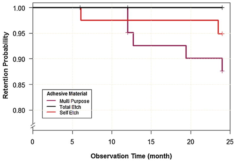
There were 5 total failed restorations, and each failure was described as “restoration missing” on the Failed Restoration Report. In the Scotchbond Multi-purpose group, 1 failure was noted at the 12 month recall and 2 failures were noted at the 24 month recall (93% 24 month retention). In the Scotchbond Universal self-etch group, 1 failure was noted at the 6 month recall and 1 failure was noted at the 12 month recall (95% 24 month retention). There was a 100% 24 month retention for the Scotchbond Universal total-etch group. The 5 failed restorations were removed from the analysis at all time points subsequent to failure.
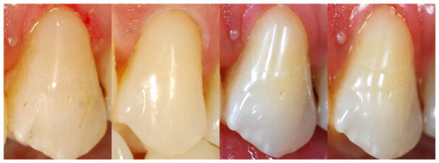
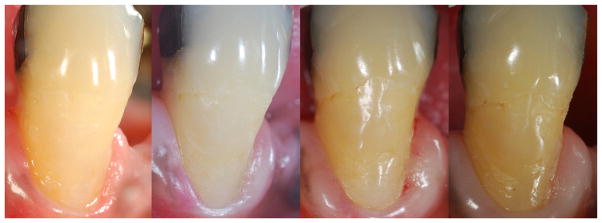
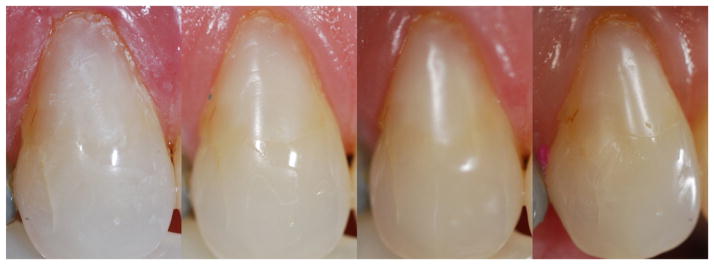
Representative images of restorations from each group at each time point are presented in Figs. 1–3. All materials show an increase in marginal discoloration over time. The restoration placed with Scotchbond self-etch (Fig. 3) shows a greater extent of marginal staining than the other materials at the 24 month photograph. At 24 months, Scotchbond Universal total-etch received the most “perfect” ratings among all materials and no restorations lost to retention. The sensitivity to cold score for Scotchbond Universal total-etch was however marginally higher than the others.
Fig. 1.
Class 5 restoration placed with Scotchbond Multi-purpose at (left to right): baseline, 6 months, 12 months, and 24 months.
Fig. 3.
Class 5 restoration placed with Scotchbond Universal self-etch at (left to right): baseline, 6 months, 12 months, and 24 months.
Regression analyses using GEE indicated that the performance of all adhesive materials decreased over time for marginal adaptation and discoloration (p <0.0001) but not secondary caries (p = 0.05) and sensitivity to cold (p = 0.41). Specifically for marginal adaptation, the odds of fair rating as opposed to perfect rating were 1.93 (95% CI = 1.62–2.32) greater per 6 months of time, given that all of the other variables in the model were held constant. Similarly for marginal discoloration, the likelihood of below perfect rating was 2.94 (95% CI = 2.18–3.97), for every additional 6 months of time. The effect of adhesive material on assessment outcome of marginal discoloration was significant (p = 0.03). This suggests that in comparison to Scotchbond Universal total-etch, both Scotchbond Universal self-etch and Scotchbond Multi-purpose materials were 3.67 (95% CI = 1.15–11.6) and 3.22 (95% CI = 1.06–9.68) times more likely to contribute to less satisfying performance in marginal discoloration over time, respectively. However, for performance in either marginal adaptation, secondary caries or sensitivity to cold, there were no significant differences between adhesive materials (p > 0.05).
While the Kaplan–Meier analysis (Fig. 4) showed no significant difference in retention between the three adhesive materials (p = 0.06), there is a trend toward increased restoration failure after 12 month follow-up for Scotchbond Multi-purpose relative to the total and self-etch modes. The probability of 12 month retention for the multi-purpose mode was 95.1% which is lower than either self (97.6%) or total-etch (100%) mode. The retention probability for multi-purpose mode reduced further to 87.6% till the end of the trial, compared with 94.9% and 100% for self and total-etch, respectively.
Fig. 4.

Kaplan-Meier survival curves illustrating retention rate with Scotchbond Multi-purpose, Scotchbond Universal total-etch, and Scotchbond Universal self-etch.
4. Discussion
The results of this study demonstrate that both adhesive materials and both etching techniques for the universal adhesive experience significant decreases in the incidence of ideal marginal adaptation and marginal discoloration over time. No changes in the incidence of secondary caries or sensitivity to cold were found. Additionally, there were no significant differences in the marginal adaptation, secondary caries or sensitivity to cold for any material. The total-etch Scotchbond Universal group had a higher percentage of restorations with ideal marginal discoloration over time. Although, no significant difference was found between the retention rates of the adhesives, Scotchbond Universal had a nominally higher probability of retention at 24 months in both self-etch and total-etch modes than Scotchbond Multi-purpose.
This is the first clinical study which has compared a universal adhesive to a two-bottle total-etch adhesive, which is often considered the gold standard of dental adhesives. A systematic review of previous clinical trials reported a higher percentage of restorations fulfilling the ADA provisional acceptance (6 months) with two-bottle total-etch (91%) and self-etch (82%) materials than “simplified” one-bottle total-etch (79%) and self-etch (68%) materials [23]. Simplified dental adhesives combine hydrophilic primers, such as HEMA, with the adhesive monomers into a single step. As a result, the adhesive layer is hydrophilic and can be permeable to water. This water permeable adhesive layer is susceptible to hydrolytic degradation from dentinal fluid, jeopardizing the dentinal bond [24]. Scotchbond Universal contains the molecule HEMA, however, it is more hydrdophobic than previous simplified adhesives. Its hydrophobicity is in part derived from the molecule MDP which is inherently hydrophobic [19]. The hydrophobic nature of the universal adhesive may help to explain its favorable comparison with the two-bottle total-etch material in this study.
Another unique feature of Scotchbond Universal is that it contains MDP and polyalkenoic-acid co-polymer, which are both capable of bonding to calcium. MDP in Scotchbond Universal has been shown to form nano-layers with calcium present in the hybrid layer. The MDP-calcium complexes can help to cross-link collagen fibers in the hybrid layer and bridge collagen in the hybrid layer with adhesive monomers in the adhesive layer [18]. An 8 year clinical study has shown a 97% clinical success rate of Clearfil SE, one of the first adhesives to contain the molecule MDP. Carboxyl groups of polyalkenoic acid have also been shown to bond to calcium in hydroxyapatite [25].
Our results demonstrate that Scotchbond Universal had similar incidence of secondary caries and non-ideal marginal adaptation in the total- and self-etch modes, however, marginal discoloration was increased in the self-etch mode. A previous study by Perdigao et al. [9] showed no difference in marginal adaptation or discoloration for Scotchbond Universal in selective-etch, self-etch or total-etch modes with the Cvar and Ryge criteria. Their study did report a significant increase in marginal discrepancies for the self-etch mode using semi-quantitative scores (SQUACE). The SQUACE evaluation accounted for the percentage of the margin with discrepancies and is a more sensitive evaluation method. The results of current clinical study and that of Perdigao et al., as well as the laboratory data reporting lower enamel bond strength of universal adhesives in the self-etch mode [10,11], suggests that longer term follow-ups may show significantly better clinical performance with the total-etch than the self-etch mode. Additionally, no difference was seen in sensitivity to cold between the total-etch group and the self-etch group, which is consistent with the findings of a previous review [26].
Limitations of this study include the small sample size, the relatively low sensitivity of the Cvar and Ryge evaluation criteria and the short length of time for which these materials have been assessed. These patients will continue to be followed for additional evaluation.
Supplementary Material
Fig. 2.
Class 5 restoration placed with Scotchbond Universal total-etch at (left to right): baseline, 6 months, 12 months, and 24 months.
Acknowledgments
This project is based upon a grant from 3 M ESPE, however, they had no role in study design, data collection, analysis or interpretation, or manuscript preparation. Statistical analysis of research reported in this publication was partially supported by National Center for Advancing Translational Sciences of the National Institutes of Health under award number UL1TR00165.
Appendix A. Supplementary data
Supplementary data associated with this article can be found, in the online version, at http://dx.doi.org/10.1016/j.jdent.2015.07.009.
Footnotes
Previously presented at the 2015 IADR meeting in Boston, MA Two-year randomized clinical trial of a universal adhesive in total-etch and self-etch mode in non-carious cervical lesions.
References
- 1.Can Say E, Ozel E, Yurdaguven H, Soyman M. Three-year clinical evaluation of a two-step self-etch adhesive with or without selective enamel etching in non-carious cervical sclerotic lesions. Clin Oral Investig. 2014;18:1427–1433. doi: 10.1007/s00784-013-1123-z. [DOI] [PubMed] [Google Scholar]
- 2.Can Say E, Yurdaguven H, Ozel E, Soyman M. A randomized five-year clinical study of a two-step self-etch adhesive with or without selective enamel etching. Dent Mater J. 2014;33:757–763. doi: 10.4012/dmj.2014-106. [DOI] [PubMed] [Google Scholar]
- 3.Ozel E, Say EC, Yurdaguven H, Soyman M. One-year clinical evaluation of a two-step self-etch adhesive with and without additional enamel etching technique in cervical lesions. Aust Dent J. 2010;55:156–161. doi: 10.1111/j.1834-7819.2010.01218.x. [DOI] [PubMed] [Google Scholar]
- 4.Van Meerbeek B, Kanumilli P, De Munck J, Van Landuyt K, Lambrechts P, Peumans M. A randomized controlled study evaluating the effectiveness of a two-step self-etch adhesive with and without selective phosphoric-acid etching of enamel. Dent Mater. 2005;21:375–383. doi: 10.1016/j.dental.2004.05.008. [DOI] [PubMed] [Google Scholar]
- 5.Torii Y, Itou K, Nishitani Y, Ishikawa K, Suzuki K. Effect of phosphoric acid etching prior to self-etching primer application on adhesion of resin composite to enamel and dentin. Am J Dent. 2002;15:305–308. [PubMed] [Google Scholar]
- 6.Van Landuyt KL, Kanumilli P, De Munck J, Peumans M, Lambrechts P, Van Meerbeek B. Bond strength of a mild self-etch adhesive with and without prior acid-etching. J Dent. 2006;34:77–85. doi: 10.1016/j.jdent.2005.04.001. [DOI] [PubMed] [Google Scholar]
- 7.Prati C, Chersoni S, Mongiorgi R, Pashley DH. Resin-infiltrated dentin layer formation of new bonding systems. Oper Dent. 1998;23:185–194. [PubMed] [Google Scholar]
- 8.Mena-Serrano A, Kose C, De Paula EA, Tay LY, Reis A, Loguercio AD, Perdigão J. A new universal simplified adhesive: 6-month clinical evaluation. J Esthet Restor Dent. 2013;25:55–69. doi: 10.1111/jerd.12005. [DOI] [PubMed] [Google Scholar]
- 9.Perdigao J, Kose C, Mena-Serrano AP, De Paula EA, Tay LY, Reis A, Loguercio AD. A new universal simplified adhesive: 18-month clinical evaluation. Oper Dent. 2014;39:113–127. doi: 10.2341/13-045-C. [DOI] [PubMed] [Google Scholar]
- 10.de Goes MF, Shinohara MS, Freitas MS. Performance of a new one-step multi-mode adhesive on etched vs non-etched enamel on bond strength and interfacial morphology. J Adhes Dent. 2014;16:243–250. doi: 10.3290/j.jad.a32033. [DOI] [PubMed] [Google Scholar]
- 11.Takamizawa T, Barkmeier W, Tsujimoto A, Scheidel D, Erickson R, Latta M, Miyazaki M. Effect of phosphoric acid pre-etching on fatigue limits of self-etching adhesives. Oper Dent. 2015 doi: 10.2341/13-252-L. Epub ahead of print. [DOI] [PubMed] [Google Scholar]
- 12.Perdigao J, Sezinando A, Monteiro PC. Laboratory bonding ability of a multi-purpose dentin adhesive. Am J Dent. 2012;25:153–158. [PubMed] [Google Scholar]
- 13.Wagner A, Wendler M, Petschelt A, Belli R, Lohbauer U. Bonding performance of universal adhesives in different etching modes. J Dent. 2014;42:800–807. doi: 10.1016/j.jdent.2014.04.012. [DOI] [PubMed] [Google Scholar]
- 14.Marchesi G, Frassetto A, Mazzoni A, Apolonio F, Diolosa M, Cadenaro M, Di Lenarda R, Pashley DH, Tay F, Breschi L. Adhesive performance of a multimode adhesive system: 1-Year in vitro study. J Dent. 2014;42:603–612. doi: 10.1016/j.jdent.2013.12.008. [DOI] [PubMed] [Google Scholar]
- 17.Van Landuyt KL, Yoshida Y, Hirata I, Snauwaert J, De Munck J, Okazaki M, Suzuki K, Lambrechts P, Van Meerbeek B. Influence of the chemical structure of functional monomers on their adhesive performance. J Dent Res. 2008;87:757–761. doi: 10.1177/154405910808700804. [DOI] [PubMed] [Google Scholar]
- 18.Yoshida Y, Yoshihara K, Nagaoka N, Hayakawa S, Torii Y, Ogawa T, Osaka A, Meerbeek BV. Self-assembled Nano-layering at the adhesive interface. J Dent Res. 2012;91:376–381. doi: 10.1177/0022034512437375. [DOI] [PubMed] [Google Scholar]
- 19.Su B. Principles of Adhesion Dentistry. AEGIS Publications; India: 2013. [Google Scholar]
- 20.Cvar JF, Ryge G. Reprint of criteria for the clinical evaluation of dental restorative materials. 1971. Clin Oral Investig. 2005;9:215–232. doi: 10.1007/s00784-005-0018-z. [DOI] [PubMed] [Google Scholar]
- 21.Pan W. Akaike’s information criterion in generalized estimating equations. Biometrics. 2001;57:120–125. doi: 10.1111/j.0006-341x.2001.00120.x. [DOI] [PubMed] [Google Scholar]
- 22.Schulz KF, Altman DG, Moher D. CONSORT 2010 statement: updated guidelines for reporting parallel group randomised trials. Int J Surg. 2011;9:672–677. doi: 10.1016/j.ijsu.2011.09.004. [DOI] [PubMed] [Google Scholar]
- 23.Peumans M, Kanumilli P, De Munck J, Van Landuyt K, Lambrechts P, Van Meerbeek B. Clinical effectiveness of contemporary adhesives: a systematic review of current clinical trials. Dent Mater. 2005;21:864–881. doi: 10.1016/j.dental.2005.02.003. [DOI] [PubMed] [Google Scholar]
- 24.Tay FR, Pashley DH, Suh BI, Carvalho RM, Itthagarun A. Single-step adhesives are permeable membranes. J Dent. 2002;30:371–382. doi: 10.1016/s0300-5712(02)00064-7. [DOI] [PubMed] [Google Scholar]
- 25.Fukuda R, Yoshida Y, Nakayama Y, Okazaki M, Inoue S, Sano H, Suzuki K, Shintani H, Van Meerbeek B. Bonding efficacy of polyalkenoic acids to hydroxyapatite, enamel and dentin. Biomaterials. 2003;24:1861–1867. doi: 10.1016/s0142-9612(02)00575-6. [DOI] [PubMed] [Google Scholar]
- 26.Perdigao J, Swift EJ., Jr Critical appraisal post-op sensitivity with direct composite restorations. J Esthet Restor Dent. 2013;25:284–288. doi: 10.1111/jerd.12045. [DOI] [PubMed] [Google Scholar]
Associated Data
This section collects any data citations, data availability statements, or supplementary materials included in this article.


