Abstract
The Frank system vectorcardiogram has been studied in 61 patients with severe mitral valve disease to determine the value of the vectorcardiogram in the recognition of the relative degree of left and right ventricular hypertrophy in this situation.
The appearance of the usual evidence of right ventricular hypertrophy is delayed in mitral valve disease by the vertical electrical position of the heart which may be due to alterations in the intrathoracic electrical field produced by left atrial enlargement.
Voltage criteria for the recognition of isolated left or right ventricular hypertrophy in the vectorcardiogram are not applicable to combined ventricular hypertrophy in mitral valve disease. The present analysis is based on the spatial pattern of the QRS loop.
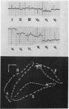
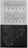
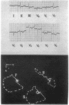
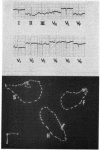
The vectorcardiograms show a continuous gradation from posterior to anterior direction, the extremes indicating dominant left and right ventricular hypertrophy, respectively. Five groups are recognized from the appearance in the horizontal plane. Long posterior loops (Fig. 1) are associated with severe left ventricular hypertrophy, open posterior loops (Fig. 2) with left ventricular dominance, and wide posterior loops (Fig. 3) with moderate hypertrophy of both ventricles. Wide crossed loops (Fig. 4) indicate right ventricular dominance, and anterior clockwise loops (Fig. 5) are found with severe right ventricular hypertrophy.
The vectorcardiogram rarely showed large QRS voltages in left ventricular hypertrophy, though these changes were often evident in the conventional electrocardiogram. The vectorcardiogram appeared to be more successful than the electrocardiogram in the recognition of severe right ventricular hypertrophy. An unusual rightwards displacement of the QRS loop was found in patients with tricuspid valve disease.
It is concluded that the vectorcardiogram gives useful additional information for the recognition of ventricular hypertrophy that is not evident in the conventional electrocardiogram in mitral valve disease.
Full text
PDF


Images in this article
Selected References
These references are in PubMed. This may not be the complete list of references from this article.
- DAVIES L. G., GOODWIN J. F., STEINE R. E., VAN LEUVEN B. D. The clinical and radiological assessment of the pulmonary arterial pressure in mitral stenosis. Br Heart J. 1953 Oct;15(4):393–400. doi: 10.1136/hrt.15.4.393. [DOI] [PMC free article] [PubMed] [Google Scholar]
- DONOSO E., JICK S., BRAUNWALD E., LAMELAS M., GRISHMAN A. The spatial vectorcardiogram in mitral valve disease. Am Heart J. 1957 May;53(5):760–766. doi: 10.1016/0002-8703(57)90344-7. [DOI] [PubMed] [Google Scholar]
- FORKNER C. E., Jr, HUGENHOLTZ P. G., LEVINE H. D. The vectorcardiogram in normal young adults. Frank lead system. Am Heart J. 1961 Aug;62:237–246. doi: 10.1016/0002-8703(61)90323-4. [DOI] [PubMed] [Google Scholar]
- FRANK E. An accurate, clinically practical system for spatial vectorcardiography. Circulation. 1956 May;13(5):737–749. doi: 10.1161/01.cir.13.5.737. [DOI] [PubMed] [Google Scholar]
- FRASER H. R., TURNER R. Electrocardiography in mitral valvular disease. Br Heart J. 1955 Oct;17(4):459–483. doi: 10.1136/hrt.17.4.459. [DOI] [PMC free article] [PubMed] [Google Scholar]
- HUGENHOLTZ P. G., GAMBOA R. EFFECT OF CHRONICALLY INCREASED VENTRICULAR PRESSURE ON ELECTRICAL FORCES OF THE HEART. A CORRELATION BETWEEN HEMODYNAMIC AND VECTROCARDIOGRAPHIC DATA (FRANK SYSTEM) IN 90 PATIENTS WITH AORTIC OR PULMONIC STENOSIS. Circulation. 1964 Oct;30:511–530. doi: 10.1161/01.cir.30.4.511. [DOI] [PubMed] [Google Scholar]
- IMPERIAL E. S., BENDEZU J., ZIMMERMAN H. A. Electrocardiographic analysis of pure mitral valvular disease: a study based on fifty-seven cases with open-heart operation. Am Heart J. 1960 Nov;60:705–715. doi: 10.1016/0002-8703(60)90353-7. [DOI] [PubMed] [Google Scholar]
- Lal S., Fletcher E., Binnion P. Frank vectorcardiogram correlated with haemodynamic measurements: quantitative analysis. Br Heart J. 1969 Jan;31(1):15–20. doi: 10.1136/hrt.31.1.15. [DOI] [PMC free article] [PubMed] [Google Scholar]
- MORRIS T. L., WHITAKER W. The ventricular patterns in the right precordial leads in mitral stenosis. Am Heart J. 1956 Nov;52(5):738–748. doi: 10.1016/0002-8703(56)90027-8. [DOI] [PubMed] [Google Scholar]
- PAGNONI A., GOODWIN J. F. The cardiographic diagnosis of combined ventricular hypertrophy. Br Heart J. 1952 Oct;14(4):451–461. doi: 10.1136/hrt.14.4.451. [DOI] [PMC free article] [PubMed] [Google Scholar]
- Romhilt D. W., Greenfield J. C., Jr, Estes E. H., Jr Vectorcardiographic diagnosis of left ventricular hypertrophy. Circulation. 1968 Jan;37(1):15–19. doi: 10.1161/01.cir.37.1.15. [DOI] [PubMed] [Google Scholar]
- SEMLER H. J., PRUITT R. D. An electrocardiographic estimation of the pulmonary vascular obstruction in 80 patients with mitral stenosis. Am Heart J. 1960 Apr;59:541–547. doi: 10.1016/0002-8703(60)90457-9. [DOI] [PubMed] [Google Scholar]
- TAYMOR R. C., HOFFMAN I., HENRY E. THE FRANK VECTORCARDIOGRAM IN MITRAL STENOSIS: A STUDY OF 29 CASES. Circulation. 1964 Dec;30:865–871. doi: 10.1161/01.cir.30.6.865. [DOI] [PubMed] [Google Scholar]
- TOOLE J. G., von der GROEBEN, SPIVACK A. P. The calculated tempero-spatial heart vector in proved isolated left ventricular overwork. Am Heart J. 1962 Apr;63:537–544. doi: 10.1016/0002-8703(62)90311-3. [DOI] [PubMed] [Google Scholar]


