

Abstract
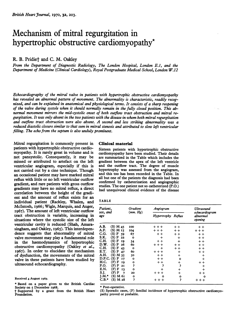
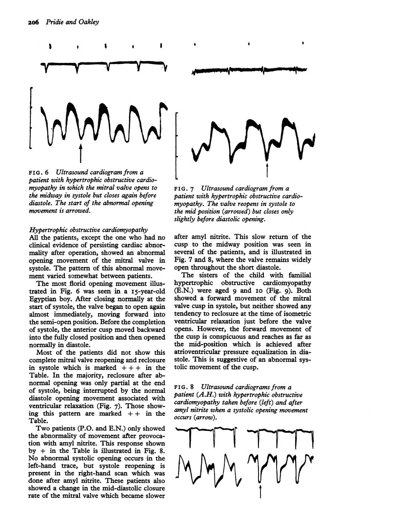
Echocardiography of the mitral valve in patients with hypertrophic obstructive cardiomyopathy has revealed an abnormal pattern of movement. The abnormality is characteristic, readily recognized, and can be explained in anatomical and physiological terms. It consists of a sharp reopening of the valve during systole when it should normally remain in the fully closed position. This abnormal movement mirrors the mid-systolic onset of both outflow tract obstruction and mitral regurgitation. It was only absent in the two patients with the disease in whom both mitral regurgitation and outflow tract obstruction were also absent. A second and less striking abnormality was a reduced diastolic closure similar to that seen in mitral stenosis and attributed to slow left ventricular filling. The echo from the septum is also unduly prominent.
Full text
PDF
Selected References
These references are in PubMed. This may not be the complete list of references from this article.
- Pridie R. B., Turnbull T. A. Diagnosis of pericardial effusion by ultrasound. Br Med J. 1968 Aug 10;3(5614):356–357. doi: 10.1136/bmj.3.5614.340-a. [DOI] [PMC free article] [PubMed] [Google Scholar]
- Rackley C. E., Whalen R. E., McIntosh H. D. Ventricular volume studies in a patient with hypertrophic subaortic stenosis. Circulation. 1966 Oct;34(4):579–584. doi: 10.1161/01.cir.34.4.579. [DOI] [PubMed] [Google Scholar]
- Wigle E. D., Marquis Y., Auger P. Pharmacodynamics of mitral insufficiency in muscular subaortic stenosis. Can Med Assoc J. 1967 Aug 5;97(6):299–301. [PMC free article] [PubMed] [Google Scholar]