Abstract
Oryza meyeriana, a wild species of rice from China, shows high resistance to Xanthomonas oryzae pv. oryzae (Xoo), the cause of rice bacterial blight, one of the most serious rice pathogens. To better understand the resistance mechanism, a proteomic study was conducted to identify changes in the proteins secreted in embryo cell suspension cultures in response to Xoo. After two-dimensional difference gel electrophoresis (2D-DIGE), 72 differentially expressed protein spots corresponding to 34 proteins were identified by Matrix-Assisted Laser Desorption/ Ionization Time of Flight Mass Spectrometry. Of the 34 proteins, 10 were up regulated and 24 down regulated. The secreted proteins identified were predicted to be involved in various biological processes, including signal transduction, defense, ROS and cell wall modification. 77% of the 34 proteins were predicted to have a signal peptide by Signal P. Quantitative Real-Time PCR showed that transcript levels of 14 secreted proteins were not well correlated with secreted protein levels. Peroxidase activity was up regulated in both O. meyriana and susceptible rice but was about three times higher in O. meyeriana. This suggests that peroxidases may play an important role in the early response to Xoo in O. meyeriana. These results not only provide a better understanding of the resistance mechanism of O. meyeriana, but have implications for studies of the interactions between other plants and their pathogens.
1 Introduction
Bacterial blight (BB) of rice (Oryza sativa) caused by Xanthomonas oryzae pv. oryzae (Xoo), a member of the γ-proteobacteria, is one of the most serious rice pathogens worldwide [1,2]. Xoo invades rice xylem tissue through water or wounds resulting in systemic infection [3]. Planting resistant lines is the most economical and effective way to control this vascular disease [4]. To date, 38 bacterial-blight resistance genes have been reported in rice, eight of which are from wild species [5]: Xa10 (O. Cas209) [6,7], Xa21 (O. longistaminata) [8], Xa23 (O. rufipogon) [9], Xa27 (O. minuta) [10], Xa29 (O. officinalis) [11], Xa30 (O. rufipogon) [12], Xa32 (O. australiensis) [13] and xa32 (O. meyeriana) [14].
O. meyeriana has strong resistance to all Xoo strains [15] but no resistance gene has yet been cloned from it [14]. There is hybrid sterility between O. meyeriana and cultivated rice [16] but the BB resistance of O. meyeriana has been introduced into cultivated rice by asymmetric somatic hybridization [16,17] leading to the development of some highly resistant rice lines including Y73 [18] and SH76 [19]. Some insights have been obtained into these highly resistant phenotypes by a microarray analysis to examine transcription in Y73 and a proteomics study of SH76. 115 genes had altered RNA expression in Y73 in response to Xoo, and they involved in oxidant redox, signal transduction and transcription. Seven of them were up- regulated more than fivefold, including two transcription factors (TFs) and one ubiquitination protein [19]. 34 proteins changed significantly in concentration in response to Xoo in SH76, and they relate to signal transduction, photosynthesis, antioxidant defense and metabolism [19]. A small auxin RNA was up in Y73 [18], and a auxin–regualted protein up regulated in SH76. Besides, a Rubisco Large subunit (RcbL) was degraded [19]. Rubisco activity is regulated by Rubisco activase (RCA). RCA moved to the thylakoid membrane in O. meyeriana 12h to 16h after inoculation with Xoo coupled with an oxidative burst, while RCA remained in chloroplast stroma in the susceptible line [20].
Secreted proteins play an important role in the rice-Xoo interaction [21–23], and studies of the rice secretome have identified some proteins in the plasma membrane [24], xylem sap [25] and leaves [26] that are involved in the early defense responses to Xoo. Cell suspension cultures have been used to study the secretome of many plants, including Arabidopsis thaliana [27], maize [28], tobacco [29], medicago [30] and rice [31] but there have been no reported studies using this method to investigate changes in the secretome of resistant rice in response to Xoo infection.
Here, we used two-dimensional difference gel electrophoresis (2D-DIGE) coupled with Mass Spectrometry (MS) to study secretome changes in an O. meyeriana embryo cell suspension in response to inoculation with Xoo. A total of 34 differentially expressed proteins were identified and their possible roles in response to Xoo are discussed.
2 Materials and Methods
2.1 Plant material
Sterile Oryza meyriana seedlings were cut into 1–1.5 cm pieces and placed on MS callus induction medium (3 mg/L 2,4-D, 0.3 mg/L 6-BA) [18]. After incubation in the dark at 28°C for 2 wk, they were transferred to a 16h/8h-light/dark regime at 28°C for 3 wk to induce calli. Growing calli (0.5–1.0g) were transferred into liquid MS medium (2.5 mg/L 2,4-D, 0.3 mg/L 6-BA) and shaken (150 rpm) at 28°C in the dark [32]. The suspension culture was sub-cultured weekly until the cells appeared dense, uniform and light yellow. Xoo strain PXO124 (race P10) was cultured on PSA liquid medium (1% tryptone, 0.1% yeast extract, 1% sucrose, 0.3% peptone, 1.5% agar) at 28°C for 48 h and adjusted to 108 CFU ml-1 before inoculation to the rice suspension culture 3 days after sub-culturing. For qQRT-PCR and peroxidase activity assays, leaves from 21 day old O. meyeriana and Nipponbare (susceptible control) seedlings were inoculated with Xoo (OD ~0.8) using the leaf-cutting method [33].
2.2 Isolation secreted and total proteins from Oryza meyriana suspension—cultured cells
After sub-culturing for three days, the rice culture medium was harvested 0 h and 24 h after inoculation with Xoo. Calli were collected through a filter (0.18 mm), washed with ddH2O and stored at -80°C. The filtered medium was centrifuged at 20,000 g for 20 min to remove the residual cells, and the supernatant was concentrated using Sartorius Slice 200 (Sartorius, Germany). Secreted and total proteins were extracted by a modified phenol-methanol method [34]. Four biological replicates were prepared. Secreted protein and total protein were further purified using the 2-D Clean Up Kit (GE healthcare, USA) and their concentrations determined using a 2-D Quant kit (GE healthcare, USA).
2.3 Western blot analysis
Approximately 15 μg of secreted proteins (S) and total proteins (T) were separated on a 12.5% (w/v) SDS-PAGE gel overlaid with a 4% (w/v) sticking gel, and electrotransferred onto a PVDF membrane. After blocking in blocking buffer [20 mM Tris-HCl, pH 7.6, 0.5 mM NaCl, 0.005% v/v Tween 20, 1% (w/v) BSA] overnight, the membrane was incubated with mouse anti-actin antibody (1:1000, Abmart, Shanghai, China) followed by the secondary antibody, mouse anti-mouse IgG conjugated with alkaline phosphatase (1:10000, Sigma, Missouri, USA). Binding of the enzyme conjugated antibodies was detected with the NBI/BCIP (Amresco, Ohio, USA) system. For western blotting of the peroxidase, extracts were prepared from 0.1 g leaves of O. meyeriana and cultivated rice Nipponbare (susceptible control) at 0 h (mock-control) and 24 h post-inoculation with Xoo. They were homogenized in 1 ml Extract Buffer [20mM Tris-HCl, 200mM NaCl, 1mM EDTA, 1mM DTT, β-mercaptoethanol (10%, v/v)], samples of approximately 10 μl (~4.5 μg/μl) were separated on SDS-PAGE gels and the blot membrane was incubated with rabbit anti- peroxidase antibody (1:10,000, Agrisera, Sweden).
2.4 2D-DIGE, Image Scanning and Analysis
Prepared secreted proteins were separated by 2D-DIGE as described previously [34]. The Cy2, Cy3 and Cy5 labeled samples (50 μg) were mixed and loaded on the strips (linear, 24cm, pI 4–7, GE Healthcare, USA) for the first dimension separation. Next, the strips were placed on top of 12.5% SDS-PAGE gels for the second dimension electrophoresis. Protein spots on gels were scanned using an Ettan DIGE Scanner (GE Healthcare, USA) and the images were analyzed using Decyder 2D software (Version 7.0, GE Healthcare, USA). Finally, spots from different gels were matched using Biological Variation Analysis. Only spots present in all gels and which exhibited statistically significant changes in intensity (≥1.5 fold or ≤-1.5 fold, p <0.05) were considered to be differentially expressed proteins.
2.5 In gel protein digestion and MS analysis
About 500 μg secreted proteins were loaded on the strips, separated by 2-DE and stained with Coomasie Brilliant Blue (CBB) R-250. Differentially expressed protein spots were manually excised from the stained 2-D gel and transferred to a sterilized tube (1.5 ml) with destaining solution [30% (w/v) acetonitrile (ACN), 100 mmol NH4HCO3]. After being vacuum freeze-dried, the spots were digested in 30 μl enzyme buffer (50 mmol NH4HCO3, 50 ng/μl trypsin) at 37°C overnight. Then, the small peptides were back-extracted in 60% ACN buffer [0.1% trifluoroacetic acid (TFA)] and dried under a stream of nitrogen. Finally, peptides were re-suspended in 50% ACN buffer [0.1% w/v TFA, 5 mg/ml α- cyano -4-hydroxycinnamic acid (CHCA)], analyzed using an ABI 4800 Proteomics Analyzer MALDI-TOF/TOF (Applied Biosystems, Foster City), and MALDI-TOF spectra were searched. All MALDI-TOF spectra were searched against the National Center for Biotechnology Information non-redundant (NCBInr) database using the GPS ExplorerTM software (v3.6, Applied Biosystems) and MASCOT search program (v2.1 Matrix Science). Finally, based on the MALDI-TOF-MS, only protein scores > 95 (p <0.05) were accepted for the identification of protein samples.
2.6 Bioinformatics analysis
Homologues of the identified proteins were searched in the RAGP (http://rice.plantbiology.msu.edu/) for matching against the NCBI database. The UniProt (http://www.uniprot.org/) database was used to determine their functions. Next, SignalP (version 4.1, http://www.cbs.dtu.dk/services/SignalP/) was used to predict their secretion pathways and PSORT II (http://psort.hgc.jp/form2.html) to predict their subcellular location. The identified proteins were annotated and grouped according to their predicted function and biological processes.
2.7 RNA extraction and Quantitative real time PCR for gene transcriptional expression analysis
Primers were designed using primer 6 (version 6.0), to be specific to the corresponding gene sequences of the proteins identified by MS in the RAGP database (S1 and S2 Tables). Total RNAs were extracted from inoculated leaves of O. meyeriana and Nipponbare (4–5 leaf stage) using TRizol reagent (Invitrogen, Germany). Residual DNA was removed by RNase-free DNase (Fermentas, Canada). The first strand cDNAs were synthesized by Synthesis kit for qRT-PCR (Bio-RAD, USA). qRT-PCR with Eva Green (Biotium, USA) was performed on the light Cycler 480 real-time PCR system (Roche, Basle, Switzerland), and the PCR program consisted of: 95°C for 3 min, 45 cycles of 95°C for 30s, 55°C for 30s and 72°C for 30s. Dissociation curves were generated at the end of PCR cycles, and relative gene expression was calculated by the 2- Ä (Ä Cp) method. The reference gene was OsActin gene (AK 060893), and all experiments were repeated three times with cDNA prepared from different biological samples.
2.8 POD activity detection
POD activity was detected in O. meyeriana and 21-day-old Nipponbare seedling leaves at 0h, 6h, 12h and 24h after inoculation with Xoo. Peroxidase activity was assayed as described in the POD detection kit (Keming, China) with some modifications. 0.1 g rice leaves were homogenized in 1ml cold extraction buffer, and centrifuged at 8,000g for 10 min at 4°C. 3.75 ul samples were mixed with 263.75 ul reaction buffer and the absorbance was measured by Spectra Max M5 (Molecular Devices, USA) at 470 nm for 30s (A1) and 60s (A2). Calculation of POD was based on the following formula: POD(U/g) = 7133×(A2-A1)÷Sample weight. One enzyme unit is defined as the absorbance of 1g sample in reaction buffer at 470 nm for a 60s change of 0.01. All experiments were repeated three times prepared from different biological samples
3 Results
3.1 Western blot analysis indicates that the secreted proteins preparation may be free from contamination with non- secreted proteins
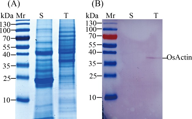
Preparation of secreted proteins is a challenging work [31]. Previously, we tested four protein isolation methods, including TCA/acetone [20], molecular weight cutoff filters, Ammonium sulfate precipitation and the phenol method [34] and found that the phenol method was suitable for extracting and isolating secreted proteins from O. meyeriana suspension culture. In this study, Western blot showed that actin (45 kDa) was detected only in the total proteins, but was absent from the secreted proteins (Fig 1). This suggests that the secreted proteins prepared from suspension culture may be free from contamination with non-secreted proteins.
Fig 1. Western blot of the secretome and total proteins from Oryza meyeriana.
A: Secretome and total protein were separated by 1D SDS-PAGE; B: Secretome and total protein were analyzed by immunoblotting with a mouse anti-actin antibody. Samples of 15 μg protein were loaded onto 12% SDS-PAGE gels, and stained with Coomassie Brilliant Blue (CBB) R-250 solution. For western blot, actin was used a representative intracellular protein. Mr: marker; S: secretome; T: total protein.
3.2 Identification of differentially expressed proteins
Secreted proteins from O. meyriana suspension culture were isolated using the phenol method, separated by 2D-DIGE and identified by MS. More than 1500 protein spots were detected reproducibly on gels by DeCyder 2D software of which 110 changed in intensity significantly (p < 0.05) and by more than 1.5 fold when compared with the control (Fig 2). After analysis by MS, 72 proteins corresponding to 34 genes were matched to the NCBI database. Among the identified proteins, 10 were up regulated and 24 down regulated (Table 1).
Fig 2. 2D-DIGE analysis of the Xoo-induced secretome in O.meyeriana suspension cultured medium.
Secreted-proteins were extracted from the medium and separated by 2D-DIGE using pH 4-7/24cm linear IPG strips and 12.5% SDS-PAGE gels. 110 protein spots showed significant changes (> 1.5 fold; p< 0.05), corresponding to 34 proteins identified by MS.
Table 1. Differential proteins identified by MS.
| Noa | Accession No. | Locus | Unique Peptideb | SC c % | Average fold changed | Molecular function and/or property | SignalPe | Cell location |
|---|---|---|---|---|---|---|---|---|
| Signal transduction | ||||||||
| M18 | gi|326515056 | Os12g44020.1 | 3 | 10 | 1.73±0.016 | Ser/Thr protein phosphatase family protein, expressed | - | extracellular |
| M30 | gi|115477980 | Os09g02729.1 | 6 | 15 | -1.78±0.0058 | phospholipase C, expressed | Y | extracellular |
| M43 | gi|357128757 | Os05g44200.1 | 7 | 15 | 2.37±0.045 | GDSL-like lipase/acyl hydrolase, expressed | Y | extracellular |
| M76 | gi|115483362 | Os08g42580.1 | 11 | 29 | 1.71±0.034 | protein kinase domain containing protein, expressed | - | cytoplasmic |
| Protein metabolism | ||||||||
| M11 | gi|315797642 | Os12g17910 | 4 | 26 | 12.98±0.042 | T-complex protein, expressed | - | ER |
| M55 | gi|149392557 | Os05g24550.1 | 7 | 20 | -3.77±0.00017 | Papain family cysteine protease domain containing protein, expressed | - | cytoplasmic |
| M101 | gi|62546209 | Os11g09280.1/2 | 9 | 13 | 1.63±0.035 | OsPDIL1-1 protein disulfide isomerase PDIL1-1, expressed | Y | ER |
| PRs and defense proteins | ||||||||
| M8 | gi|62733152 | Os11g47820.2 | 8 | 25 | -1.64±0.039 | glucan endo-1,3-beta-glucosidase | Y | mitochondrial |
| M16 | gi|242032727 | Os03g57880.3 | 6 | 12 | 3.52±0.0052 | glucan endo-1,3-beta-glucosidase, expressed | Y | extracellular |
| M40 | gi|116309669 | Os04g51460.1 | 9 | 28 | 3.54±0.000027 | glycosyl hydrolases family 16, expressed | Y | extracellular |
| M41 | gi|218195513 | Os04g51460.1 | 8 | 24 | 3.58±0.00067 | glycosyl hydrolases family 16, expressed | Y | extracellular |
| M42 | gi|49387539 | Os02g42310.1 | 5 | 12 | -1.69±0.023 | OsSCP8—Putative Serine Carboxypeptidase homologue, expressed | - | extracellular |
| M49 | gi|125560666 | Os08g13920.1 | 7 | 25 | 2.57±0.0051 | glycosyl hydrolases family 16, expressed | Y | cytoplasmic |
| M66 | gi|151935395 | Os05g31140.3 | 2 | 14 | -1.85±0.003 | glycosyl hydrolases family 17, expressed | - | extracellular |
| M28 | gi|125526903 | Os01g43490.1 | 4 | 12 | -5.96±0.0011 | polygalacturonase, expressed | Y | cytoplasmic |
| M90 | gi|304301588 | Os07g04560 | 1 | 12 | -9.82±0.025 | no apical meristem protein, expressed | - | mitochondrial |
| M106 | gi|115452789 | Os03g21040.2 | 11 | 27 | 1.53±0.048 | stress responsive protein, expressed | Y | cytoplasmic |
| M107 | gi|125586117 | Os03g21040.2 | 12 | 31 | 1.61±0.025 | stress responsive protein, expressed | Y | cytoplasmic |
| Redox | ||||||||
| M4 | gi|293336711 | Os03g57880.3 | 5 | 12 | 2.07±0.036 | monocopper oxidase, expressed | Y | extracellular |
| M31 | gi|55700999 | Os04g51460.1 | 2 | 7 | -2.57±0.0028 | Peroxidase expressed | Y | extracellular |
| M32 | gi|242089641 | Os04g51460.1 | 6 | 14 | -1.75±0.0047 | Peroxidase expressed | Y | mitochondrial |
| M33 | gi|115477493 | Os02g42310.1 | 8 | 25 | -5.13±0.0011 | hypothetical protein | ER | |
| M34 | gi|45685281 | Os08g13920.1 | 6 | 16 | -1.72±0.032 | peroxidase expressed | Y | extracellular |
| M52 | gi|55701087 | Os05g31140.3 | 4 | 10 | -2.2±0.000038 | peroxidase expressed | Y | extracellular |
| M99 | gi|115473931 | Os01g43490.1 | 6 | 40 | -1.64±0.049 | copper/zinc superoxide dismutase, expressed | N | cytoplasmic |
| M105 | gi|115474057 | Os07g04560 | 4 | 14 | -2.03±0.0019 | hypothetical protein | Y | extracellular |
| M111 | gi|357473921 | Os03g21040.2 | 3 | 15 | -1.93±0.0045 | peroxidase expressed | Y | nuclear |
| Cell wall structure proteins | ||||||||
| M23 | gi|125524938 | Os01g12070.1 | 6 | 9 | -2.67±0.0062 | endoglucanase, expressed | Y | ER |
| M10 | gi|326491047 | Os11g05760.1 | 4 | 13 | 7.34±0.0069 | OsProCP5—Putative Lysosomal Pro-x Carboxypeptidase homologue, expressed | Y | mitochondrial |
| M27 | gi|57899795 | Os01g66830.1 | 4 | 13 | -1.71±0.007 | pectin acetylesterase domain containing protein, expressed | Y | Golgi |
| M78 | gi|162458364 | Os10g40720.1 | 7 | 12 | -1.75±0.0019 | expansin expressed | - | vesicles of secretory system |
| M89 | gi|125532912 | Os10g40700.1 | 9 | 32 | -7.12±0.00083 | Expansine expressed | Y | extracellular |
| M94 | gi|115462831 | Os05g15690.1 | 2 | 13 | -32.47±0.001 | expansin expressed | Y | extracellular |
| M95 | gi|116310398 | Os04g46630.1 | 1 | 5 | -38.52±0.0002 | expansin expressed | Y | cytoplasmic |
| M96 | gi|125551519 | Os05g15690.1 | 1 | 10 | -32.99±0.00059 | expansin expressed | Y | extracellular |
| M97 | gi|125549272 | Os04g46630.1 | 3 | 17 | -13.57±0.000058 | expansin expressed | Y | cytoplasmic |
| M104 | gi|115483362 | Os10g40720.1 | 10 | 28 | -5.55±0.0034 | expansin expressed | Y | extracellular |
a spot number as given in Fig 2. Some proteins from different spots correspond to the same gene suggesting they may have been post-translationally modified.
b number of matched peptides
c sequence coverage
d Fold change with p <0.05
e “Yes” means protein was predicted to have a signal peptide.
3.3 Bioinformatics analysis of differentially expressed proteins
Thirty four proteins from rice were identified by BLAST in the RAGP and Uniprot database, and classified into five functional groups (Table 1). These include proteins with signaling roles, proteins involved in redox and defense, cell wall proteins and proteins involved in metabolism. Twenty six proteins were predicted to have signal peptides and more than 17 were predicted to be located in the extracellular space. Three proteins without signal peptides (M18, M42, M78) were predicted to be located in the extracellular space. Proteins can be transported across membranes through the classical or non classical secretory pathways [35]. For example, the key gluconeogenic enzyme in the Calvin Cycle, fructose-1,6-bisphosphatase [36] does not contain signal sequences but is secreted via a non-classical secretory pathway similar to our results.
3.4 Correlation between protein and mRNA expression
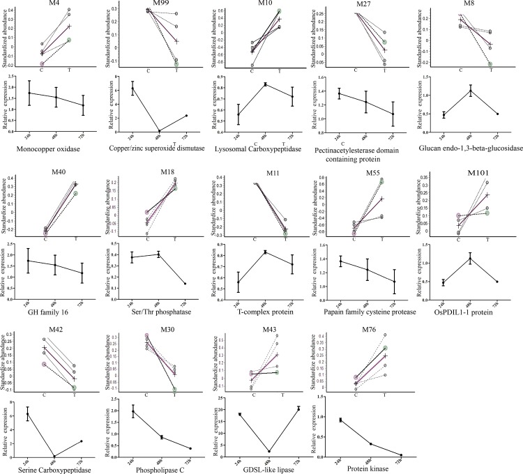
To investigate the relationship between the identified proteins and their transcription, Quantitative real time PCR was performed to analyze the transcriptional activities of 14 randomly selected rice genes corresponding to the rice proteins identified. Samples were tested at 0 h, 24 h, 48 h, and 72 h post inoculation with Xoo. Three up-and three down regulated proteins showed good correlation between protein and RNA expression at 24 hpi, including a monocopper oxidase, a GDSL-like lipase, a pectinacetylesterase, a glucan endo-1,3-beta-glucosidase, a protein kinase and glycosyl hydrolases (Fig 3). Transcription of the remaining eight proteins was not well correlated with their protein expression at 24 hpi. The mRNA transcriptional level does not always correlate well with the protein expression levels [37]. For example, it was reported that Phospholipase C was down regulated, but the transcription was up regulated [38], similar to our results. The transcription of many chilling- related proteins was first up and then down regulated after cold stress [39]. It should be noted that suspension cultured cells were used to measure secreted protein expression levels, while transcript levels were determined from seedling leaves. In addition, gene expression in infected leaves may also be influenced by the circadian rhythm.
Fig 3. Xoo-responsive rice proteins identified by 2-D DIGE.
Changes in protein abundance of the 14 rice proteins in the secreted protein fraction of suspension-cultured cells 24 h after inoculation (top panel); mRNA expression of the 14 rice genes in the leaves of rice seedlings 24 h, 48 h and 72 h after inoculation (bottom panel).
3.5 Quantitative Real Time PCR and Enzyme activity assay for Peroxidases
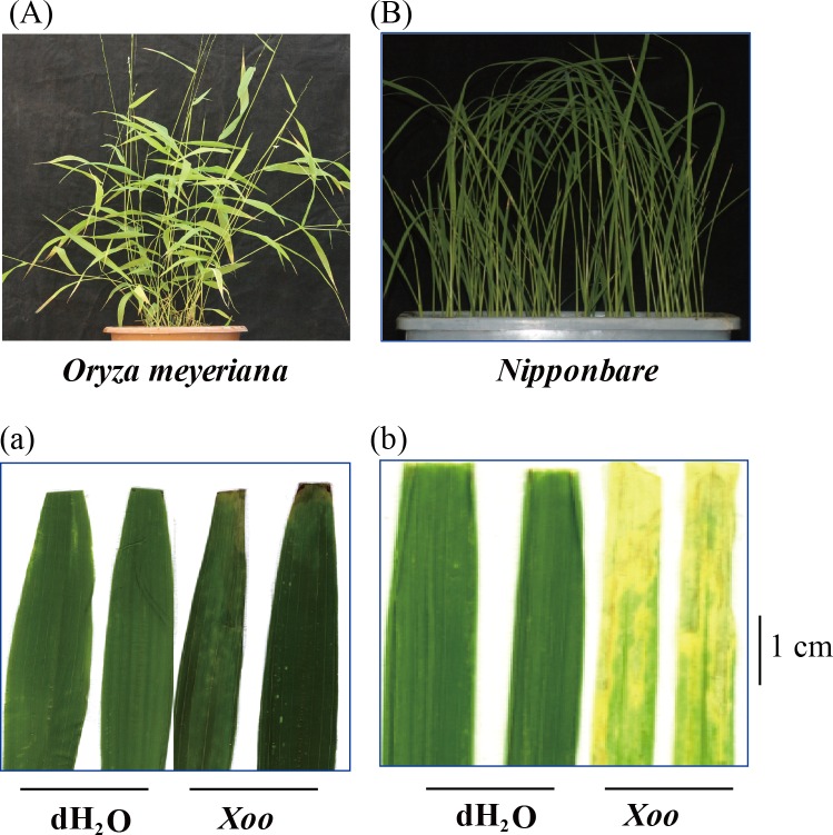
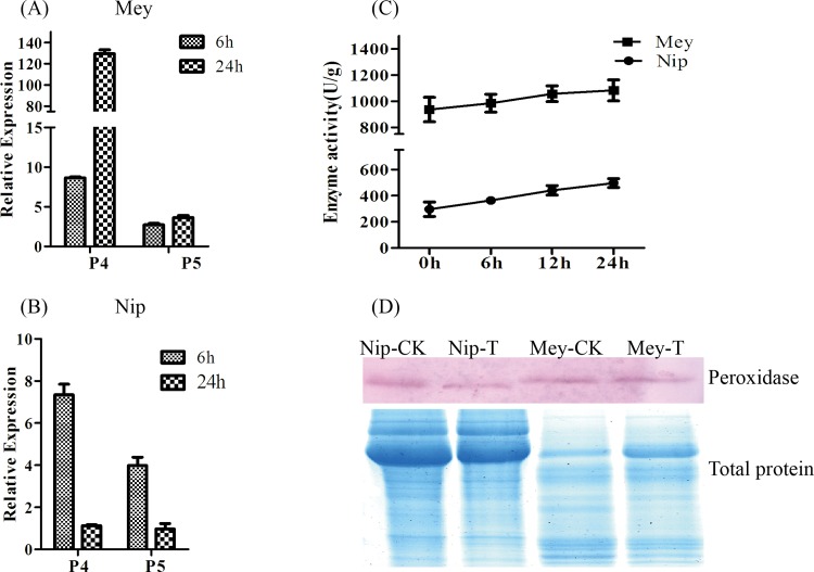
Many peroxidase proteins were down regulated in O. meyeriana (Table 1). Quantitative Real Time PCR was performed to analyze the transcriptional activities of two randomly selected peroxidase genes, corresponding to the rice proteins identified (P4 and P5; for primers see S2 Table). Cultivated rice Nipponbare is high susceptible to Xoo (Fig 4). The transcription of P4 and P5 was up regulated in O. meyeriana in response to Xoo, while they were both down regulated in Nipponbare (Fig 5A and 5B). Peroxidase enzyme activity was up regulated in both O. meyeriana and Nipponbare, with the activity in O. meyeriana consistently about three times higher than in Nipponbare (Fig 5C). Expression of the peroxidase proteins was slightly down regulated in Nipponbare, consistent with their transcription, but they were unchanged in O. meyeriana (Fig 5D).
Fig 4. The effect of Xoo (PXO124) on O. meyeriana and susceptible cultivated rice Nipponbare.
Upper panels show the plant phenotypes; lower panels show leaves 14 days after inoculation with Xoo using the clipping method. Controls were inoculated with water.
Fig 5. qQRT-PCR and enzyme activity assay of peroxidases in O. meyeriana and Nipponbare.
(A and B) qQRT-PCR of peroxidase 4 (P4) and peroxidase 5 (P5) in O. meyeriana (A) or Nipponbare (B) 6h and 24h after inoculation with Xoo. (C) Peroxidase activity 0h, 6h, 12h and 24h after inoculation with Xoo; OsActin was used as a mock gene. (D) Western blot of peroxidase in O. meyeriana and Nipponbare 0h and 24h after inoculation with Xoo (top panel); total protein stained with CBB (bottom panel). CK: Control Mock; T: Treated with Xoo.
4 Discussion
Secreted proteins play important roles in a number of pathological processes, such as signal transduction [40] and defense [41]. Suspension-culture is a model system for studying the secreted proteins [42] and has been widely used for secretome analysis of interactions between plants and their pathogens including bacteria [43] and fungi [44]. In this study, we used this method to investigate the altered protein expression in resistant rice cells in response to Xoo inoculation.
4.1 Signal transduction secreted proteins
Signal transduction plays important roles in many signal networks in activating the plant immune response [45]. GDSL has a crucial role in signal transduction [46] and can enhance resistance through the ethylene signaling pathway [47]. Overexpression of GDSL can increase disease resistance and abiotic stress tolerance [48,49]. Ser/Thr protein phosphatases (PP2C) can negatively regulate the snRK2 signal pathway by terminating signal transduction cascades [50]. Lysin motif (LysM) receptor-like kinase is a cell membrane protein that can enhance host disease resistance through the LysM signaling pathway [51]. Here, we observed that a GDSL-like lipase (Spot M43), a PP2C (Spot M18) and a LysM receptor-like kinase (Spot M76) were up regulated in O. meyeriana suspension culture in response to Xoo inoculation. Phospholipase C (Spot M30) and carboxypeptidase D (Spot M44) were both down regulated. Plant resistance to pathogens is very complex and may include recognition, signal transduction and activation of host defense systems [52]. Further investigation is needed to determine whether the three up regulated signal proteins identified in this study are the main reason for the resistance of O. meyeriana to Xoo.
4.2 Defense proteins
Systemic induction of pathogenesis-related (PR) proteins is important in defense against plant pathogens [53]. Glucan endo-1,3-beta glucosidase, Glycoside hydrolase (GH) 16 and GH17 all belong to the OsPR2 family. Glucan endo- 1,3-beta glucosidase can inhibit the growth of fungi by degrading their cell walls [54]. GH proteins are important enzymes in chemical defense [55]. Here, we observed a Glucan endo-1,3-beta glucosidase (Spot M8) and a GH16 (M40) protein that were up- regulated under the stress of Xoo infection, while a GH17 (M66) protein and a polygalacturonase (M68) were down regulated. GH 45 was reported to be down regulated in response to a pathogen [56] similar to our results. GH16 may be involved in chemical defense and Glucan endo-1,3-beta glucosidase may be involved in degrading the cell wall [57]. Further experimental work is needed to determine whether these two up regulated OsPR2 proteins make it more difficult for Xoo to infect cells and thus strengthen host resistance.
4.3 Cell wall structural proteins
The plant cell wall plays an important role as the first defensive barrier against pathogen invasion [58–60]. Expansins are cell wall glycoproteins that accumulate more quickly in resistant than in susceptible cotton after inoculation with its pathogen [61,62]. However, indole-3-acetic acid-amino synthetase (GH3–8) can inhibit the expression of Expansins and enhance rice resistance to Xoo [63]. Pectin acetylesterase can degrade pectin and soften the primary cell wall [64]. Here, we observed that several Expansins and one pectin acetylesterase were down regulated in O. meyeriana suspension culture in response to Xoo infection. It would be interesting to investigate whether the down regulated Expansins are inhibited by GH3-8 that activates the immune response through the Salicylate-(SA) and Jasmonate (JA) signaling pathway.
4.4 Oxidative stress proteins
It is well known that an oxidative burst is one of the first responses of plants when attacked by pathogens, and that it is always accompanied by activation of PR genes [65]. ROS is a signaling molecule that is involved in triggering the hypersensitive response (HR) inducing systemic acquired resistance, but also has roles in many other processes including PCD (programmed cell death) and in responses to biotic and abiotic stress [66]. However, regulation of ROS is extremely complex. In Arabidopsis, at least 152 genes form a complex gene network controlling ROS toxicity [67]. Peroxidases play crucial roles in regulating ROS [68] and extracellular peroxidases (POXs) can be induced by JA, SA, ABA, thus participating in plant defense [69]. In our results, peroxidase was down regulated but its transcription was up regulated similar to a previous report [38]. Peroxidase enzyme activity was up regulated in O. meyeriana in response to Xoo infection. Enzyme activity can be regulated by other factors, such as hormones and metal ions. Hydrogen peroxide can rapidly accumulate in mesophyll cell gaps in the early stages of Xoo infection [70]. Expression of peroxidase and HR happens simultaneously during pathogen invasion [71]. We suggest that there may be a rapid and dramatic HR induced by peroxidase in O. meyeriana during the early stages of Xoo invasion that needs to be further studied.
4.5 Comparison with experiment using susceptible rice
Plants possess a complex defense mechanisms to fend off infection by pathogens [72], and the host resistance is determined by the genotypes of both the plant and its pathogen [73]. Host responses to their pathogens are classified into two groups: Incompatible (resistance) and Compatible (susceptibility) [74]. In previous work, we used proteomics to analyze the secreted proteins of a susceptible rice suspension culture challenged by Xoo [75] and comparisons with this study show a different pattern of secreted proteins (S1 Fig). In susceptible rice culture, seven rice proteins responded to Xoo and four Xoo proteins also changed in intensity. Three of the Xoo proteins were localized in the cell membrane and one protein, Xoo3654, is likely a negative regulator of Xoo virulence. In contrast, we did not detect any Xoo proteins in the O. meyeriana suspension culture. The seven proteins that responded to Xoo in susceptible rice are involved in cell wall modification, the TCA cycle, glycolysis and redox. Many more proteins involved in redox and cell wall modification were detected in O. meyeriana along with expansins (which were not detected in susceptible rice). These results suggest that the defense mechanism of O. meyeriana is not merely an amplification of the response of susceptible rice.
Concluding Remarks
This study has established a secreted protein extraction and purification procedure which can enrich the lower-concentration secreted protein from the suspension cultures. We used 2D-DIGE coupled with MS to analyze differentially expressed secreted proteins in resistant rice suspension culture in response to Xoo infection. The identified proteins are involved in various biological processes, including signal transduction, defense, reactive oxygen species and cell wall modification. We observed that peroxidase activity in O. meyeriana is about three time higher than that in susceptible rice. Together, those results not only help us better understand the interaction between resistance rice and Xoo, but also serve as a reference for studying the interaction between other plants and their pathogens.
Supporting Information
A: classification of Xoo-responsive proteins in Meyeriana; B: Xoo- responsive proteins in Nipponbare (Susceptible rice) [73]. ↑: up regulated protein; ↓: down regulated protein.
(PDF)
(PDF)
(PDF)
Acknowledgments
This research was supported by grants from State Basic Research Program of China (2014CB160309, 2014CB138403), the Hi-Tech Program (‘863’ Program) of China, Ministry of Science and Technology (Grant 2014AA10A603-15) and the Zhejiang Provincial Foundation for Natural Science (Grant 2014C140001, LZ16C130002, 2013C11009), and Key Subject Construction Program of Zhejiang for Modern Agricultural Biotechnology and Crop Disease Control. We thank Professor M. J. Adams (Stevenage, UK) for his correction of the English manuscript.
Data Availability
All relevant data are within the paper and its Supporting Information files.
Funding Statement
This research was supported by grants from State Basic Research Program of China (2014CB160309, 2014CB138403), the Hi-Tech Program (‘863’ Program) of China, Ministry of Science and Technology (Grant 2014AA10A603-15) and the Zhejiang Provincial Foundation for Natural Science (Grants 2014C140001, LZ16C130002, 2013C11009), and Key Subject Construction Program of Zhejiang for Modern Agricultural Biotechnology and Crop Disease Control.
References
- 1.Sundaram RM, Chatterjee S, Oliva R, Laha GS, Cruz CV, Leach JE, et al. Update on Bacterial Blight of Rice: Fourth International Conference on Bacterial Blight. Rice. 2014;7(1):12 10.1186/s12284-014-0012-7 [DOI] [PMC free article] [PubMed] [Google Scholar]
- 2.Swings J, Van Den Mooter M, Vauterin L, Hoste B, Gillis M, Mew TW, et al. Reclassification of the Causal Agents of Bacterial Blight (Xanthomonas campestris pv. oryzae) and Bacterial Leaf Streak (Xanthomonas campestris pv. oryzicola) of Rice as Pathovars of Xanthomonas oryzae (ex Ishiyama 1922) sp. nov., nom. rev. Int J Syst Evol Microbiol. 1990;40(3):309–11. 10.1099/00207713-40-3-309 [DOI] [Google Scholar]
- 3.White FF, Yang B. Host and pathogen factors controlling the rice-Xanthomonas oryzae interaction. Plant Physiol. 2009;150(4):1677–86. 10.1104/pp.109.139360 [DOI] [PMC free article] [PubMed] [Google Scholar]
- 4.Verdier V, Vera Cruz C, Leach JE. Controlling rice bacterial blight in Africa: needs and prospects. J Biotechnol. 2012;159(4):320–8. 10.1016/j.jbiotec.2011.09.020 [DOI] [PubMed] [Google Scholar]
- 5.Liu W, Liu J, Triplett L, Leach JE, Wang GL. Novel insights into rice innate immunity against bacterial and fungal pathogens. Annu Rev Phytopathol. 2014;52:213–41. 10.1146/annurev-phyto-102313-045926 [DOI] [PubMed] [Google Scholar]
- 6.Gu K, Sangha JS, Li Y, Yin Z. High-resolution genetic mapping of bacterial blight resistance gene Xa10. Theor Appl Genet. 2008;116(2):155–63. 10.1007/s00122-007-0655-5 [DOI] [PubMed] [Google Scholar]
- 7.Tian D, Wang J, Zeng X, Gu K, Qiu C, Yang X, et al. The rice TAL effector-dependent resistance protein XA10 triggers cell death and calcium depletion in the endoplasmic reticulum. Plant Cell. 2014;26(1):497–515. 10.1105/tpc.113.119255 [DOI] [PMC free article] [PubMed] [Google Scholar]
- 8.Song WY, Wang GL, Chen LL, Kim HS, Pi LY, Holsten T, et al. A receptor kinase-like protein encoded by the rice disease resistance gene, Xa21. Science. 1995;270(5243):1804–6. [DOI] [PubMed] [Google Scholar]
- 9.Wang C, Zhang X, Fan Y, Gao Y, Zhu Q, Zheng C, et al. XA23 is an executor R protein and confers broad-spectrum disease resistance in rice. Mol Plant. 2015;8(2):290–302. 10.1016/j.molp.2014.10.010 [DOI] [PubMed] [Google Scholar]
- 10.Gu K, Yang B, Tian D, Wu L, Wang D, Sreekala C, et al. R gene expression induced by a type-III effector triggers disease resistance in rice. Nature. 2005;435(7045):1122–5. 10.1038/nature03630 [DOI] [PubMed] [Google Scholar]
- 11.Tan GX, Ren X, Weng QM, Shi ZY, Zhu LL, He GC. Mapping of a new resistance gene to bacterial blight in rice line introgressed from Oryza officinalis. J Genet Genomics. 2004;31(7):724–9. [PubMed] [Google Scholar]
- 12.Jin X, Wang C, Yang Q, Jiang Q, Fan Y, Liu G, et al. Breeding of near-isogenic line CBB30 and molecular mapping of Xa30 (t), a new resistance gene to bacterial blight in rice. Zhongguo nongye kexue. 2007;40(6):1094–100. [Google Scholar]
- 13.Zheng C, Wang C, Yu Y, Liang Y, Zhao K. Identification and Molecular Mapping of Xa32(t), a Novel Resistance Gene for Bacterial Blight (Xanthomonas oryzae pv. oryzae) in Rice. Acta Agronomica Sinica. 2009;35(7):1173–80. 10.1016/S18752780(08)60089-9. [DOI] [Google Scholar]
- 14.Ruan HH, Yan CQ, An DR, Liu RH, Chen JP. Identifying and Mapping New Gene xa32 (t) for Resistance to Bacterial Blight (Xanthomonas oryzae pv. oryzae, Xoo) from Oryza meyeriana L. Xi Bei Nong Ye Xue Bao. 2008;6:035. [Google Scholar]
- 15.Han X, Yang Y, Wang X, Zhou J, Zhang W, Yu C, et al. Quantitative trait Loci mapping for bacterial blight resistance in rice using bulked segregant analysis. Int J Mol Sci. 2014;15(7):11847–61. 10.3390/ijms150711847 [DOI] [PMC free article] [PubMed] [Google Scholar]
- 16.Yan CQ, Qian KX, Xue GP, Wu ZC, Chen YL, Yan QS, et al. Production of bacterial blight resistant lines from somatic hybridization between Oryza sativa L. and Oryza meyeriana L. J Zhejiang Univ Sci. 2004;5(10):1199–205. 10.1631/jzus.2004.1199 [DOI] [PMC free article] [PubMed] [Google Scholar]
- 17.Yan CQ, Qian KX, Yan QS, Zhang XQ, Xue GP, Huangfu WG, et al. Use of asymmetric somatic hybridization for transfer of the bacterial blight resistance trait from Oryza meyeriana L. to O. sativa L. ssp. japonica. Plant Cell Rep. 2004;22(8):569–75. 10.1007/s00299-003-0732-4 [DOI] [PubMed] [Google Scholar]
- 18.Wang XM, Zhou J, Yang Y, Yu FB, Chen J, Yu CL, et al. Transcriptome analysis of a progeny of somatic hybrids of cultivated rice (Oryza sativa L.) and wild rice (Oryza meyeriana L.) with high resistance to bacterial blight. J Phytopathol 2013;161(5):324–34. [Google Scholar]
- 19.Yu CL, Yan SP, Wang CC, Hu HT, Sun WN, Yan CQ, et al. Pathogenesis-related proteins in somatic hybrid rice induced by bacterial blight. Phytochemistry. 2008;69(10):1989–96. 10.1016/j.phytochem.2008.04.006 [DOI] [PubMed] [Google Scholar]
- 20.Yang Y, Yu CL, Wang XM, Yan CQ, Cheng Y, Chen JP. Inoculation with Xanthomonas oryzae pv. oryzae induces thylakoid membrane association of Rubisco activase in Oryza meyeriana. J Plant Physiol. 2011;168(14):1701–4. 10.1016/j.jplph.2011.02.012 [DOI] [PubMed] [Google Scholar]
- 21.Chatterjee S, Sankaranarayanan R, Sonti RV. PhyA, a secreted protein of Xanthomonas oryzae pv. oryzae, is required for optimum virulence and growth on phytic acid as a sole phosphate source. Mol Plant Microbe Interact. 2003;16(11):973–82. 10.1094/mpmi.2003.16.11.973 [DOI] [PubMed] [Google Scholar]
- 22.Akimoto-Tomiyama C, Furutani A, Tsuge S, Washington EJ, Nishizawa Y, Minami E, et al. XopR, a type III effector secreted by Xanthomonas oryzae pv. oryzae, suppresses microbe-associated molecular pattern-triggered immunity in Arabidopsis thaliana. Mol Plant Microbe Interact. 2012;25(4):505–14. 10.1094/mpmi-06-11-0167 [DOI] [PubMed] [Google Scholar]
- 23.Guo W, Cai LL, Zou HS, Ma WX, Liu XL, Zou LF, et al. Ketoglutarate transport protein KgtP is secreted through the type III secretion system and contributes to virulence in Xanthomonas oryzae pv. oryzae. Appl Environ Microbiol. 2012;78(16):5672–81. 10.1128/aem.07997-11 [DOI] [PMC free article] [PubMed] [Google Scholar]
- 24.Chen F, Yuan Y, Li Q, He Z. Proteomic analysis of rice plasma membrane reveals proteins involved in early defense response to bacterial blight. Proteomics. 2007;7(9):1529–39. 10.1002/pmic.200500765 [DOI] [PubMed] [Google Scholar]
- 25.Gonzalez JF, Degrassi G, Devescovi G, De Vleesschauwer D, Hofte M, Myers MP, et al. A proteomic study of Xanthomonas oryzae pv. oryzae in rice xylem sap. J Proteomics. 2012;75(18):5911–9. 10.1016/j.jprot.2012.07.019 [DOI] [PubMed] [Google Scholar]
- 26.Wang Y, Kim SG, Wu J, Huh HH, Lee SJ, Rakwal R, et al. Secretome analysis of the rice bacterium Xanthomonas oryzae (Xoo) using in vitro and in planta systems. Proteomics. 2013;13(12–13):1901–12. 10.1002/pmic.201200454 [DOI] [PubMed] [Google Scholar]
- 27.Tran HT, Plaxton WC. Proteomic analysis of alterations in the secretome of Arabidopsis thaliana suspension cells subjected to nutritional phosphate deficiency. Proteomics. 2008;8(20):4317–26. 10.1002/pmic.200800292 [DOI] [PubMed] [Google Scholar]
- 28.Chivasa S, Simon WJ, Yu XL, Yalpani N, Slabas AR. Pathogen elicitor-induced changes in the maize extracellular matrix proteome. Proteomics. 2005;5(18):4894–904. 10.1002/pmic.200500047 [DOI] [PubMed] [Google Scholar]
- 29.Lippmann R, Kaspar S, Rutten T, Melzer M, Kumlehn J, Matros A, et al. Protein and metabolite analysis reveals permanent induction of stress defense and cell regeneration processes in a tobacco cell suspension culture. Int J Mol Sci. 2009;10(7):3012–32. 10.3390/ijms10073012 [DOI] [PMC free article] [PubMed] [Google Scholar]
- 30.Kusumawati L, Imin N, Djordjevic MA. Characterization of the secretome of suspension cultures of Medicago species reveals proteins important for defense and development. J Proteome Res. 2008;7(10):4508–20. 10.1021/pr800291z [DOI] [PubMed] [Google Scholar]
- 31.Jung YH, Jeong SH, Kim SH, Singh R, Lee JE, Cho YS, et al. Systematic secretome analyses of rice leaf and seed callus suspension-cultured cells: workflow development and establishment of high-density two-dimensional gel reference maps. J Proteome Res. 2008;7(12):5187–210. 10.1021/pr8005149 [DOI] [PubMed] [Google Scholar]
- 32.Lin ZL, Bai XG, Lei LG, Wei Wei LI, Yin FY, Huang XQ, et al. Establishment of Embryogenic Cell Suspension Cultures and Plants Regeneration from Protoplast Culture of Wild Rice Oryza meyeriana. Zhi Wu Sheng Li Xue Tong Xun. 2008;44(6):1181–4. [Google Scholar]
- 33.Kauffman H, Reddy A, Hsieh S, Merca S. Improved technique for evaluating resistance of rice varieties to Xanthomonas oryzae. Plant Dis Rep 1973. [Google Scholar]
- 34.Deng Z, Zhang X, Tang W, Oses-Prieto JA, Suzuki N, Gendron JM, et al. A proteomics study of brassinosteroid response in Arabidopsis. Mol Cell Proteomics. 2007;6(12):2058–71. 10.1074/mcp.M700123-MCP200 [DOI] [PMC free article] [PubMed] [Google Scholar]
- 35.Porter BW, Yuen CY, Christopher DA. Dual protein trafficking to secretory and non-secretory cell compartments: clear or double vision? Plant Sci. 2015;234:174–9. 10.1016/j.plantsci.2015.02.013 [DOI] [PubMed] [Google Scholar]
- 36.Giardina BJ, Chiang HL. The key gluconeogenic enzyme fructose-1,6-bisphosphatase is secreted during prolonged glucose starvation and is internalized following glucose re-feeding via the non-classical secretory and internalizing pathways in Saccharomyces cerevisiae. Plant Signal Behav. 2013;8(8). 10.4161/psb.24936 [DOI] [PMC free article] [PubMed] [Google Scholar]
- 37.Vogel C, Marcotte EM. Insights into the regulation of protein abundance from proteomic and transcriptomic analyses. Nat Rev Genet. 2012;13(4):227–32. 10.1038/nrg3185 [DOI] [PMC free article] [PubMed] [Google Scholar]
- 38.Mostertz J, Scharf C, Hecker M, Homuth G. Transcriptome and proteome analysis of Bacillus subtilis gene expression in response to superoxide and peroxide stress. Microbiology. 2004;150(Pt 2):497–512. 10.1099/mic.0.26665-0 [DOI] [PubMed] [Google Scholar]
- 39.Yan SP, Zhang QY, Tang ZC, Su WA, Sun WN. Comparative proteomic analysis provides new insights into chilling stress responses in rice. Mol Cell Proteomics. 2006;5(3):484–96. 10.1074/mcp.M500251-MCP200 [DOI] [PubMed] [Google Scholar]
- 40.Drakakaki G, Dandekar A. Protein secretion: how many secretory routes does a plant cell have? Plant Sci. 2013;203–204:74–8. 10.1016/j.plantsci.2012.12.017 [DOI] [PubMed] [Google Scholar]
- 41.Spoel SH, Dong X. How do plants achieve immunity? Defence without specialized immune cells. Nat Rev Immunol. 2012;12(2):89–100. 10.1038/nri3141 [DOI] [PubMed] [Google Scholar]
- 42.Kim JY, Wu J, Kwon SJ, Oh H, Lee SE, Kim SG, et al. Proteomics of rice and Cochliobolus miyabeanus fungal interaction: insight into proteins at intracellular and extracellular spaces. Proteomics. 2014;14(20):2307–18. 10.1002/pmic.201400066 [DOI] [PubMed] [Google Scholar]
- 43.Lonjon F, Turner M, Henry C, Rengel D, Lohou D, van de Kerkhove Q, et al. Comparative Secretome Analysis of Ralstonia solanacearum Type 3 Secretion-Associated Mutants Reveals a Fine Control of Effector Delivery, Essential for Bacterial Pathogenicity. Mol Cell Proteomics. 2015. 10.1074/mcp.M115.051078 [DOI] [PMC free article] [PubMed] [Google Scholar]
- 44.Fernandes I, Alves A, Correia A, Devreese B, Esteves AC. Secretome analysis identifies potential virulence factors of Diplodia corticola, a fungal pathogen involved in cork oak (Quercus suber) decline. Fungal Biol. 2014;118(5–6):516–23. 10.1016/j.funbio.2014.04.006 [DOI] [PubMed] [Google Scholar]
- 45.Yang Y, Shah J, Klessig DF. Signal perception and transduction in plant defense responses. Genes Dev. 1997;11(13):1621–39. [DOI] [PubMed] [Google Scholar]
- 46.Ling H. Sequence analysis of GDSL lipase gene family in Arabidopsis thaliana. Pak J Biol Sci. 2008;11(5):763–7. [DOI] [PubMed] [Google Scholar]
- 47.Kwon SJ, Jin HC, Lee S, Nam MH, Chung JH, Kwon SI, et al. GDSL lipase-like 1 regulates systemic resistance associated with ethylene signaling in Arabidopsis. Plant J. 2009;58(2):235–45. 10.1111/j.1365-313X.2008.03772.x [DOI] [PubMed] [Google Scholar]
- 48.Naranjo MA, Forment J, Roldan M, Serrano R, Vicente O. Overexpression of Arabidopsis thaliana LTL1, a salt-induced gene encoding a GDSL-motif lipase, increases salt tolerance in yeast and transgenic plants. Plant Cell Environ. 2006;29(10):1890–900. 10.1111/j.1365-3040.2006.01565.x [DOI] [PubMed] [Google Scholar]
- 49.Lee DS, Kim BK, Kwon SJ, Jin HC, Park OK. Arabidopsis GDSL lipase 2 plays a role in pathogen defense via negative regulation of auxin signaling. Biochem Biophys Res Commun. 2009;379(4):1038–42. 10.1016/j.bbrc.2009.01.006 [DOI] [PubMed] [Google Scholar]
- 50.Pereira SR, Vasconcelos VM, Antunes A. The phosphoprotein phosphatase family of Ser/Thr phosphatases as principal targets of naturally occurring toxins. Crit Rev Toxicol. 2011;41(2):83–110. 10.3109/10408444.2010.515564 [DOI] [PubMed] [Google Scholar]
- 51.Wan J, Zhang XC, Neece D, Ramonell KM, Clough S, Kim SY, et al. A LysM receptor-like kinase plays a critical role in chitin signaling and fungal resistance in Arabidopsis. Plant Cell. 2008;20(2):471–81. 10.1105/tpc.107.056754 [DOI] [PMC free article] [PubMed] [Google Scholar]
- 52.Bernoux M, Ellis JG, Dodds PN. New insights in plant immunity signaling activation. Curr Opin Plant Biol. 2011;14(5):512–8. 10.1016/j.pbi.2011.05.005 [DOI] [PMC free article] [PubMed] [Google Scholar]
- 53.Stintzi A, Heitz T, Prasad V, Wiedemann-Merdinoglu S, Kauffmann S, Geoffroy P, et al. Plant 'pathogenesis-related' proteins and their role in defense against pathogens. Biochimie. 1993;75(8):687–706. [DOI] [PubMed] [Google Scholar]
- 54.Mauch F, Mauch-Mani B, Boller T. Antifungal Hydrolases in Pea Tissue: II. Inhibition of Fungal Growth by Combinations of Chitinase and beta-1,3-Glucanase. Plant Physiol. 1988;88(3):936–42. [DOI] [PMC free article] [PubMed] [Google Scholar]
- 55.Xu Z, Escamilla-Trevino L, Zeng L, Lalgondar M, Bevan D, Winkel B, et al. Functional genomic analysis of Arabidopsis thaliana glycoside hydrolase family 1. Plant Mol Biol. 2004;55(3):343–67. 10.1007/s11103-004-0790-1 [DOI] [PubMed] [Google Scholar]
- 56.Gkarmiri K, Finlay RD, Alstrom S, Thomas E, Cubeta MA, Hogberg N. Transcriptomic changes in the plant pathogenic fungus Rhizoctonia solani AG-3 in response to the antagonistic bacteria Serratia proteamaculans and Serratia plymuthica. BMC genomics. 2015;16:630 10.1186/s12864-015-1758-z [DOI] [PMC free article] [PubMed] [Google Scholar]
- 57.Minic Z, Jouanin L. Plant glycoside hydrolases involved in cell wall polysaccharide degradation. Plant Physiol Biochem. 2006;44(7–9):435–49. 10.1016/j.plaphy.2006.08.001 [DOI] [PubMed] [Google Scholar]
- 58.Lagaert S, Belien T, Volckaert G. Plant cell walls: Protecting the barrier from degradation by microbial enzymes. Semin Cell Dev Biol. 2009;20(9):1064–73. 10.1016/j.semcdb.2009.05.008 [DOI] [PubMed] [Google Scholar]
- 59.Huckelhoven R. Cell wall-associated mechanisms of disease resistance and susceptibility. Annu Rev Phytopathol. 2007;45:101–27. 10.1146/annurev.phyto.45.062806.094325 [DOI] [PubMed] [Google Scholar]
- 60.Hematy K, Cherk C, Somerville S. Host-pathogen warfare at the plant cell wall. Curr Opin Plant Biol. 2009;12(4):406–13. 10.1016/j.pbi.2009.06.007 [DOI] [PubMed] [Google Scholar]
- 61.Josè-Estanyol M, Puigdomènech P. Plant cell wall glycoproteins and their genes. Plant Physiol Biochem. 2000;38(1–2):97–108. 10.1016/S0981-9428(00)00165-0. [DOI] [Google Scholar]
- 62.Feng J, Chen QY, Shi LY. Studies on Extensin on Cotton Cell Wall Related to the Resistance to Fusarium Wilt. Cotton Science. 1993;05(02):94–5. [Google Scholar]
- 63.Ding X, Cao Y, Huang L, Zhao J, Xu C, Li X, et al. Activation of the indole-3-acetic acid-amido synthetase GH3-8 suppresses expansin expression and promotes salicylate- and jasmonate-independent basal immunity in rice. Plant Cell. 2008;20(1):228–40. 10.1105/tpc.107.055657 [DOI] [PMC free article] [PubMed] [Google Scholar]
- 64.Vercauteren I, de Almeida Engler J, De Groodt R, Gheysen G. An Arabidopsis thaliana pectin acetylesterase gene is upregulated in nematode feeding sites induced by root-knot and cyst nematodes. Mol Plant Microbe Interact. 2002;15(4):404–7. 10.1094/mpmi.2002.15.4.404 [DOI] [PubMed] [Google Scholar]
- 65.Wojtaszek P. Oxidative burst: an early plant response to pathogen infection. Biochem J. 1997;322 (Pt 3):681–92. [DOI] [PMC free article] [PubMed] [Google Scholar]
- 66.Kovtun Y, Chiu WL, Tena G, Sheen J. Functional analysis of oxidative stress-activated mitogen-activated protein kinase cascade in plants. Proc Natl Acad Sci USA. 2000;97(6):2940–5. [DOI] [PMC free article] [PubMed] [Google Scholar]
- 67.Mittler R, Vanderauwera S, Gollery M, Van Breusegem F. Reactive oxygen gene network of plants. Trends Plant Sci. 2004;9(10):490–8. 10.1016/j.tplants.2004.08.009 [DOI] [PubMed] [Google Scholar]
- 68.Passardi F, Longet D, Penel C, Dunand C. The class III peroxidase multigenic family in rice and its evolution in land plants. Phytochemistry. 2004;65(13):1879–93. 10.1016/j.phytochem.2004.06.023 [DOI] [PubMed] [Google Scholar]
- 69.Kawano T. Roles of the reactive oxygen species-generating peroxidase reactions in plant defense and growth induction. Plant Cell Rep. 2003;21(9):829–37. 10.1007/s00299-003-0591-z [DOI] [PubMed] [Google Scholar]
- 70.Young SA, Guo A, Guikema JA, White FF, Leach JE. Rice cationic peroxidase accumulates in xylem vessels during incompatible interactions with Xanthomonas oryzae pv oryzae. Plant Physiol. 1995;107(4):1333–41. [DOI] [PMC free article] [PubMed] [Google Scholar]
- 71.Delannoy E, Jalloul l A, Assigbetse K, Marmey P, Geiger JP, Lherminier J, et al. Activity of class III peroxidases in the defense of cotton to bacterial blight. Mol Plant Microbe Interact. 2003;16(11):1030–8. 10.1094/mpmi.2003.16.11.1030 [DOI] [PubMed] [Google Scholar]
- 72.Jones JD, Dangl JL. The plant immune system. Nature. 2006;444(7117):323–9. 10.1038/nature05286 [DOI] [PubMed] [Google Scholar]
- 73.Bruns E, Carson M, May G. Pathogen and host genotype differently affect pathogen fitness through their effects on different life-history stages. BMC evolutionary biology. 2012;12:135 10.1186/1471-2148-12-135 [DOI] [PMC free article] [PubMed] [Google Scholar]
- 74.Stael S, Kmiecik P, Willems P, Van Der Kelen K, Coll NS, Teige M, et al. Plant innate immunity—sunny side up? Trends in plant science. 2015;20(1):3–11. 10.1016/j.tplants.2014.10.002 [DOI] [PMC free article] [PubMed] [Google Scholar]
- 75.Chen X, Deng Z, Yu C, Yan C, Chen J. Secretome analysis of rice suspension-cultured cells infected by Xanthomonas oryzae pv.oryza (Xoo). Proteome Sci. 2016;14:2 10.1186/s12953-016-0091-z [DOI] [PMC free article] [PubMed] [Google Scholar]
Associated Data
This section collects any data citations, data availability statements, or supplementary materials included in this article.
Supplementary Materials
A: classification of Xoo-responsive proteins in Meyeriana; B: Xoo- responsive proteins in Nipponbare (Susceptible rice) [73]. ↑: up regulated protein; ↓: down regulated protein.
(PDF)
(PDF)
(PDF)
Data Availability Statement
All relevant data are within the paper and its Supporting Information files.


