Abstract
Purpose
We conducted an in vivo study using Dutch pigmented rabbit eyes to test the usefulness of polyethylene glycol (PEG) sealant for the closure of sutureless sclerotomies in microincisional vitrectomy surgery (MIVS).
Methods
Three-port, 23-gauge vitrectomy was performed on rabbit eyes. After air leakage was confirmed by the application of 0.625% povidone–iodine at the sclerotomy site, PEG sealant was subconjunctivally injected using a 27-gauge needle through conjunctival incisions to cover the sclerotomy wounds, following which it was polymerized by the application of xenon light for 60 seconds. Ophthalmological examinations and intraocular pressure measurements were conducted the day before and 1, 3, 5, and 7 days after surgery. The eyes were enucleated for histological evaluation 7 days after surgery.
Results
PEG sealant was rapidly polymerized by the application of xenon light after subconjunctival injection, and it firmly sealed the sclerotomies without air leakage, as confirmed by povidone–iodine dropping, in all cases. Conjunctival and scleral wounds closed with PEG sealant were successfully attached and remained intact till the end of the follow-up period. There was no sign of postoperative hypotony or infection in any eye, and no adverse effects of PEG sealant were found. In histological examination, linear scar formation and eosinophilic staining of collagen fibers were observed at the sclerotomy sites, while the sclerotomy tunnels appeared tightly closed.
Conclusions
PEG sealant can be useful for the closure of sutureless 23-gauge vitrectomy incisions in rabbits.
Translational Relevance
The PEG sealant may become an effective option for closing vitrectomy incisions including pediatric cases.
Keywords: vitrectomy, sclerotomy, sutureless, sealing
Introduction
Microincisional vitrectomy surgery (MIVS) has gained acceptance by vitreoretinal surgeons worldwide since its introduction. The indications for MIVS have expanded to include conditions involving the most posterior segment, because the instrumentation for this procedure has been improved to rival that of traditional 20-gauge vitrectomy. Sutureless MIVS offers clear advantages to the patient, including a decreased procedural duration,1,2 quicker visual recovery from decreased corneal astigmatism and higher-order abberations,3 less anterior segment inflammation,2 less postoperative discomfort, and early patient rehabilitation.1,2 However, potential adverse postoperative events such as early hypotony and endophthalmitis remain issues of concern.4–6
Endophthalmitis and hypotony are attributed to the entry of ocular surface fluid or leakage of intraocular fluid via the vitrectomy incisions in the early postoperative period. Surgeons recognize this as a possible limitation of MIVS. According to the 2009 American Society of Retina Specialists (ASRS) Preferences and Trends survey, 36% of the retinal surgeon respondents sutured at least one of the sclerotomies in 23-gauge vitrectomy 1% to 25% of the time, while 15% respondents placed a suture 26% to 50% of the time. Duval et al.7 reviewed 589 eyes that were subjected to primary 23-gauge vitrectomy and found that at least one sclerotomy was sutured in 227 eyes (38.5%). However, suture placement prolongs the surgical duration, induces corneal astigmatism, and possibly delays visual recovery. Furthermore, the foreign body sensation provided by sutures compromises patient comfort. In pediatric vitrectomy, postoperative hypotony due to wound leakage is a matter of concern, and wound closure with sutures is required in pediatric eyes because of a more elastic sclera compared with that in adults. If the wound shows postoperative leakage, the child will have to undergo an additional wound repair surgery under general anesthesia.
In order to overcome potential complications related to wound leakage, wound closure with sutures, cyanoacrylate glue, tissue fibrin glue, and other complex adhesives has been attempted.8,9 It seems reasonable to use tissue adhesives as an alternative to sutures for scleral closure in order to avoid the abovementioned suture-related problems. Although cyanoacrylate and fibrin glue have been used, each has its drawbacks such as heat production, friability, theoretical risk of anaphylaxis, and disease transmission.8,9
In situ polymerizing synthetic polymers containing synthetic polyethylene glycol (PEG) have been approved for use as dura mater sealants, lung sealants, and abdominopelvic adhesion barriers. These compounds can be engineered to form adherent hydrogel coatings with varying absorption abilities, consistency, and flexibility depending on the indication for use. We designed and conducted an in vivo study using Dutch pigmented rabbit eyes to test a PEG-based synthetic hydrogel (PEG sealant) as a sealant to close sutureless sclerotomies in MIVS.
Materials and Methods
PEG Sealant
FocalSeal (Genzyme Corporation, Cambridge, MA), which has been approved by the US Food and Drug Administration (FDA) as a sealant to limit air leakage following pulmonary resection, is an absorbable PEG-based synthetic hydrogel that can be polymerized by visible illumination from a xenon arc lamp (450–500 nm, blue green) for 40 to 60 seconds to form a clear, flexible, and firmly adherent hydrogel that can seal air and fluid leaks.10,11 This material is absorbed over a 1- to 6-month period into water-soluble substances that are subsequently excreted by the body.10,12,13
Vitrectomy
Six pigmented Dutch rabbits (weight, 2.0–3.0 kg; Kitayama Labes Ltd., Nagano, Japan) were used in our study. We followed all applicable institutional and governmental regulations concerning the ethical use of animals. The study conformed to the ARVO Statement for the Use of Animals in Ophthalmic and Vision Research. All procedures were performed in the right eye of the animals using sterile techniques. The animals were anesthetized with intramuscular injection of ketamine hydrochloride (35 mg/kg) and xylazine (5 mg/kg). Topical anesthesia (0.4% oxybuprocaine hydrochloride drops) was also applied to the eyes. The pupils were dilated with topical 0.5% phenylephrine hydrochloride, 0.5% tropicamide, and 1% atropine.
An experienced vitreoretinal surgeon (FO) performed lensectomy and three-port, 23-gauge vitrectomy in the right eye of the study animals. The 23-gauge two-step Eckardt vitrectomy system (DORC, Zuidland, the Netherlands) uses a microvitreoretinal (MVR) blade to place a scleral incision, following which the trocar cannula is inserted.
The conjunctiva was prepared using 5% povidone–iodine solution. Following lens removal (phacoemulsification technique) through an approximately 3-mm sclerocorneal incision, the wound was sutured with 10-0 nylon suture (Mani Nylon; Mani, Tochigi, Japan). For vitrectomy, the incision site was marked on the conjunctiva at a distance of 1 mm from the corneal limbs. The conjunctiva was laterally displaced by forceps, and the MVR blade was applied on the globe at an angle of 45° to the surface of the eye. It was then advanced in an oblique direction, 1 mm from the bevel margin, and perpendicularly lifted to enter the vitreous in a direction pointing toward the center of the globe. The trocar cannula of the two-step system was inserted after the sclerotomies were complete. Three sclerotomies were created using the above described procedure.
A vitreous cutter, endoilluminator optical fiber, and infusion tip were respectively placed in the three sclerotomies. A flat-concave contact lens was placed on the cornea and vitrectomy was performed under a surgical microscope. Triamcinolone acetonide was used to visualize the vitreous body, and posterior vitreous detachment was induced. The vitreous body around the trocar cannulae was carefully removed to eliminate the possibility of vitreous incarceration. After the vitreous body was removed, sterilized air at a pressure of 30 to 35 mm Hg was continuously pumped into the eye via the infusion tip to exchange the intraocular equilibrium liquid.
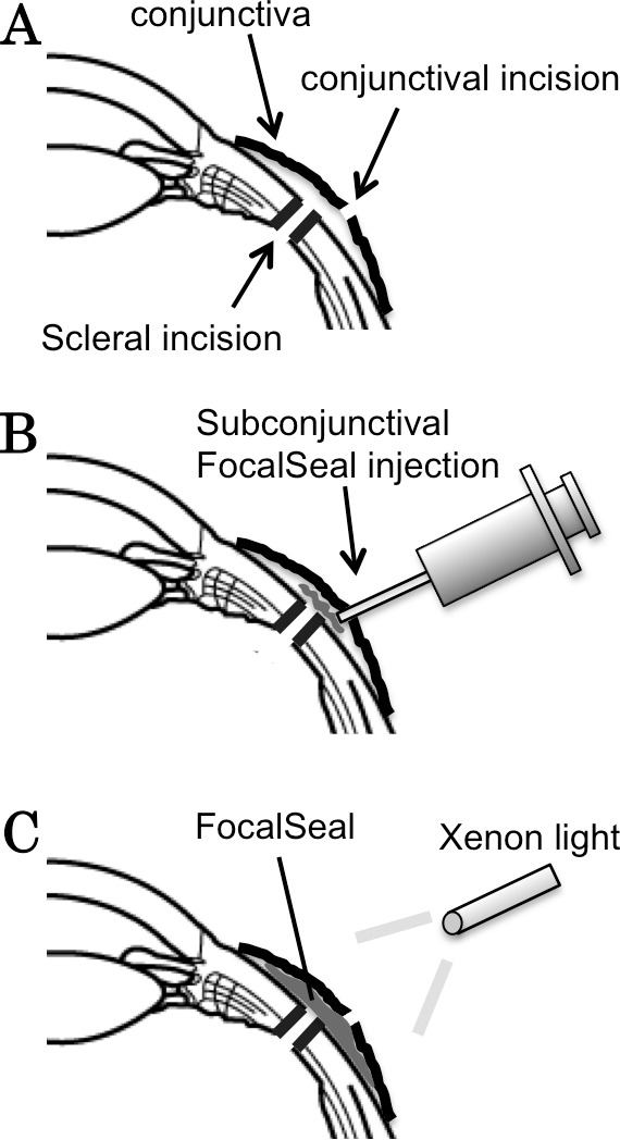
The three sclerotomies were termed infusion sclerotomy (involving the use of an infusion tip), experimental sclerotomy, and contralateral sclerotomy. The cannulae for experimental sclerotomy were removed and air leakage confirmed by the application of 0.625% povidone–iodine at the sclerotomy site. Then, 0.05 to 0.1 mL of PEG sealant was subconjunctivally injected through conjunctival incisions using a 27-gauge needle to cover the experimental sclerotomy wounds, following which it was polymerized by the application of light (420- to 700-nm wavelength) from a xenon light source (Accurus Xenon Illuminator; Alcon, Fort Worth, TX) for 60 seconds. The experimental sclerotomy wound was checked again for leakage at the sclerotomy site by dropping 0.625% povidone–iodine (Fig. 1). Then, the cannula at the contralateral sclerotomy site was removed and the wound was closed using 8-0 vicryl sutures (Coated VICRYL [polyglactin 910]; Ethicon Inc., Somerville, NJ). The air pressure via the infusion tip was gradually increased until air leakage was observed by dropping 0.625% povidone–iodine at the experimental or contralateral sclerotomy sites; this was done in only one of the six rabbits to evaluate how tightly the scleral wound was closed after PEG sealant application.
Figure 1.

Diagram of the process for sealing scleral incisions with PEG sealant. (A, B) After cannula removal and confirmation of air leakage at a sclerotomy site, PEG sealant is subconjunctivally injected using a 27-gauge needle through conjunctival incisions to cover the sclerotomy. (C) PEG sealant is polymerized by 60-second application of xenon light over the conjunctiva.
The infusion sclerotomy wound was then closed using 8-0 vicryl sutures after removal of the cannula connected to the infusion tip. Following the surgery, 0.1% betamethasone and 1.5% levofloxacin were applied three times a day for 7 days.
Ophthalmologic Examination
Slit-lamp microscopy and funduscopy by indirect ophthalmoscopy were performed after pupil dilatation the day before and 1, 3, 5, and 7 days after surgery. The bilateral intraocular pressure was measured using a rebound tonometer (Icare Pro; Icare Finland Oy, Helsinki, Finland) the day before and 1, 3, 5, and 7 days after surgery. A two-tailed paired t-test was used for statistical analysis to examine the relationship between the operated and contralateral eyes.
Histology
Rabbits were sacrificed with an overdose of pentobarbital 7 days after surgery, and their eyes were enucleated. After enucleation, the sclerotomy sites covered with PEG sealant were excised and fixed in 2% paraformaldehyde and 2.5% gluteraldehyde solution, dehydrated with serial alcohols, and embedded in paraffin. Sections were cut with a thickness of 4 μm, stained with hematoxylin and eosin, and examined under a light microscope.
Results
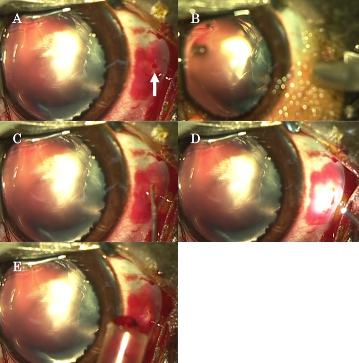
Before sealing the experimental sclerotomies with PEG sealant, air bubbles from the scleral incisions at all sites were observed when povidone–iodine was dropped on the wounds, confirming air leakage. After application, PEG sealant was rapidly polymerized by the application of xenon light after subconjunctival injection. Following this, no air leakage was observed when povidone-iodine was dropped in all cases (Fig. 2). When the air pressure was increased to 90 mm Hg, air leakage was observed at the contralateral sclerotomy site sutured with 8-0 vicryl, while the experimental sclerotomy site closed with PEG sealant remained secure.
Figure 2.
Photographs showing closure of a scleral incision using PEG sealant in a rabbit vitrectomy model. (A) After vitreous body removal and fluid–air exchange, the cannula is removed. The arrow indicates the conjunctival incision placed after cannula removal. (B) Air leakage, evident by the appearance of air bubbles from the scleral incisions, is confirmed by pouring 0.625% povidone-iodine at the sclerotomy site. (C) PEG sealant is subconjunctivally injected using a 27-gauge needle through conjunctival incisions to cover the sclerotomy. (D) PEG sealant is rapidly polymerized by the application of xenon light. The sclerotomy is closed. (E) No air leakage is observed when povidone-iodine is poured at the sclerotomy site.
In anterior segment examination, conjunctival hyperemia was observed in all eyes on the first postoperative day. Conjunctival and scleral wounds closed with PEG sealant were successfully attached and remained intact till the end of the follow-up period. Conjunctival bleb formation was not observed on the conjunctival surface at the sclerotomy sites during and after surgery. Mild intraocular inflammation was present on the first postoperative day, which resolved within 7 days. There was no sign of postoperative hypotony or infection in any eye, and no adverse effects of PEG sealant were encountered.
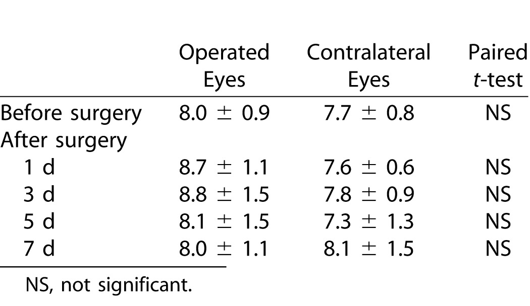
Table 1 shows the mean intraocular pressure in the operated and contralateral eyes before and 1, 3, 5, and 7 days after surgery; there were no significant differences between the two sets of eyes throughout the observation period.
Table 1.
The Mean Intraocular Pressure (mm Hg) in the Operated and Contralateral Eyes before and 1, 3, 5, and 7 Days after Surgery

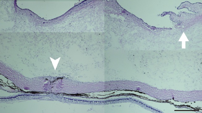
In histological examination, three of six sclerotomies were identified. Linear scar formation and eosinophilic staining of collagen fibers were observed at the sclerotomy sites, while the sclerotomy tunnels appeared tightly closed (Fig. 3). PEG sealant was not identified in any specimen.
Figure 3.

Photomicrographs of sections obtained from a sclerotomy site closed with PEG sealant. Connective tissue and collagen fibers can be observed in the scleral gap (arrowhead). The arrow indicates the conjunctival incision. There is no evidence of excessive inflammation, foreign body reaction, or toxic effects. Bar: 500 μm.
Discussion
PEG sealant is an absorbable PEG-based synthetic hydrogel. The liquid is polymerized under visible xenon illumination and forms a clear, flexible, and firmly adherent hydrogel. In our in vivo study, PEG sealant was easily delivered into the subconjunctival sclerotomy sites using a 27-gauge needle through conjunctival incisions and rapidly photopolymerized in a flexible mass by the application of xenon light, resulting in the successful sealing of sutureless 23-gauge vitrectomy incisions in rabbits.
In order to overcome potential complications related to wound leakage from sclerotomies, wound closure with cyanoacrylate glue, tissue fibrin glue, and other complex adhesives has been suggested.8,9 Cyanoacrylate glue is used during corneal procedures in some cases; however, it produces heat and settles rapidly into an inflexible and friable material. It should be recognized that patients may experience discomfort caused by the rough and dry surface of the glue on the ocular surface when it used for sealing sclerotomies. Furthermore, cyanoacrylate was reported to cause direct tissue toxicity, which is another limitation.14
Tissue fibrin glue (Tisseel; Baxter AG, Vienna, Austria) was used as an alternative to sutures in a small series of patients who underwent surgery for the closure of MIVS and 20-gauge vitrectomy incisions.15 Unlike cyanoacrylate glue, fibrin glue forms a smooth seal rather than a hard mass, thus providing greater postoperative comfort with fewer complications.16 Although it is nontoxic to the tissue and is less likely to cause a foreign body reaction, fibrin glue carries the theoretical risk of anaphylaxis and disease transmission.15
In this experiment, we tested the ability of PEG sealant to close 23-gauge vitrectomy incisions. PEG is widely used in the pharmaceutical industry because of its low toxicity and impressive safety profile, although a PEG-induced anaphylatic reaction has been reported.17 Many ophthalmic products, including lubricant eye drops, ophthalmic corticosteroids, ocular decongestants, and artificial tears, also contain this chemical. The hydrogel comprises approximately 95% water, with PEG cross-linked with trilysine as solid components. The link between each PEG and trilysine molecule contains a hydrolysable segment. Therefore, in the months following implantation, the hydrogel gradually weakens and liquefies, releasing the individual PEG and trilysine molecules. Hydrolysable linkages between the PEG molecules cause the hydrogel to liquefy within approximately 2 months. This gel degradation process is solely dependent on the presence of water and is not affected by enzymes. Trilysine is a product of L-lysine synthesis, which plays an important role in the formation of collagen and is a naturally occurring essential amino acid. The PEG and trilysine molecules are rapidly absorbed and cleared from the body via renal filtration.
Singh et al.18 conducted a laboratory study to evaluate the ability of the hydrogel bandage (ReSure Ocular Bandage; Ocular Therapeutix, Inc., Bedford, MA) to seal sutureless pars plana vitrectomy sclerotomies performed on human globes procured from an eye bank.18 The incisions received either a hydrogel device, a suture, or neither and were evaluated for the ingress of India ink. The bandage prevented the entry of ink particles in all covered incisions (11 of 11). One sutured eye (1 of 5) and four control eyes (4 of 5) permitted the ingress of ink through the incision. In a separate study with a different set of human cadaveric eyes, hydrogel sealant resulted in a secure, watertight seal for sutureless 23-gauge vitrectomy incisions.19 In that in vitro experiment, the outcomes were considered equivalent to those achieved with sutures. Laboratory experiments showed that the hydrogel seal was sufficiently strong and maintained ocular integrity at intraocular pressures beyond those experienced by the globe during eyelid blinking, eye rubbing, coughing, or eyelid squeezing.20 However, these experiments are limited by their in vitro nature, and it remains unclear whether PEG-based synthetic hydrogel bandage successfully closes sutureless 23-gauge vitrectomy incisions in vivo. Furthermore, the hydrogel sealant used by the previous authors requires mixing of two phases to initiate a cross-linking reaction before application. Also, the sealants were applied to the sclerotomies using a foam-tipped applicator and in the absence of conjunctiva; this does not simulate the actual clinical setting, where conjunctiva will also be present.18,19
To the best of our knowledge, this is the first in vivo experiment on the effectiveness of PEG-based synthetic hydrogel as a sealant for sclerotomy in MIVS. Because PEG sealant is a photocurable and single-phase gel that does not require prior mixing, it was easily delivered using a 27-gauge needle through conjunctival incisions and photopolymerized with xenon intraocular illumination, which is used in routine opthalmological surgeries. Our in vivo experiment indicated that PEG sealant was sufficiently strong and maintained ocular integrity throughout the observation period.
In ophthalmologic examination and histological evaluation, no adverse effects of PEG sealant on the ocular tissue were observed, with no signs of increased intraocular inflammation. It only induced a mild to moderate local inflammatory reaction, which is considered acceptable. We previously described that PEG sealant was nontoxic to rabbit eyes in vivo.21
In our histological examination, PEG sealant tightly closed the sclerotomy through fibrosis and collagen formation. Histological evaluations have shown that PEG sealant effectively adheres to tissues in other organs. Ranger et al.10 used PEG sealant for pulmonary air leaks in dog models and observed complete healing with a fibrotic layer overlying the area of the pulmonary defect in their histological examination. Alleyne et al.13 used PEG sealant as a dural sealing material in a canine craniotomy model and showed healing of the cut dural edges by the formation of a bridging fibrous and collagenous scar in their histological examination.
The limitations of this study include the relatively short observation period, because of which the long-term effects of the sealant remain unknown. Further studies with long-term observation periods are thus necessary. Because the thin sclera of rabbits was assessed in the current study, further studies with suitable models such as the pig eye or the human cadaveric eye are required to more precisely evaluate the effectiveness of PEG sealant in vitrectomy wound closure. In addition, the irritant effects of PEG sealant application to scleral wounds remain unknown in this animal study, as do the effects of blinking on the adherence of the sealant. It remains unknown whether PEG sealant will have similar disadvantages when sutures are used in humans. Further studies should clarify these issues.
In conclusion, we demonstrated the ability of PEG sealant, which is a PEG-based synthetic hydrogel, to successfully close sutureless 23-gauge vitrectomy incisions in rabbits. The material can be useful for pediatric vitrectomy because it precludes the requirement for suturing.
Acknowledgments
The authors thank Genzyme Corporation for providing FocalSeal. They also thank Editage (www.editage.jp) for English language editing.
Supported by Japan Society for the Promotion of Science (JSPS) KAKENHI (Grants-in-Aid for Scientific Research) Grant Number 23592551.
References
- 1. Sandali O,, El Sanharawi M,, Lecuen N,, et al. 25-, 23-, and 20-gauge vitrectomy in epiretinal membrane surgery: a comparative study of 553 cases. Graefes Arch Clin Exp Ophthalmol. 2011; 249: 1811–1819. [DOI] [PubMed] [Google Scholar]
- 2. Rizzo S,, Genovesi-Ebert F,, Murri S,, et al. 25-gauge, sutureless vitrectomy and standard 20-gauge pars plana vitrectomy in idiopathic epiretinal membrane surgery: a comparative pilot study. Graefes Arch Clin Exp Ophthalmol. 2006; 244: 472–479. [DOI] [PubMed] [Google Scholar]
- 3. Okamoto F,, Okamoto C,, Sakata N,, et al. Changes in corneal topography after 25-gauge transconjunctival sutureless vitrectomy versus after 20-gaugestandard vitrectomy. Ophthalmology. 2007; 114: 2138–2141. [DOI] [PubMed] [Google Scholar]
- 4. Kunimoto DY,, Kaiser RS;, Wills Eye Retina Service. Incidence of endophthalmitis after 20- and 25-gauge vitrectomy. Ophthalmology. 2007; 114: 2133–2137. [DOI] [PubMed] [Google Scholar]
- 5. Scott IU,, Flynn HW,, Jr,, Dev S,, et al. Endophthalmitis after 25-gauge and 20-gauge pars plana vitrectomy: incidence and outcomes. Retina. 2008; 28: 138–142. [DOI] [PubMed] [Google Scholar]
- 6. Gupta OP,, Ho AC,, Kaiser PK,, et al. Short-term outcomes of 23-gauge pars plana vitrectomy. Am J Ophthalmol. 2008; 146: 193–197. [DOI] [PubMed] [Google Scholar]
- 7. Duval R,, Hui JM,, Rezaei KA. Rate of sclerotomy suturing in 23-gauge primary vitrectomy. Retina. 2014; 34: 679–683. [DOI] [PubMed] [Google Scholar]
- 8. Batman C,, Ozdamar Y,, Aslan O,, Sonmez K,, Mutevelli S,, Zilelioglu G. Tissue glue in sutureless vitreoretinal surgery for the treatment of wound leakage. Ophthalmic Surg Lasers Imaging. 2008; 39: 100–106. [DOI] [PubMed] [Google Scholar]
- 9. Chan SM,, Boisjoly H. Advances in the use of adhesives in ophthalmology. Curr Opin Ophthalmol. 2004; 15: 305–310. [DOI] [PubMed] [Google Scholar]
- 10. Ranger WR,, Halpin D,, Sawhney AS,, Lyman M,, Locicero J. Pneumostasis of experimental air leaks with a new photopolymerized synthetic tissue sealant. Am Surg. 1997; 63: 788–795. [PubMed] [Google Scholar]
- 11. Macchiarini P,, Wain J,, Almy S,, Dartevelle P. Experimental and clinical evaluation of a new synthetic, absorbable sealant to reduce air leaks in thoracic operations. J Thorac Cardiovasc Surg. 1999; 117: 751–758. [DOI] [PubMed] [Google Scholar]
- 12. White JK,, Titus JS,, Tanabe H,, Aretz HT,, Torchiana DF. The use of a novel tissue sealant as a hemostatic adjunct in cardiac surgery. Heart Surg Forum. 2000; 3: 56–61. [PubMed] [Google Scholar]
- 13. Alleyene CH,, Jr, Cawley CM,, Barrow DL,, et al. Efficacy and biocompatibility of a photopolymerized, synthetic, absorbable hydrogel as a dural sealant in a canine craniotomy model. J Neurosurg. 1998; 88: 308–313. [DOI] [PubMed] [Google Scholar]
- 14. Carlson AN,, Wilhelmus KR. Giant papillary conjunctivitis associated with cyanoacrylate glue. Am J Ophthalmol. 1987; 104: 437–438. [DOI] [PubMed] [Google Scholar]
- 15. Panda A,, Kumar S,, Kumar A,, Bansal R,, Bhartiya S. Fibrin glue in ophthalmology. Indian J Ophthalmol. 2009; 57: 371–379. [DOI] [PMC free article] [PubMed] [Google Scholar]
- 16. Sharma A,, Kaur R,, Kumar S,, et al. Fibrin glue versus N-butyl-2-cyanoacrylate in corneal perforations. Ophthalmology. 2003; 110: 291–298. [DOI] [PubMed] [Google Scholar]
- 17. Gachoka D. Polyethylene glycol (PEG)-induced anaphylactic reaction during bowel preparation. ACG Case Rep J. 2015; 4: 216–217. [DOI] [PMC free article] [PubMed] [Google Scholar]
- 18. Singh A,, Hosseini M,, Hariprasad SM. Polyethylene glycol hydrogel polymer sealant for closure of sutureless sclerotomies: a histologic study. Am J Ophthalmol. 2010; 150: 346–351. [DOI] [PubMed] [Google Scholar]
- 19. Hariprasad SM,, Singh A. Polyethylene glycol hydrogel polymer sealant for vitrectomy surgery: an in vitro study of sutureless vitrectomy incision closure. Arch Ophthalmol. 2011; 129: 322–325. [DOI] [PubMed] [Google Scholar]
- 20. Coleman DJ,, Trokel S. Direct-recorded intraocular pressure variations in a human subject. Arch Ophthalmol. 1969; 82: 637–640. [DOI] [PubMed] [Google Scholar]
- 21. Hoshi S,, Okamoto F,, Arai M,, Hirose T,, Sugiura Y,, Kaji Y,, Oshika T. In vivo and in vitro feasibility studies of intraocular use of polyethylene glycol-based synthetic sealant to close retinal breaks in porcine and rabbit eyes. Invest Ophthalmol Vis Sci. 2015; 56: 4705–4711. [DOI] [PubMed] [Google Scholar]


