Abstract
Supernumerary breast is an uncommon disease seen in Asian population at an incidence of 2–6 % of general population. The pathology despite being present since birth remains asymptomatic till hormonal stimulation viz. puberty, pregnancy, and lactation. The management for such benign ectopic breast tissue is excision. However, one should always rule out malignancy in such lumps.
Keywords: Supernumerary breast, Ectopic breast, Kajava classification, Mammary glands
Supernumerary Breast on the Back
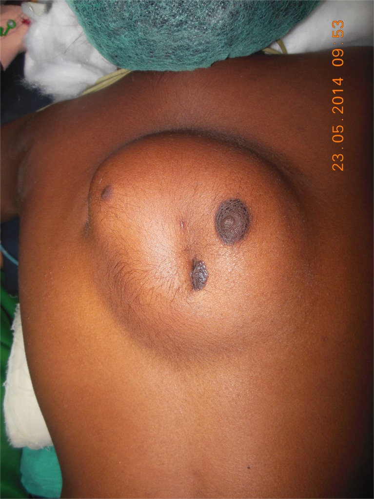
A 15-year-old female presented with progressively increasing lump in the back, coinciding with her thelarche. On examination, she had two normally placed breasts, with two accessory ectopic supernumerary breasts in interscapular region with two nipples and seven areolas (Fig. 1). NCCT chest revealed soft tissue density lesion in the subcutaneous plane of posterior chest wall in paramedian location bilaterally, from D4 to D10 (Fig. 2). She underwent excision of the accessory ectopic breast tissue, which on histopathological examination showed normal breast tissue with normal development as per age.
Fig. 1.
Interscapular bilateral polymastia with polythelia
Fig. 2.
NCCT chest of the patient showing subcutaneous posterior chest wall swelling
Discussion
Mammary glands are modified sweat glands which develop from mammary ridges that extend from the axilla to groin [1]. Failure of any portion of this ridge to involute leads to ectopic breast and usually lies along milk line (67 %). Rarely, it may lie outside these lines like the face, ears, neck, buttocks, and vulva [2]. It is usually sporadic; however, a hereditary and familial predisposition has also been reported [3].
In 1915, the Kajava classification was developed to describe supernumerary breasts: (1) complete breast with nipple, areola and glandular tissue; (2) supernumerary breast without areola; (3) supernumerary breast without nipple; (4) aberrant glandular tissue only; (5) nipple and areola with fat (pseudomamma); (6) nipple only; (7) areola only; and (8) hair only [4].
Although present at birth, it stays dormant until puberty, pregnancy, or lactation. It presents as a soft tissue swelling with or without a surrounding areola or nipple. It has been associated with many malformations, e.g., cardiac conduction disturbances, pyloric stenosis, Becker nevus, epilepsy, vertebral, and ear abnormalities. Excision is the recommended treatment for this pathology; however, liposuction has also been used [5].
Compliance with Ethical Standards
Conflict of Interest
The author declares that she has no competing interests.
References
- 1.Moore KL, Persaud TVN. The integumentary system. In: Moore KL, Persaud TVN, editors. The Developing Human: Clinically Oriented Embryology. Philadelphia, PA: W.B. Saunders Co; 1998. pp. 513–530. [Google Scholar]
- 2.Duvvur S, Sotres M, Lingam K, et al. Ectopic breast tissue of the vulva. J Obstet Gynaecol. 2007;27:530–531. doi: 10.1080/01443610701467333. [DOI] [PubMed] [Google Scholar]
- 3.Loukas M, Clarke P, Tubbs RS. Accessory breasts: a historical and current perspective. Am Surg. 2007;73:525–528. [PubMed] [Google Scholar]
- 4.Newman M. Supernumerary nipples. Am Fam Physician. 1988;38:183–188. [PubMed] [Google Scholar]
- 5.Aydogan F, Baghaki S, Celik V, et al. Surgical treatment of axillary accessory breasts. Am Surg. 2010;76:270–2. [PubMed] [Google Scholar]


