Abstract
Background
The search for intrinsic factors, which account for a protein's capability to act as an allergen, is ongoing. Fold stability has been identified as a molecular feature that affects processing and presentation, thereby influencing an antigen's immunologic properties.
Objective
We assessed how changes in fold stability modulate the immunogenicity and sensitization capacity of the major birch pollen allergen Bet v 1.
Methods
By exploiting an exhaustive virtual mutation screening, we generated mutants of the prototype allergen Bet v 1 with enhanced thermal and chemical stability and rigidity. Structural changes were analyzed by means of x-ray crystallography, nuclear magnetic resonance, and molecular dynamics simulations. Stability was monitored by using differential scanning calorimetry, circular dichroism, and Fourier transform infrared spectroscopy. Endolysosomal degradation was simulated in vitro by using the microsomal fraction of JAWS II cells, followed by liquid chromatography coupled to mass spectrometry. Immunologic properties were characterized in vitro by using a human T-cell line specific for the immunodominant epitope of Bet v 1 and in vivo in an adjuvant-free BALB/c mouse model.
Results
Fold stabilization of Bet v 1 was pH dependent and resulted in resistance to endosomal degradation at a pH of 5 or greater, affecting presentation of the immunodominant T-cell epitope in vitro. These properties translated in vivo into a strong allergy-promoting TH2-type immune response. Efficient TH2 cell activation required both an increased stability at the pH of the early endosome and efficient degradation at lower pH in the late endosomal/lysosomal compartment.
Conclusions
Our data indicate that differential pH-dependent fold stability along endosomal maturation is an essential protein-inherent determinant of allergenicity.
Key words: Allergic sensitization, Bet v 1, structural stability, endolysosomal degradation, antigen processing and presentation, molecular allergology
Abbreviations used: CD, Circular dichroism; DSC, Differential scanning calorimetry; FTIR, Fourier transform infrared spectroscopy; moDC, Monocyte-derived dendritic cell; NMR, Nuclear magnetic resonance; RBL, Rat basophil leukemia; SEC-MALS, Size exclusion chromatography coupled to multiangle light scattering
Among the plethora of environmental antigens to which human subjects are regularly exposed, only a few display a high propensity to act as allergens triggering type I allergic diseases (ie, TH2-biased immune responses characterized by aberrant levels of IgE production). Allergens encompass a diverse group of molecules that address innate immune pathways through protease activity, lipid binding, engagement of pattern recognition receptors, molecular mimicry of Toll-like receptor signaling molecules, or provision of complex carbohydrate structures.1 Interestingly, allergens can be found in only 2% of all known protein families, and the distribution of allergens is highly biased toward a few of these protein families.2 Hence it is obvious to speculate that beyond the abovementioned particular intrinsic biological functions, common structural and biochemical determinants of allergenicity might exist that have yet to be determined.
Bet v 1.0101 (Bet v 1), the major allergen found in birch pollen, belongs to family 10 of the plant pathogenesis-related proteins and is the most intensely studied allergen. Its structure,3 as well as its T- and B-cell epitopes,4, 5, 6, 7 are known. The hydrophobic cavity of Bet v 1 has been demonstrated to bind a variety of ligands,8 including pollen-derived quercetin,9 an iron-binding catechol derivative. Recently, the apo-form of Bet v 1 (loaded with catechol in the absence of iron) has been suggested to have a TH2-promoting potential.10 Moreover, binding of the model ligand deoxycholate has been implicated in compaction and rigidification of the Bet v 1 molecule11 and stabilization of its conformational epitopes.12 These data indicate that the structural stability of Bet v 1 represents a determinant of its allergenicity.
To investigate solely the effect of conformational stability on the sensitization capacity of Bet v 1, we used an approach lacking any immunologically relevant confounders, such as artificial ligands or immunization with adjuvants. We generated variants of Bet v 1 with enhanced fold stability by means of introducing 1, 2, 3, or 4 sequential point mutations. Mutations were selected based on a recently developed in silico approach that uses normalized knowledge-based energy potentials for predicting the influence of single point mutations or combinations thereof on protein stability.13 In-depth comparison of the Bet v 1 mutants with the wild-type protein revealed increased thermal and chemical stability, reduced backbone flexibility, and enhanced resistance to degradation by proteases. In the absence of adjuvants, the wild-type molecule induced only marginal immune responses, whereas the stabilized mutants triggered significant serum IgG and IgE titers and IL-4 production. Allergenicity and immunogenicity of the mutant proteins strongly correlated with protease resistance at slightly acidic pH and efficient proteolytic processing at lower pH, resembling the early and late endosomal environment, respectively. Based on our findings, we propose that differential, pH-dependent fold stability in the early versus late endosomal compartment is a protein-inherent key determinant of allergenicity.
Methods
Expression and purification of recombinant proteins
Wild-type Bet v 1 and the 4 mutants were expressed from pET28b constructs in Escherichia coli strain BL21 Star (DE3; Invitrogen, Carlsbad, Calif), as described in the Methods section in this article's Online Repository at www.jacionline.org.
Protein characterization and structure determination
Protein folding in solution and thermal and chemical denaturation were monitored by using differential scanning calorimetry (DSC), circular dichroism (CD), and Fourier transform infrared spectroscopy (FTIR). The monomeric state was measured by using size exclusion chromatography coupled to multiangle light scattering (SEC-MALS). Structure was determined by means of x-ray crystallography. Protein flexibility was assessed by using molecular dynamics simulations, nuclear magnetic resonance (NMR) spectroscopy, and hydrogen/deuterium exchange. These methods are described in detail in the Methods section in this article's Online Repository.
Mice and immunizations
Female 6- to 10-week-old BALB/c mice were obtained from Charles River Laboratories (Sulzfeld, Germany) and maintained at the animal facility of the University of Salzburg according to local guidelines for animal care. Animal experiments were approved by the Austrian Ministry of Science (permit no. BMWFW-66.012/0008-WF/II/3b/2014). Mice (n = 5) were immunized 5 times with 20 μg of Bet v 1 or one of the mutant proteins in sterile PBS by means of intradermal injection into the ear pinnae on days 0, 14, 28, 42, and 81. A total volume of 80 μL was divided between the 2 sides. Blood samples were taken at regular intervals. Mice were killed on day 88 for preparation of splenocytes.
Antibodies and cytokines
Levels of IgG1 and IgG2a subclass antibodies in sera measured on day 88 were evaluated by using a luminometric ELISA14 at serum dilutions of 1:300 and 1:100, respectively, lying within the linear range of the assay.
The IgE cross-linking capacity of the mutant proteins was assessed by using a β-hexosaminidase release assay, as described in the Methods section in this article's Online Repository.
The amount of cell-bound IgE was measured by using a basophil activation test, as previously described.15 Heparinized whole blood taken on day 84 of the experimental schedule was diluted 1:2 with RPMI 1640 and incubated with 10 ng/mL Bet v 1 or the respective mutant or left untreated as a control for 2 hours at 37°C and 5% CO2. Cells were washed, and surface staining was performed with anti-CD200 R (clone OX110; eBioscience, San Diego, Calif), anti-IgE (clone RME-1; BioLegend, San Diego, Calif), anti-CD4 (clone GK1.5, BioLegend), and anti-CD19 (clone 6D5, BioLegend). Subsequently, cells were washed with PBS/1% BSA/2 mmol/L EDTA and analyzed on a FACSCanto II flow cytometer (BD Biosciences, San Jose, Calif).
Splenocytes from immunized mice were prepared and cultured, as previously described,14 in the presence of Bet v 1 or the mutants (20 μg/mL) to determine the number of IL-4 and IFN-γ producers per 2 × 10⁵ cells by using ELISpot (Millipore, Bedford, Mass).
Endolysosomal degradation simulations
Endolysosomal degradation assays were performed, as previously described.16 Briefly, 5 μg of protein substrates (Bet v 1 and Bet_mut1 to Bet_mut4) was mixed with 8 μg of isolated microsomal fraction from the JAWS II cell line in 50 mmol/L citrate buffer (pH 5.9, pH 5.2, or pH 4.5) and 2 mmol/L dithiothreitol.
The pool of peptides generated from Bet v 1 and the mutants in the degradation assay at pH 5.2 and pH 4.5 was assessed by using mass spectrometry with a Q-Exactive Orbitrap Mass Spectrometer (Thermo Fisher Scientific, Waltham, Mass) with nanoelectrospray and nano-HPLC (Dionex Ultimate 3000, Thermo Fisher Scientific). For details, see the Methods section in this article's Online Repository.
Uptake and processing of Bet v 1 and Bet variants by monocyte-derived dendritic cells and stimulation of Bet v 1–specific T cells
Experiments involving human cells were conducted in accordance with the guidelines of the World Medical Association's Declaration of Helsinki. Samples of human origin were obtained from blood donations, and use of residual cells was approved by the Institutional Review Board of Upper Austria. Monocyte-derived dendritic cells (moDCs) were generated from buffy coats from healthy anonymous donors (provided by the Red Cross Transfusion Service of Upper Austria), as described in the Methods section in this article's Online Repository. For investigating the uptake kinetics of Bet v 1 and its variants, moDCs were incubated with DyLight488-labeled proteins at 20 μg/mL for 30, 60, and 120 minutes, respectively, and uptake was determined by means of flow cytometry.
moDCs from HLA-DRB1*07:01–restricted donors were incubated for 30 to 480 minutes with Bet v 1 or the mutants at 20 μg/mL. Subsequently, moDCs were washed and cocultured with Jurkat T cells expressing a Bet v 1142-153–specific T-cell receptor and harboring a human IL-2 promoter/enhancer controlling luciferase expression17 for 6 hours. Cells were lysed in 50 μL of lysis buffer (100 mmol/L potassium phosphate, 0.1% Triton X-100, and 1 mmol/L dithiothreitol), and luciferase activity was measured in a Tecan Infinite 200 Pro microplate reader with 50 μL of luciferase substrate (Promega, Madison, Wis).
Results
Generation of Bet v 1 mutants with increased fold stability
We have previously demonstrated the use of knowledge-based potentials for generating destabilized allergens.13 Knowledge-based potentials are used to investigate sequence-structure relations in known 3-dimensional protein structures, describing the interactions between amino acid residues and the interaction of the residues with the surrounding solvent. By using this method, a z score is generated, which indicates either stabilization (decrease of z score) or destabilization (increase of z score).18 Here we applied this approach to generate fold-stabilized mutants of Bet v 1. By using in silico permutation of every amino acid, mutants were selected, which predicted the strongest stabilizing effect. The immunodominant region of Bet v 1 (Bet142-156)4 was excluded from the mutation process to avoid changes in immunogenicity because of destruction of T-cell epitopes. Likewise, solvent-exposed residues (accessible surface area >30%), which can contribute to IgE binding,19 were left untouched. In an iterative manner, up to 4 point mutations were introduced, resulting in the generation of Bet_mut1, Bet_mut2, Bet_mut3, and Bet_mut4 (Table I). All selected mutations were located in regions representing minor T-cell epitopes recognized by only 3.5% to 8.8% of allergic donors4 or 0% to 3.3% of sensitized BALB/c mice,20 respectively.
Table I.
Bet v 1 mutants used in the study
| Protein | Mutations | Combined z score |
|---|---|---|
| Bet v 1 | — | −8.34 |
| Bet_mut1 | D69I | −8.9 |
| Bet_mut2 | D69I, K97I | −9.34 |
| Bet_mut3 | D69I, K97I, P90L | −9.80 |
| Bet_mut4 | D69I, K97I, P90L, G26L | −10.18 |
Proteins were expressed tag-free in E coli and purified by using hydrophobic interaction chromatography and anion exchange chromatography, followed by an endotoxin removal step. The final purity was greater than 99%, as determined by using SDS-PAGE (see Fig E1, A, in this article's Online Repository at www.jacionline.org), and preparations were essentially LPS free (<0.4 pg/μg protein). Identity was confirmed by means of mass spectroscopy (see Fig E1, B), and the correct conformation (see Fig E1, C) and monomeric state (see Fig E1, D) of the proteins were demonstrated by using CD spectroscopy and SEC-MALS, respectively.
A more detailed analysis with x-ray crystallography showed that the 3-dimensional structures of the mutants were remarkably similar to that of the wild-type protein. The superimposition of the crystal structures of the quadruple mutant Bet_mut4 with Bet v 1 revealed that the overall fold is identical, with a total root-mean-square deviation of 0.7 Å (Fig 1).
Fig 1.
Crystal structure of the quadruple mutant Bet_mut4 (blue) in comparison with Bet v 1 (gray, PDB: 4A88). Superimposition of Bet_mut4 with Bet v 1 revealed that the overall fold is conserved. A, Mutated residues D69I, K97I, P90L, and G26L are shown as sticks. B, An enlarged view of the mutated residue D69I shows that it is stabilized by several hydrophobic residues, including Ile56, Phe58, Val67, Tyr83, and Val85. C, The surface-exposed mutation K97I is likely to entropically stabilize the protein by releasing coordinated solvent molecules. D, Mutation P90L induced an alternative conformation, as indicated. E, Mutation G26L fills a hydrophobic cavity formed by Phe21, Ile22, and Phe30. All mutated residues are shown as blue sticks compared with sticks from wild-type Bet v 1 (gray). The electron density map (2Fobs-Fcalc) is contoured at 1 σ over the mean.
Overall fold identity was also confirmed by using an IgE-binding assay with rat basophil leukemia (RBL) cell degranulation (see Fig E2, A, in this article's Online Repository at www.jacionline.org), which demonstrated that all mutants had retained the capacity to cross-link cell-bound IgE. This indicates that surface epitopes were mainly left intact by the introduction of stabilizing mutations. Degranulation was slightly lower in the mutants compared with the wild-type protein, suggesting the possibility of subtle changes to conformational epitopes. Likewise, an RBL assay and a basophil activation test were performed with human sera from donors with birch pollen allergy. All mutant proteins showed very similar IgE-binding capacity compared with the wild-type Bet v 1 (see Fig E2, B and C). This confirms that the surface structure of the recombinant mutant proteins resembles that of naturally occurring Bet v 1.
Introduction of the point mutations effectively enhanced the thermal and chemical fold stability of Bet v 1, as assessed by using CD spectroscopy and DSC. All proteins were analyzed in phosphate buffer at pH 7.4. The first mutation (D69I) was sufficient to enhance chemical stability (ie, the amount of urea required to induce 50% unfolding of the protein) from 2.8 to 5.2 mol/L urea, corresponding to a shift in Gibbs-free energy (ΔG) of approximately 4 to 5 kcal/mol (Fig 2, A). Interestingly, the additional mutations showed no additive effect. This was confirmed by results of thermal denaturation analysis in which D69I induced a shift of greater than 20°C in melting temperature (Fig 2, B-D). Inclusion of the P90L mutation in mutants 3 and 4 led to evident aggregation at temperatures of greater than 80°C, as indicated by the distorted baseline after the transition peak (marking the transition from native to unfolded state) in the DSC diagram (Fig 2, C). Despite this complexity, the same pattern of stabilization for these mutants over wild-type protein was observed at different protein concentrations (0.15-0.5 mg/mL; Fig 2, C). In the case of mutant 4, there was also an additional reproducible decrease in baseline before the transition peak of unknown origin. A similar stabilization for the mutants of approximately 5 kcal/mol could be estimated from the DSC data. Thermal denaturation was not repeatable, indicating a nonequilibrium mechanism and thus preventing detailed interpretation of the DSC data. However, the data indicated the exact same pattern and magnitude of significant fold stabilization seen in chemical and thermal denaturation by using CD (Fig 2, A and B).
Fig 2.
Thermal stability of Bet v 1 and mutant proteins 1 to 4 (Bet_mut1-4). A, Chemical denaturation was assessed by means of CD spectroscopy, and the fraction of denatured protein (Fdenatured) is displayed. B and C, Thermal denaturation was measured by using CD (Fig 2, B) and DSC (Fig 2, C). DSC data for wild-type Bet v 1 and mutants were run at a 60°/h scan rate and at 0.15 mg/mL (lower traces) and 0.5 mg/mL (upper traces) offset in the y-axis for clarity. D, Melting points calculated from DSC data.
Increased fold stability enhances allergenicity and immunogenicity
BALB/c mice were immunized intradermally into the ear pinnae with the respective protein dissolved in PBS to assess immune response in the absence of immunomodulatory confounders, such as adjuvants. As shown in Fig 3, A, Bet v 1 was nonimmunogenic in this adjuvant-free setting. In contrast, mutants 2, 3, and 4 induced significantly higher titers of Bet v 1–specific IgG1 and low levels of IgG2a. Notably, antibody responses against the homologous mutant protein in the individual groups (with few exceptions) closely resembled those observed against the wild-type protein, underscoring the high degree of conformational homology of the mutant proteins (see Fig E3 in this article's Online Repository at www.jacionline.org). Introducing fold-stabilizing mutations not only increased the immunogenicity in terms of IgG1 production but also resulted in the formation of allergen-specific IgE, which was mainly cell bound (Fig 3, B). This was most prominent for mutant 4, which also induced high levels of IL-4–secreting splenocytes (Fig 3, C) and detectable levels of free, allergen-specific serum IgE (Fig 3, B). No significant levels of IFN-γ were detected in any group (Fig 3, C). In summary, allergenicity and immunogenicity were increased with every additional introduction of a fold-stabilizing mutation, with the most striking effects observed for the mutant harboring 4 point mutations.
Fig 3.
Influence of fold-stabilizing mutations on the immunogenicity and sensitization capacity of Bet v 1. A, Bet v 1–specific IgG1 and IgG2a titers of BALB/c mice (n = 5) immunized intradermally with the different proteins were measured by using a luminometric ELISA at a serum dilution of 1:300 for IgG1 and 1:100 for IgG2a. Data are shown as relative light units (RLU). B, Bet v 1–specific IgE levels in serum were measured with an RBL degranulation assay, and results are presented as the percentage of total release. Cell-bound IgE was assessed by using a basophil activation test, and data are shown as stimulation indices (SI). C, Numbers of IL-4– and IFN-γ–secreting splenocytes after restimulation with Bet v 1 and the respective mutants were determined by using the ELISpot assay. SFU, Spot-forming units. Statistical significance compared with Bet v 1 was assessed by using 1-way ANOVA, followed by the Dunnett post hoc test. *P < .05, **P < .01, and ***P < .001.
Fold-stabilizing mutations reduce flexibility
The data shown above clearly point to an effect of the number of fold-stabilizing mutations on allergenicity and immunogenicity. However, none of the 4 mutants differed in their thermal stability. Because the flexibility of a protein is known to influence the accessibility of lysosomal proteases and hence presentation of peptides on MHC molecules,21, 22 we studied the molecular dynamics of the wild-type molecule and the 4 mutants with simulations over 100 ns for each system by using AMBER12.23 To capture residue-wise flexibility, we calculated B factors for Cα atoms from the resulting conformational ensembles of the proteins after global alignment. Furthermore, we calculated dihedral entropies based on the distributions of torsion angles of the protein backbone.24 Simulation data indicate that all systems are rigidified considerably by the introduction of the point mutations. Local dynamics of residues around the mutation sites are especially reduced, thus pointing to a gain in stability (see Fig E4 in this article's Online Repository at www.jacionline.org).
To experimentally confirm changes in flexibility, we used hydrogen/deuterium exchange measurement with FTIR. As shown in Fig 4, A, all mutants showed an overall slower exchange rate compared with the wild-type protein, indicating reduced molecular dynamics. However, at this much slower time scale, Bet_mut4 turned out to be the most rigid molecule. These findings could be further substantiated by using relaxation dispersion NMR,25 which confirmed the substantial decrease in flexibility on the microsecond-to-millisecond time scale in Bet_mut4 (Fig 4, D) compared with Bet_mut1 (Fig 4, C) and the wild-type molecule (Fig 4, B).
Fig 4.
A-D, Influence of fold-stabilizing mutations on the molecular flexibility of the protein, as indicated by hydrogen deuterium exchange (Fig 4, A) and relaxation dispersion NMR (Fig 4, B-D). In Fig 4, B-D, the 11 residues with the largest backbone amide 15N relaxation dispersion profiles are displayed.
Protease-mediated degradation is reduced in fold-stabilized mutants
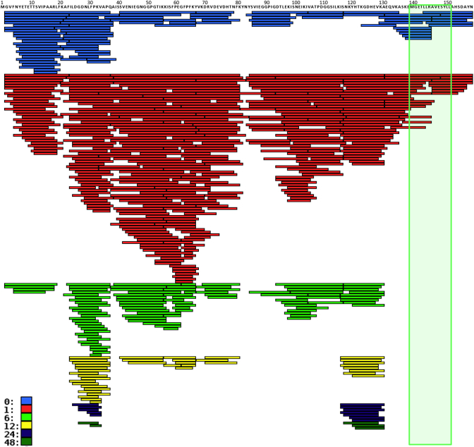
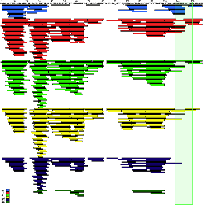
To assess whether the increased thermal stability and reduced flexibility of fold-stabilized mutants indeed affects the degradation of the protein in the endolysosome, we performed a degradation assay using microsomal enzyme preparations of JAWS II cells at different pH values, thus simulating the pH decrease during maturation from early endosome to late endolysosome.
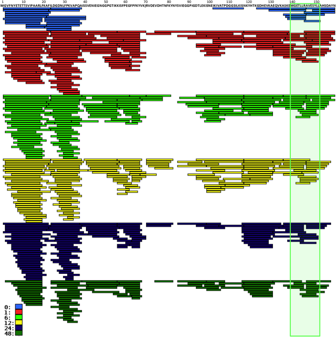
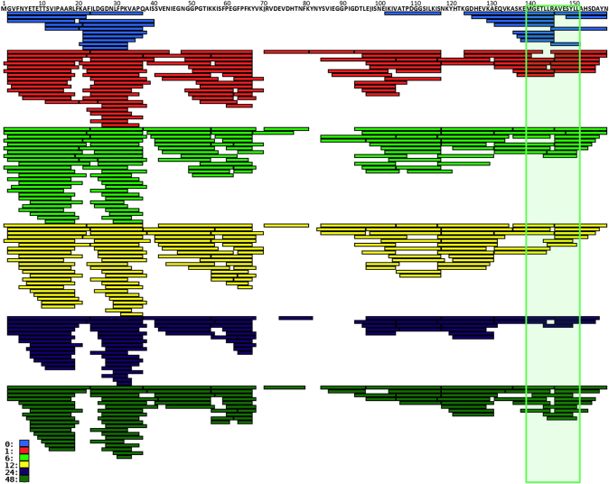
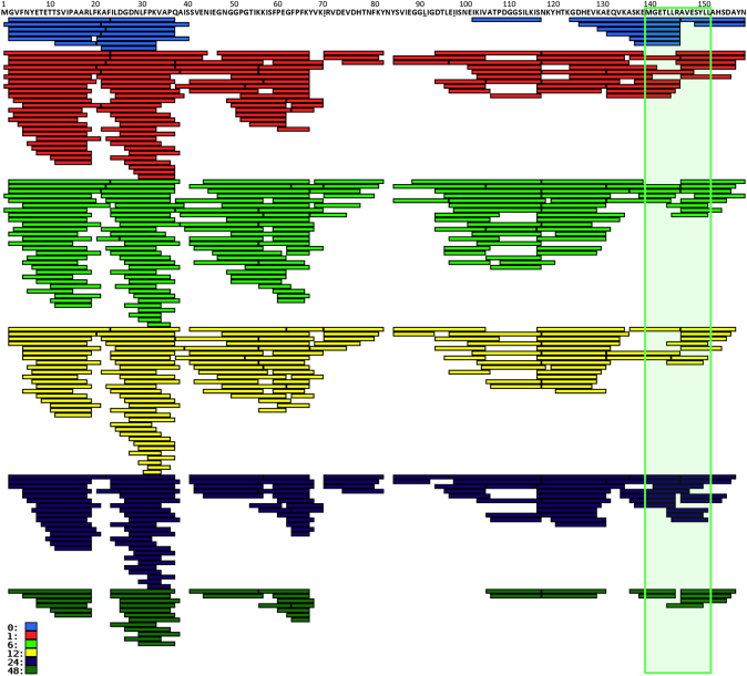
The degradation assay revealed no detectable proteolysis of Bet v 1 at pH 5.9, which might reflect an intrinsic stability of wild-type Bet v 1, as well as a reduced proteolytic activity at this pH, but a complete degradation at a pH of 5.2 within 12 hours, as assessed by using SDS-PAGE (Fig 5, A). In contrast, all mutants were resistant to proteolysis under these conditions. However, at lower pH, Bet_mut4 suddenly became susceptible to degradation. Interestingly, this change in protease susceptibility at lower pH correlated with a reduced thermal stability, as assessed by using CD spectroscopy (monitoring protein unfolding; Fig 5, B) and FTIR (here used for monitoring aggregation by unfolding; Fig 5, C). Analysis of the resulting peptides with liquid chromatography coupled to mass spectrometry revealed that processing of Bet v 1 progresses from the N- and C-termini of the molecule to the inner core. Proteolysis of the mutants followed a similar pattern, although at lower speed (Fig 5, D). No differences in the population of generated peptides were observed between Bet v 1 and its variants, indicating that no cleavage sites were introduced or removed by the inserted point mutations (full degradation patterns at different pH can be found in Figs E5-E14 in this article's Online Repository at www.jacionline.org). It must be taken into account that these degradation processes will happen much faster in vivo because of the extremely high local concentration of proteolytic enzymes in the endolysosome. For example, cathepsin concentrations in lysosomes can well exceed 1 mmol/L,26 which is at least 106-fold higher compared with the concentrations used in our in vitro assays. Nevertheless, the assay qualitatively resembles the course of in vivo processing because the full proteolytic machinery is present in the endolysosomal preparations.16
Fig 5.
A-D, Proteolytic degradation of Bet v 1 and the mutants Bet_mut1 to Bet_mut4 (Fig 5, A and D) and their thermal stability (Fig 5, B and C) at different pH values. Proteolytic degradation was assessed by using 15% SDS-PAGE and Coomassie staining (Fig 5, A). Thermal stability was followed by using CD spectroscopy (Fig 5, B), and thermally induced aggregation was followed by using attenuated total reflectance FTIR (Fig 5, C). FTIR data are presented as the fraction of aggregated protein versus temperature. Protein degradation patterns over time were analyzed by using liquid chromatography–mass spectroscopy (Fig 5, D): peptides generated earlier during the proteolytic processing are colored in white, whereas peptides that were not detected during the whole experiment (48 hours) were colored in black. Amino acid positions are labeled at the top. The immunodominant epitope of Bet v 1 is framed in green.
Presentation of the immunodominant epitope Bet142-153 is enhanced in Bet_mut4
Uptake of Bet v 1 and the mutant proteins by moDCs was monitored by pulsing cells with DyLight488-labeled proteins. Flow cytometric analysis revealed no differences with regard to the percentage of antigen-positive cells (Fig 6, A) and their mean fluorescence intensity (Fig 6, B).
Fig 6.
A and B,In vitro antigen processing and presentation. Uptake of DyLight488-labeled Bet v 1 and Bet_mut1-4 by moDCs. The percentage of positive cells (Fig 6, A) and their mean fluorescence intensity (MFI; Fig 6, B) were analyzed by using flow cytometry. moDCs from HLA-DRB1*07:01+ donors were preincubated with Bet v 1 or Bet_mut1-4 for the indicated time span and subsequently cocultured with Bet142-153–specific Jurkat T cells. C, Luciferase reporter expression under control of the IL-2 promoter was measured after 6 hours of coculture. Data are shown as means ± SEMs of relative light units (RLU) of 2 independent experiments by using 2 different donors.
To rule out that the increase in immunogenicity observed for Bet_mut2-4 might be due to the generation of new T-cell epitopes by the mutation process, we investigated the activation of a transgenic HLA-DRB1*07:01–restricted Jurkat T-cell line specific for the immunodominant peptide Bet142-153. moDCs from a HLA-DRB1*07:01–positive donor were cultured with Bet v 1 and the mutant proteins for 30 to 1440 minutes before adding Jurkat cells expressing the specific T-cell receptor. Six hours later, T-cell activation was determined by measuring a luciferase reporter under control of the IL-2 promoter.17
Although antigen uptake was similar for the wild-type protein and the 4 mutants, Bet_mut4 (and, to a lesser degree, Bet_mut3) showed enhanced T-cell stimulatory capacity, especially at the early time points (Fig 6, C). This indicates that larger amounts of the immunodominant peptide are presented by moDCs when processing the fold-stabilized mutants Bet_mut3 and Bet_mut4. Notably, both variants were highly stable at a pH of greater than 5.2 while becoming destabilized at lower pH. These in vitro data from human cells remarkably resemble in vivo data from mouse experiments (Fig 3), supporting a mechanism for immunogenicity being dependent on high stability in the early endosome and protease susceptibility in the late endosome.
Discussion
Recent concepts link protein-intrinsic features, such as protease activity, molecular mimicry, and ligand binding, to allergic immune responses.1, 27 However, there are very few data dealing with the role of protein fold stability concerning induction of a TH2 immune response.28
Antigen processing and presentation consists of several steps, including uptake/internalization of proteins, reduction of disulfide bonds and unfolding, endosomal/lysosomal proteolysis, and loading of peptides onto MHC class II molecules. Because proteases preferentially degrade proteins in a denatured state, unfolding represents an indispensable step for intracellular processing of protein antigens.29 Interestingly, modifying proteins to increase their stability has produced contradictory results. Some studies reported decreased T-cell or antibody responses when immunizing with stabilized antigens,30, 31, 32 whereas others could correlate increased stability/slower proteolytic degradation of internalized antigens with prolonged retention of antigen in secondary lymphoid organs and enhanced presentation of T-cell epitopes33 and increased humoral and long-lasting cellular immunity.34, 35, 36 Reduced susceptibility to degradation might not only enhance the preservation of CD4+ T-cell epitopes but also prolong the persistence of the proteins for sustained processing and presentation by DCs and even allow for transfer of intact antigen to B cells.37, 38
In the present study we used an unbiased computational approach and generated Bet v 1 mutants with a striking increase in their fold stability while preserving the natural conformation of the wild-type molecule. The stabilizing effects of the mutations D69I, K97I, and G26L can be explained by the local environment within the crystal structure (Fig 1, A). Contrasting the original Asp69, the isoleucine matches the hydrophobic nature of the cavity formed by Ile56, Phe58, Val67, Tyr83, and Val85. Additionally, several solvent molecules were released in the mutated structure compared with the wild-type structure, leading to an entropically driven stabilization (Fig 1, B). A similar effect was also observed for K97I, which is located on the protein surface. Furthermore, the conformation of the neighboring residue Glu95 was slightly different, thus stabilizing the hydrophobic residue with its aliphatic chain (Fig 1, C). The mutation P90L induced an alternative conformation in the main chain of the surrounding residues as indicated, which is more difficult to interpret with respect to its effect on stability (Fig 1, D). Mutation G26L fills an internal hydrophobic cavity formed by Phe21, Ile22, and Phe30 (Fig 1, E).
Taken together, all the introduced mutations were aliphatic residues oriented toward the hydrophobic cavity of Bet v 1 (Fig 1). These mutations act like anchors responsible for the dramatic rigidification of the molecular structure predicted by molecular dynamics (see Fig E4) and further confirmed by hydrogen/deuterium exchange and NMR (Fig 4). Interestingly, binding of model ligands to the hydrophobic cavity of Bet v 1 has been shown to result in rigidification11 and reduced proteolytic degradation12 of the protein, which is similar to what we observed in our experimental system. Ligand binding to Bet v 1 has also been linked to its allergenic potential.10, 12 However, in our ligand-free system we could demonstrate for the first time that not the ligand itself but the change of the structural stability of the protein represents the underlying general principle of immunomodulation. Also, we can exclude aggregation39 as a biologically relevant mechanism for the observed effects because aggregation events only took place at temperatures of greater than 60°C (Fig 2, C).
In a recent publication Ohkuri et al31 have suggested that a certain energy threshold exists that solely defines whether a protein is immunogenic. Our data clearly indicate that this is an oversimplification not reflecting the complex effect of pH-dependent protein stability in the course of degradation, starting in the early and moving to the late endosomal compartment. It has to be taken into account that antigens face a continuous decrease in pH from 7.4 (in the extracellular environment) to 4.0 in terminal lysosomes. Indeed, we could demonstrate that the mutant proteins Bet_mut1 to Bet_mut4 show similarly high stability at neutral pH; however, they strongly differ in their stability and proteolytic susceptibility at acidic pH, and this affects their immunogenicity. Interestingly, the dominant T-cell epitope of Bet v 1 was found to be highly susceptible to degradation independent of pH or time point in in vitro degradation assays (Fig 5, D, and see Figs E5-E14); indeed, we never identified the full-length peptide Bet142-153. This suggests that in vivo this epitope has to be protected from proteolytic degradation, possibly by binding to class II molecules.40
On the basis of these results, we propose a model (Fig 7) in which immunogenicity/allergenicity is determined by a pH-dependent balance of protease accessibility of allergens, resulting in optimal loading of MHC class II. It is known that the early endosome (approximately pH 6) and the terminal lysosome (approximately pH 4.0-4.5) are poor in MHC class II, whereas late endosomes (approximately pH 4.5-5.5) display high quantities of MHC class II organized in multivesicular bodies.41 The poor immunogenicity of Bet v 1 after vaccinating mice in the absence of adjuvant can be explained by the low proteolytic stability of the molecule along the endosome maturation, a process highly conserved between species.42, 43 This results in low peptide abundance in the late endosome, in which the majority of MHC loading takes place.41 Interestingly, a similarly low immunogenicity can be observed for Bet_mut1, although it is extremely stable to pH-mediated unfolding. The resulting poor accessibility to proteases leads to the same effect as low proteolytic stability (ie, low peptide abundance in the late endosome and suboptimal MHC loading). In contrast, Bet_mut4 is highly stable at a pH of greater than 5, suggesting that it accumulates during the early endosomal maturation and can then be processed efficiently in the late endosome, where it becomes susceptible to degradation. Our in vivo mouse data are further substantiated by in vitro data from human moDCs (Fig 6). Thus the differential stability of Bet_mut4 at different pH values (Fig 5) results in efficient presentation of T-cell epitopes in the context of MHC-II on human moDCs (Fig 6) and promotes TH2-biased immune responses in mice (Fig 3).
Fig 7.
Model for immunogenicity/allergenicity of an allergen depending on differential fold stability at different endosomal pH. During antigen uptake and processing, allergens face a broad range of different pH values, ranging from approximately 7.4 in the extracellular matrix to approximately 4.0 in the terminal lysosome. Proteins with low fold stability in the early endosome are proteolytically processed prematurely, resulting in poor antigen presentation because of low abundance of MHC class II in this compartment (“blue” protein). Proteins with high fold stability at low pH (“black” protein) remain structurally intact until they reach the terminal lysosome, where again MHC class II levels are low and antigen presentation is inefficient. Only proteins that survive the early endosome but are efficiently processed at a more acidic pH can accumulate in the late endosome, where they are finally degraded and loaded on highly abundant MHC II molecules (“red” protein).
In summary, our data clearly show that the pH-dependent stability of Bet v 1 is a key factor for allergenicity and immunogenicity in vivo and that epitope-specific T-cell activation is highly influenced by the fold stability of the protein. Our results also provide a mechanism for how real-life ligand binding to the hydrophobic cavity of Bet v 1 could render the molecule highly allergenic (ie, by inducing a change in fold stability similar to that observed for Bet_mut4). This has direct implications for the use of recombinant Bet v 1.0101 for allergen-specific immunotherapy because in the absence of a ligand, the recombinant protein might not be optimal in stimulating potent T-cell responses. Thus in addition to increasing the understanding of structural determinants of allergenicity, our findings have broader implications for a rational design of hyperimmunogenic antigens for vaccination, as well as hypoimmunogenic molecules for use as biological agents.
Key messages.
-
•
Fold stability is a key determinant of the sensitizing potential of allergen molecules.
-
•
Efficient TH2 cell activation requires both an increased stability at the pH of the early endosome and efficient degradation at a lower pH resembling the late endosomal/lysosomal compartment.
Acknowledgments
We thank Winfried Pickl and Alina Neunkirchner (University of Vienna) for supplying Bet v 1 T-cell receptor transgenic Jurkat cell line and for their help in establishing the culture, as well as Michael Wallner (University of Salzburg), who provided sera from patients with birch pollen allergy used in the RBL assay.
Footnotes
Disclosure of potential conflict of interest: Y. Machado, R. Freier, and J. Thalhamer have received research support from the Austrian Science Fund FWF (W01213). S. Scheiblhofer, K. R. Liedl, and R. Weiss have received research support from the Austrian Science Fund FWF (P26849). The rest of the authors declare that they have no relevant conflicts of interest.
Supplementary data
Fig E1.
Fig E2.
Fig E3.

Fig E4.
Fig E5.
Fig E6.
Fig E7.
Fig E8.
Fig E9.
Fig E10.
Fig E11.
Fig E12.
Fig E13.
Fig E14.
References
- 1.Wills-Karp M., Nathan A., Page K., Karp C.L. New insights into innate immune mechanisms underlying allergenicity. Mucosal Immunol. 2010;3:104–110. doi: 10.1038/mi.2009.138. [DOI] [PMC free article] [PubMed] [Google Scholar]
- 2.Radauer C., Bublin M., Wagner S., Mari A., Breiteneder H. Allergens are distributed into few protein families and possess a restricted number of biochemical functions. J Allergy Clin Immunol. 2008;121:847–852.e7. doi: 10.1016/j.jaci.2008.01.025. [DOI] [PubMed] [Google Scholar]
- 3.Gajhede M., Osmark P., Poulsen F.M., Ipsen H., Larsen J.N., Joost van Neerven R.J. X-ray and NMR structure of Bet v 1, the origin of birch pollen allergy. Nat Struct Biol. 1996;3:1040–1045. doi: 10.1038/nsb1296-1040. [DOI] [PubMed] [Google Scholar]
- 4.Jahn-Schmid B., Radakovics A., Luttkopf D., Scheurer S., Vieths S., Ebner C. Bet v 1142-156 is the dominant T-cell epitope of the major birch pollen allergen and important for cross-reactivity with Bet v 1-related food allergens. J Allergy Clin Immunol. 2005;116:213–219. doi: 10.1016/j.jaci.2005.04.019. [DOI] [PubMed] [Google Scholar]
- 5.Mutschlechner S., Egger M., Briza P., Wallner M., Lackner P., Karle A. Naturally processed T cell-activating peptides of the major birch pollen allergen. J Allergy Clin Immunol. 2010;125:711–718.e2. doi: 10.1016/j.jaci.2009.10.052. [DOI] [PubMed] [Google Scholar]
- 6.Kitzmuller C., Zulehner N., Roulias A., Briza P., Ferreira F., Fae I. Correlation of sensitizing capacity and T-cell recognition within the Bet v 1 family. J Allergy Clin Immunol. 2015;136:151–158. doi: 10.1016/j.jaci.2014.12.1928. [DOI] [PMC free article] [PubMed] [Google Scholar]
- 7.Klinglmayr E., Hauser M., Zimmermann F., Dissertori O., Lackner P., Wopfner N. Identification of B-cell epitopes of Bet v 1 involved in cross-reactivity with food allergens. Allergy. 2009;64:647–651. doi: 10.1111/j.1398-9995.2008.01844.x. [DOI] [PubMed] [Google Scholar]
- 8.Kofler S., Asam C., Eckhard U., Wallner M., Ferreira F., Brandstetter H. Crystallographically mapped ligand binding differs in high and low IgE binding isoforms of birch pollen allergen bet v 1. J Mol Biol. 2012;422:109–123. doi: 10.1016/j.jmb.2012.05.016. [DOI] [PMC free article] [PubMed] [Google Scholar]
- 9.Seutter von Loetzen C., Hoffmann T., Hartl M.J., Schweimer K., Schwab W., Rosch P. Secret of the major birch pollen allergen Bet v 1: identification of the physiological ligand. Biochem J. 2014;457:379–390. doi: 10.1042/BJ20130413. [DOI] [PubMed] [Google Scholar]
- 10.Roth-Walter F., Gomez-Casado C., Pacios L.F., Mothes-Luksch N., Roth G.A., Singer J. Bet v 1 from birch pollen is a lipocalin-like protein acting as allergen only when devoid of iron by promoting Th2 lymphocytes. J Biol Chem. 2014;289:17416–17421. doi: 10.1074/jbc.M114.567875. [DOI] [PMC free article] [PubMed] [Google Scholar]
- 11.Grutsch S., Fuchs J.E., Freier R., Kofler S., Bibi M., Asam C. Ligand binding modulates the structural dynamics and compactness of the major birch pollen allergen. Biophys J. 2014;107:2972–2981. doi: 10.1016/j.bpj.2014.10.062. [DOI] [PMC free article] [PubMed] [Google Scholar]
- 12.Asam C., Batista A.L., Moraes A.H., de Paula V.S., Almeida F.C., Aglas L. Bet v 1–a Trojan horse for small ligands boosting allergic sensitization? Clin Exp Allergy. 2014;44:1083–1093. doi: 10.1111/cea.12361. [DOI] [PubMed] [Google Scholar]
- 13.Thalhamer T., Dobias H., Stepanoska T., Proll M., Stutz H., Dissertori O. Designing hypoallergenic derivatives for allergy treatment by means of in silico mutation and screening. J Allergy Clin Immunol. 2010;125:926–934.e10. doi: 10.1016/j.jaci.2010.01.031. [DOI] [PubMed] [Google Scholar]
- 14.Hartl A., Weiss R., Hochreiter R., Scheiblhofer S., Thalhamer J. DNA vaccines for allergy treatment. Methods. 2004;32:328–339. doi: 10.1016/j.ymeth.2003.08.014. [DOI] [PubMed] [Google Scholar]
- 15.Weinberger E.E., Himly M., Myschik J., Hauser M., Altmann F., Isakovic A. Generation of hypoallergenic neoglycoconjugates for dendritic cell targeted vaccination: a novel tool for specific immunotherapy. J Control Release. 2013;165:101–109. doi: 10.1016/j.jconrel.2012.11.002. [DOI] [PMC free article] [PubMed] [Google Scholar]
- 16.Egger M., Jurets A., Wallner M., Briza P., Ruzek S., Hainzl S. Assessing protein immunogenicity with a dendritic cell line-derived endolysosomal degradome. PLoS One. 2011;6:e17278. doi: 10.1371/journal.pone.0017278. [DOI] [PMC free article] [PubMed] [Google Scholar]
- 17.Neunkirchner A., Leb-Reichl V.M., Schmetterer K.G., Mutschlechner S., Kueng H.J., Haiderer D. Human TCR transgenic Bet v 1-specific Th1 cells suppress the effector function of Bet v 1-specific Th2 cells. J Immunol. 2011;187:4077–4087. doi: 10.4049/jimmunol.1003220. [DOI] [PubMed] [Google Scholar]
- 18.Wiederstein M., Sippl M.J. Protein sequence randomization: efficient estimation of protein stability using knowledge-based potentials. J Mol Biol. 2005;345:1199–1212. doi: 10.1016/j.jmb.2004.11.012. [DOI] [PubMed] [Google Scholar]
- 19.Gepp B., Lengger N., Bublin M., Hemmer W., Breiteneder H., Radauer C. Chimeras of Bet v 1 and Api g 1 reveal heterogeneous IgE responses in patients with birch pollen allergy. J Allergy Clin Immunol. 2014;134:188–194. doi: 10.1016/j.jaci.2013.12.1073. [DOI] [PMC free article] [PubMed] [Google Scholar]
- 20.Bauer L., Bohle B., Jahn-Schmid B., Wiedermann U., Daser A., Renz H. Modulation of the allergic immune response in BALB/c mice by subcutaneous injection of high doses of the dominant T cell epitope from the major birch pollen allergen Bet v 1. Clin Exp Immunol. 1997;107:536–541. doi: 10.1046/j.1365-2249.1997.d01-953.x. [DOI] [PMC free article] [PubMed] [Google Scholar]
- 21.Dai G., Steede N.K., Landry S.J. Allocation of helper T-cell epitope immunodominance according to three-dimensional structure in the human immunodeficiency virus type I envelope glycoprotein gp120. J Biol Chem. 2001;276:41913–41920. doi: 10.1074/jbc.M106018200. [DOI] [PubMed] [Google Scholar]
- 22.Carmicle S., Steede N.K., Landry S.J. Antigen three-dimensional structure guides the processing and presentation of helper T-cell epitopes. Mol Immunol. 2007;44:1159–1168. doi: 10.1016/j.molimm.2006.06.014. [DOI] [PubMed] [Google Scholar]
- 23.Case D.A., Darden T.A., Cheatham T.E., III, Simmerling C.L., Wang J., Duke R.E. University of California; San Francisco: 2012. AMBER, version 12. [Google Scholar]
- 24.Huber R.G., Eibl C., Fuchs J.E. Intrinsic flexibility of NLRP pyrin domains is a key factor in their conformational dynamics, fold stability, and dimerization. Protein Sci. 2015;24:174–181. doi: 10.1002/pro.2601. [DOI] [PMC free article] [PubMed] [Google Scholar]
- 25.Mittermaier A., Kay L.E. New tools provide new insights in NMR studies of protein dynamics. Science. 2006;312:224–228. doi: 10.1126/science.1124964. [DOI] [PubMed] [Google Scholar]
- 26.Turk V., Turk B., Turk D. Lysosomal cysteine proteases: facts and opportunities. EMBO J. 2001;20:4629–4633. doi: 10.1093/emboj/20.17.4629. [DOI] [PMC free article] [PubMed] [Google Scholar]
- 27.Traidl-Hoffmann C., Jakob T., Behrendt H. Determinants of allergenicity. J Allergy Clin Immunol. 2009;123:558–566. doi: 10.1016/j.jaci.2008.12.003. [DOI] [PubMed] [Google Scholar]
- 28.So T., Ito H., Hirata M., Ueda T., Imoto T. Contribution of conformational stability of hen lysozyme to induction of type 2 T-helper immune responses. Immunology. 2001;104:259–268. doi: 10.1046/j.1365-2567.2001.01314.x. [DOI] [PMC free article] [PubMed] [Google Scholar]
- 29.Thai R., Moine G., Desmadril M., Servent D., Tarride J.L., Menez A. Antigen stability controls antigen presentation. J Biol Chem. 2004;279:50257–50266. doi: 10.1074/jbc.M405738200. [DOI] [PubMed] [Google Scholar]
- 30.So T., Ito H.O., Koga T., Watanabe S., Ueda T., Imoto T. Depression of T-cell epitope generation by stabilizing hen lysozyme. J Biol Chem. 1997;272:32136–32140. doi: 10.1074/jbc.272.51.32136. [DOI] [PubMed] [Google Scholar]
- 31.Ohkuri T., Nagatomo S., Oda K., So T., Imoto T., Ueda T. A protein's conformational stability is an immunologically dominant factor: evidence that free-energy barriers for protein unfolding limit the immunogenicity of foreign proteins. J Immunol. 2010;185:4199–4205. doi: 10.4049/jimmunol.0902249. [DOI] [PubMed] [Google Scholar]
- 32.Liu W., Onda M., Kim C., Xiang L., Weldon J.E., Lee B. A recombinant immunotoxin engineered for increased stability by adding a disulfide bond has decreased immunogenicity. Protein Eng Des Sel. 2012;25:1–6. doi: 10.1093/protein/gzr053. [DOI] [PMC free article] [PubMed] [Google Scholar]
- 33.Delamarre L., Pack M., Chang H., Mellman I., Trombetta E.S. Differential lysosomal proteolysis in antigen-presenting cells determines antigen fate. Science. 2005;307:1630–1634. doi: 10.1126/science.1108003. [DOI] [PubMed] [Google Scholar]
- 34.Arancibia S., Del Campo M., Nova E., Salazar F., Becker M.I. Enhanced structural stability of Concholepas hemocyanin increases its immunogenicity and maintains its non-specific immunostimulatory effects. Eur J Immunol. 2012;42:688–699. doi: 10.1002/eji.201142011. [DOI] [PubMed] [Google Scholar]
- 35.Lecoq A., Moine G., Bellanger L., Drevet P., Thai R., Lajeunesse E. Increasing the humoral immunogenic properties of the HIV-1 Tat protein using a ligand-stabilizing strategy. Vaccine. 2008;26:2615–2626. doi: 10.1016/j.vaccine.2008.02.057. [DOI] [PubMed] [Google Scholar]
- 36.Turbant S., Martinon F., Moine G., Le Grand R., Leonetti M. Cynomolgus macaques immunized with two HIV-1 Tat stabilized proteins raise strong and long-lasting immune responses with a pattern of Th1/Th2 response differing from that in mice. Vaccine. 2009;27:5349–5356. doi: 10.1016/j.vaccine.2009.06.083. [DOI] [PubMed] [Google Scholar]
- 37.Delamarre L., Couture R., Mellman I., Trombetta E.S. Enhancing immunogenicity by limiting susceptibility to lysosomal proteolysis. J Exp Med. 2006;203:2049–2055. doi: 10.1084/jem.20052442. [DOI] [PMC free article] [PubMed] [Google Scholar]
- 38.Qi H., Egen J.G., Huang A.Y., Germain R.N. Extrafollicular activation of lymph node B cells by antigen-bearing dendritic cells. Science. 2006;312:1672–1676. doi: 10.1126/science.1125703. [DOI] [PubMed] [Google Scholar]
- 39.Zaborsky N., Brunner M., Wallner M., Himly M., Karl T., Schwarzenbacher R. Antigen aggregation decides the fate of the allergic immune response. J Immunol. 2010;184:725–735. doi: 10.4049/jimmunol.0902080. [DOI] [PMC free article] [PubMed] [Google Scholar]
- 40.Kim A., Hartman I.Z., Poore B., Boronina T., Cole R.N., Song N. Divergent paths for the selection of immunodominant epitopes from distinct antigenic sources. Nat Commun. 2014;5:5369. doi: 10.1038/ncomms6369. [DOI] [PMC free article] [PubMed] [Google Scholar]
- 41.van Niel G., Wubbolts R., Stoorvogel W. Endosomal sorting of MHC class II determines antigen presentation by dendritic cells. Curr Opin Cell Biol. 2008;20:437–444. doi: 10.1016/j.ceb.2008.05.011. [DOI] [PubMed] [Google Scholar]
- 42.Huotari J., Helenius A. Endosome maturation. EMBO J. 2011;30:3481–3500. doi: 10.1038/emboj.2011.286. [DOI] [PMC free article] [PubMed] [Google Scholar]
- 43.Lemmon S.K., Traub L.M. Sorting in the endosomal system in yeast and animal cells. Curr Opin Cell Biol. 2000;12:457–466. doi: 10.1016/s0955-0674(00)00117-4. [DOI] [PubMed] [Google Scholar]
Associated Data
This section collects any data citations, data availability statements, or supplementary materials included in this article.


