

Abstract
Esophageal atresia (EA) with tracheoesophageal fistula occurs in about 1:2,500 births. We report a case of persistent bronchography in a newborn with EA and distal tracheoesophageal fistula. A large amount of barium sulfate was injected for mistake by a tube directly in the right bronchus to evaluate the patency of the esophagus. The infant showed signs of respiratory distress; he was intubated and transported at children's Hospital Santobono Pausilipon. Here, it was performed a chest X-ray that confirmed the opacification of the right bronchial tree, and it was suspected an EA type 3b. The literature recommends that: evaluation of the patency of the esophagus, with an iodinate contrast medium, should be done in a pediatric specialized center for high risk of lung aspiration.
Keywords: Esophageal atresia, Barium sulfate, Iodate medium, Respiratory distress, Bronchography
Case report
A male infant weighting 3,380 g was born through elective Cesarean section at 37+ 4 of 7 weeks of gestation in a community hospital. Pregnancy was complicated by polyhydramnios. Apgar scores were 7I-9V. Based on the prenatal history and the presence of copious drooling after birth, an esophageal atresia (EA) was suspected. A catheter was inserted for a length of 10 cm from the lip to assess the interruption of the esophagus. Afterward, barium sulfate was injected to confirm the diagnosis of EA. The chest X-ray showed opacification of only the right bronchial tree so it was suspected an EA with proximal tracheoesophageal fistula (TEF; Fig. 1). Immediately, the newborn showed signs of respiratory distress due to barium sulfate aspiration. For this reason, the infant was intubated and transported at children's Hospital Santobono Pausilipon by neonatal emergency transport system. During the transport, the baby was subjected to synchronized intermittent positive pressure ventilation with peak inspiratory pressure = 22 cm H2O, positive end expiratory pressure = 25 cm H2O, respiratory rate = 40 b/min, FiO2 = 0.3, and aspirated continually. At the hospital, the baby continued mechanical ventilation with the same parameters and the chest X-ray with iopamidol did not reveal the proximal TEF (Fig. 2). Because of radiologic evidence along with the presence of air into stomach, we suspected the presence of EA with distal TEF. Also, the revaluation of the first chest X-ray obtained in community hospital by a trainee pediatrician, it was observed that the tip of the catheter was inserted directly into right bronchus. At 3 days of life, the patient underwent surgical intervention for EA. An axillary incision was executed on the right side of the chest and a thoracotomy at fourth intercostal space. It was resolved the distal TEF and executed the esophageal anastomosis. The surgery had a good outcome. The infant began to breathe spontaneously at 9 days of life. There have been no complications reported during hospitalization. The baby was discharged at 24 day of life in good clinical condition. In follow-up visit at 4 months, the infant did not show any respiratory complications but persisted the bronchoalveolar opacification of the right lung (Fig. 3).
Fig. 1.
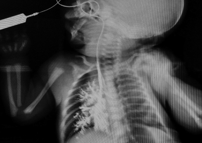
At birth, thoracoabdominal X-ray showed opacification of right bronchial tree and the tip of the catheter placed in the main right bronchus
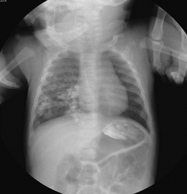
Fig. 2.

After 2 hours of life, the chest X-ray detected an atretic esophageal pouch (one arrow), the opacification of right bronchial tree, and air in the stomach.
Fig. 3.
After surgical intervention and during the 4th month of life, thoracoabdominal X-ray showed persistent unilateral bronchography with lower intensity.
Discussion
EA with or without TEF is the most common congenital anomaly of the esophagus and occurs approximately to 1:2,500 births. There are 5 types of EA, they are described more frequently according to Vogt's classification. Type 1 is TEF without EA, type 2 is EA without TEF, type 3a is EA with proximal TEF, type 3b is EA with distal TEF, and finally type 3c is EA with proximal and distal TEF [1]. The diagnosis is made in the early postnatal period in the vast majority of infants [2]. During prenatal age, polyhydramnios along with a small stomach has around a 55% predictive value for EA [2], [3]. If the diagnosis is suspected, a catheter should be inserted from the mouth into stomach. If there is ongoing doubt regarding the diagnosis, a contrast study may be helpful [2], [4]. In these cases, a small amount of water soluble contrast medium should be administered [4], [5], preferably in a pediatric specialized center [6] with extreme care for the high risk of aspiration of contrast medium during the study [2], [4]. In our case report, the infant was born with a good Apgar score but due to the evaluation of EA, it was inserted with a catheter from the lip to assess the interruption of the esophagus. Unknowingly, the catheter was inserted into right bronchus, and it was injected barium sulfate to evaluate the presence of TEF. The infant showed respiratory distress for barium sulfate aspiration, and it was suspected an EA with proximal TEF. This contrast medium was chosen because the baby was born in a community hospital where only barium sulfate is used for contrastographic examinations. Barium sulfate should be avoided if there is the suspect of obstructions, fistulas, and intestinal perforations. If it comes out on the viscera, barium sulfate becomes toxic; indeed, it can cause peritonitis or mediastinitis chemical because of its high osmolarity. It is recommended instead the use of iodinate contrast medium to evaluate the patency of esophagus because of its low osmolarity [7]. At children's Hospital Santobono Pausilipon, we performed a chest X-ray with iopamidol, and there was not proximal TEF found, instead we suspected a distal TEF. During the surgery, our diagnosis was confirmed. In a similar case report to evaluate the patency of esophagus in an infant with suspected EA, it was administered gastrografin (diatrizoate meglumine and diatrizoate sodium contrast) by orogastric tube. The infant aspirated the contrast medium that caused a tracheobronchogram, which developed chemical pneumonitis from gastrografin aspiration after surgical operation [8]. In the past, gastrografin and barium sulfate aspiration induced chemical pneumonitis and caused death both in adults and in children [9], [10]. If barium sulfate is aspired, lungs will metabolize it slowly. The real danger of barium sulfate aspiration is immediate because it causes pneumonitis chemical or pulmonary edema within few seconds or few hours from aspiration. Complications also depend on the amount ingested. Long-term effects of contrast media aspirated have been studied in an animal study on rats and dogs until 9 months. The study revealed that nonionic iodine-containing agents were better tolerated and evoked less pulmonary response than barium sulfate [11].
Footnotes
Competing Interests: The authors have declared that no competing interests exist.
References
- 1.Vogt E.C. Congenital esophageal atresia. AJR Am J Roentgenol. 1929;22:463–465. [Google Scholar]
- 2.Smith N. Oesophageal atresia and tracheo-oesophageal fistula. Early Hum Dev. 2014;90:947–950. doi: 10.1016/j.earlhumdev.2014.09.012. [DOI] [PubMed] [Google Scholar]
- 3.Pinheiro P.F., Simões e Silva A.C., Pereira R.M. Current knowledge on esophageal atresia. World J Gastroenterol. 2012;18:3662–3672. doi: 10.3748/wjg.v18.i28.3662. [DOI] [PMC free article] [PubMed] [Google Scholar]
- 4.Muensterer O.J., Berdon W.E. From Vogt to Haight and Holt to now: the history of esophageal atresia over the last century. Pediatr Radiol. 2015;45:1230–1235. doi: 10.1007/s00247-015-3276-1. [DOI] [PubMed] [Google Scholar]
- 5.Parolini F., Morandi A., Macchini F., Canazza L., Torricelli M., Zanini A. Esophageal atresia with proximal tracheoesophageal fistula: a missed diagnosis. J Pediatr Surg. 2013;48(6):E13–E17. doi: 10.1016/j.jpedsurg.2013.04.018. [DOI] [PubMed] [Google Scholar]
- 6.Atrésie de l'oesophage Protocole national de diagnostic et de soins. Haute Autoritè de Santé (Octobre 2008). http://www.has-sante.fr/portail/upload/docs/application/pdf/2008-11/pnds_atresie_oesophage_web.pdf. Accessed 14 Nov 2015.
- 7.Cova M. Mezzi di Contrasto. In: Angelelli G., Becciolini A., Biti G., Blandino A., Brunese L., Brunetti A., editors. Diagnostica per Immagini. IDELSON-GNOCCHI; Napoli: 2009. pp. 99–101. [Google Scholar]
- 8.McDuffie L.A., Wakeman D., Warner B.W. Diagnosis of esophageal atresia with tracheoesophageal fistula: is there a need for gastrointestinal contrast? J Pediatr. 2010;156:852. doi: 10.1016/j.jpeds.2009.10.030. [DOI] [PubMed] [Google Scholar]
- 9.Albeldawi M., Makkar R. Barium sspiration. N Engl J Med. 2012;366:1038. doi: 10.1056/NEJMicm1108468. [DOI] [PubMed] [Google Scholar]
- 10.McAlister W.H., Siegel M.J. Fatal aspirations in infancy during gastrointestinal series. Pediatr Radiol. 1984;14:81–83. doi: 10.1007/BF01625811. [DOI] [PubMed] [Google Scholar]
- 11.McAlister W.H., Askin F.B. The effect of some contrast agents in the lung: an experimental study in the rat and dog. AJR Am J Roentgenol. 1983;140(2):245–251. doi: 10.2214/ajr.140.2.245. [DOI] [PubMed] [Google Scholar]