Abstract
Glutathione S-Transferase Pi (GSTP1) mediates cellular defense against reactive electrophiles. Here, we report LAS17, a dichlorotriazine-containing compound that irreversibly inhibits GSTP1 and is selective for GSTP1 within cellular proteomes. Mass spectrometry and mutational studies identified Y108 as the site of modification, providing a unique mode of GSTP1 inhibition.
Glutathione S-transferase Pi (GSTP1) is the most ubiquitous member of the soluble GST protein superfamily and is primarily responsible for catalyzing the conjugation of glutathione (GSH) to exogenous and endogenous electrophiles as a mechanism of cellular detoxification.1 In addition to its catalytic function, GSTP1 has also been demonstrated to regulate the Jun N-terminal kinase (JNK)/mitogen-activated protein kinases (MAPK) signaling pathway through protein-protein interaction with JNK.2 Tyrosine phosphorylation of GSTP1 by the epidermal growth factor receptor (EGFR) kinase promotes formation of the GSTP1-JNK complex, which suppresses downstream JNK signaling.3 A variety of human cancers, including breast, colon and ovarian cancers, express high levels of GSTP1 relative to healthy tissue.4 Furthermore, human tumor cell lines that exhibit resistance to chemotherapeutic agents have elevated GSTP1 levels.5 Resistance is attributed to GSTP1-catalyzed inactivation of chemotherapeutic alkylating agents through conjugation of GSH.
Due to the confirmed roles of GSTP1 in promoting tumorigenesis and drug resistance, GSTP1 has emerged as a promising cancer therapeutic target. Many GSH analogs, small-molecules and natural products have been generated as GSTP1 inhibitors.2 Reversible GSTP1 inhibitors include 8-methoxypsoralen, piperlongumine, aloe emodin, and benstatin A,2, 6, 7 as well as Telintra (Ezatiostat), which is currently in clinical trials for the treatment of myelodysplastic syndrome.8, 9 GSTP1 contains four cysteine residues, two of which (C47 and C101) have been shown to be highly reactive and targeted by irreversible GSTP1 inhibitors.10 These cysteine-targeted covalent modifiers of GSTP1 include quercetin,11 and S-(N-benzylthio-carbamoyl) glutathione (BITC-SG).12 Quercetin targets C47, whereas BITC-SG covalently modifies C47 and C101 via an S-thiocarbamoylation reaction.11, 12
In addition to the two characterized reactive cysteines, GSTP1 has also been shown to contain reactive tyrosine residues that are susceptible to modification by sulfonyl-fluoride electrophiles. These sulfonyl fluorides have been shown to target the equivalent of Y8 and Y108 in mouse and rat GSTP1.13–15 Mutation of the equivalent of Y108 to phenylalanine in the Schistosoma japonicum GST resulted in loss of GSTP1 activity,14 indicating the functional importance of this tyrosine residue. Previous sulfonyl-fluoride probes used for tyrosine labeling are not selective for GSTP1, and covalently modify various other proteins within the proteome.14, 16 Here, we report the development of a selective, irreversible GSTP1 inhibitor, LAS17, which covalently modifies the functional tyrosine, Y108, in human GSTP1. LAS17 was discovered through screening a library of dichlorotriazine-containing compounds and shown to selectively modify GSTP1 within human cancer-cell proteomes. In addition to the dichlorotriazine electrophile for covalent modification, LAS17 contains a terminal alkyne functionality that allows for evaluating target occupancy and promiscuity of LAS17 in cellular and in vivo systems, using established copper (I)-catalyzed azide-alkyne cycloaddition (CuAAC) methods.17, 18
Aryl-halides, such as p-chloronitrobenzenes, have been shown to covalently modify proteins at cysteine residues through a nucleophilic aromatic substitution (SNAr) mechanism.19, 20 Evaluation of a panel of aryl halides for protein reactivity and site of modification revealed that dichlorotriazines predominately targeted lysine residues within a proteome, in contrast to p-chloronitrobenzenes that are highly cysteine selective.20 To further explore the unique reactivity of the dichlorotriazine electrophile and develop selective covalent modifiers to target reactive amino acids other than cysteine, we sought to synthesize a library of probes with the following functionalities: (1) a dichlorotriazine electrophile for covalent modification of the protein target; (2) a bio-orthogonal alkyne handle for CuAAC-mediated target identification; and (3) a diversity element to direct the probes toward diverse subsets of the proteome (Figure S1A).
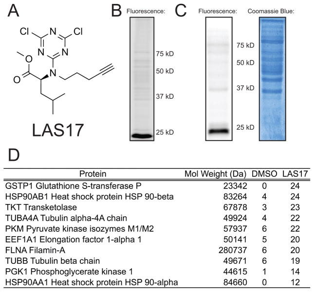
A library of 20 compounds (LAS1-20, Figure S1B) was synthesized via a 3-step synthetic scheme (Scheme S1). Briefly, 4-pent-yn-ol (1) was tosylated to generate a common intermediate (2), which was then diversified by coupling to the primary amine of 20 directing groups (3). The resulting secondary amines were then coupled to cyanuric chloride to afford the final library of 20 dichlorotriazine probes (4) (LAS1-20). LAS1-20 were evaluated for covalent protein labeling in HeLa cell lysates. Lysates were treated with 1 μM of each compound for 1 hour, subjected to CuAAC with rhodamine-azide, followed by SDS-PAGE and fluorescence visualization. These in-gel fluorescence screening studies demonstrated that several library members intensely labeled a low molecular weight protein band (~25 kD) (Figure S2). Of the 20 compounds, LAS17 (Figure 1A), containing an L-leucine methyl-ester directing group, was chosen for subsequent study due to the high intensity of labeling for the 25 kD protein and minimal labeling of other proteins within the cell lysates (Figure 1B). Furthermore, treatment of live HeLa cells with 1 μM of LAS17 demonstrated the potency and selectivity of this compound for covalent modification of the ~25 kD protein within a cellular context (Figure 1C). To identify the ~25 kD protein, HeLa cell lysates treated with either DMSO or 1 μM LAS17 were subjected to CuAAC with biotin-azide. Biotinylated proteins were enriched on streptavidin-agarose beads and subjected to LC/LC-MS/MS analysis. Spectral counts for proteins identified in three DMSO-treated samples were compared to LAS17-treated samples to identify proteins specifically labeled by LAS17 (Table S1). These data identified GSTP1 as the ~25 kD protein target of LAS17 (Figure 1D).
Figure 1.
(A) Structure of LAS17 (B) HeLa lysates (50 μL, 2 mg/mL) were treated with LAS17 (1 μM) at RT for 1 hr. Lysates were subjected to CuAAC with rhodamine-azide, separated by SDS-PAGE and analyzed by in-gel fluorescence. LAS17 labeled a ~25 kD protein with high intensity. Coomassie Blue-stained gel is shown in Figure S2B. (C) Live HeLa cells were treated with LAS17 (1 μM), lysed and subjected to CuAAC and in-gel fluorescence as described above to demonstrate the cell permeability and selectivity of LAS17. (D) HeLa lysates treated with LAS17 (1 μM) or DMSO were subjected to CuAAC with biotin-azide, enrichment on streptavidin-agarose, and LC/LC-MS/MS analysis. The top 10 proteins with the greatest difference in average spectral counts among three trials for DMSO and LAS17 treated samples are listed. The complete dataset is displayed in Table S1.
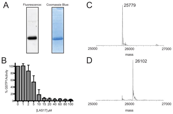
To confirm the LC/LC-MS/MS findings, human GSTP1 with an N-terminal hexahistidine tag was recombinantly expressed in E. coli and purified to homogeneity (Figure S3). The purified protein was treated with 1 μM LAS17 and analyzed by in-gel fluorescence. Significant protein labeling of purified GSTP1 was observed (Figure 2A), confirming that LAS17 covalently modifies GSTP1. To determine if LAS17 inhibits GSTP1 activity, an in vitro activity assay was performed that spectrophotometrically monitored the transfer of glutathione (GSH) to 1-bromo-2,4-dinitrobenzene (BDNB).21 LAS17 inhibited GSTP1 activity in vitro in a concentration-dependent manner (Figure 2B). Given the covalent mode of action for LAS17, time-dependent inhibition of GSTP1 was monitored, affording a second-order rate constant of inactivation (kinact/KI) of 31,200 M−1s−1 (Figure S4). To determine if the presence of other proteins affects the inhibitory activity of LAS17, inhibition studies were performed within a complex background of HeLa cell lysates, demonstrating that the presence of other cellular proteins does not affect the inhibitory function of LAS17 (Figure S5). The visualization of protein labeling after denaturing SDS-PAGE, and the time-dependent inhibition observed for LAS17, confirm that this compound functions by covalently modifying GSTP1. However, given the high reactivity observed for the dichlorotriazine electrophile,20 we sought to determine if LAS17 was covalently modifying a single amino acid or multiple sites within GSTP1. To determine the stoichiometry of modification, intact-protein mass spectrometry of purified GSTP1 was performed in the presence of 3 equivalents of LAS17. Mass-spectrometry analysis identified a single modified species corresponding to modification of GSTP1 with LAS17 at a single site (Figure 2C and D and Figure S6).
Figure 2.
(A) Recombinant, purified GSTP1 was treated with LAS17 and subjected to CuAAC, SDS-PAGE and in-gel fluorescence and Coomassie Blue staining. (B) In vitro GSTP1 activity data monitored in the presence of increasing concentrations of LAS17 (30 min pre-incubation) demonstrated a concentration-dependent decrease in GSTP1 activity.(C) Intact-protein mass spectrum of purified, recombinant GSTP1. (D) Intact-protein mass spectrum of purified, recombinant GSTP1 treated with LAS17 (3 equivalents). The mass difference between untreated and LAS17-treated GSTP1 corresponds to covalent modification of GSTP1 at a single site by LAS17.
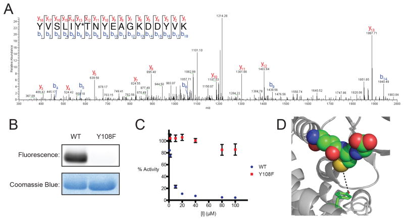
To identify the site of GSTP1 labeling by LAS17, purified wild-type GSTP1 was incubated with LAS17 and subjected to denaturation, reduction, cysteine alkylation, and trypsin digestion. The resulting tryptic-peptide mixtures were analyzed by LC-MS/MS. The generated fragmentation spectra were searched for a differential modification mass of +322.12 on all potentially nucleophilic amino acids. These analyses identified Y108 as the site of modification by LAS17 on GSTP1 (Figure 3A and Figure S7). To confirm Y108 as the site of modification, a Y108F mutant was generated, which was overexpressed and purified in tandem with wild-type (WT) GSTP1. Both the WT and the Y108F-mutant GSTP1 were treated with LAS17 and analyzed by in-gel fluorescence. The results show that replacing the nucleophilic tyrosine residue with a phenylalanine eliminated the ability of LAS17 to covalently modify GSTP1 (Figure 3B), thereby confirming that modification was selectively occurring at this residue.
Figure 3.
(A) Fragmentation (MS/MS) spectra of the YVSLIY*NYEAGKDDYVK peptide of GSTP1 confirming that the LAS17 modification (+322.12 Da) is present on Y108, which is indicated with *. (B) In-gel fluorescence analysis of purified Y108F mutant compared to wild-type GSTP1, confirms Y108 as the site of labeling. (C) The Y108F mutant is resistant to LAS17-mediated inhibition compared to the wild-type GSTP1. (D) Proximity of Y108 to the GSH binding site of GSTP1 (PBD: 6GSS).
Since the dichlorotriazine electrophile was previously shown to covalently modify lysine residues in proteomes 20, we sought to confirm that LAS17 was not modifying a lysine residue on GSTP1. This was achieved by generating lysine to alanine mutants at each of the 12 lysine residues on GSTP1. Mutation of these lysine residues did not result in a decrease in LAS17 labeling (Figure S8), further confirming that covalent modification was occurring at Y108. Lastly, activity assays with the Y108F mutant showed that LAS17 had negligible effect on the activity of the mutant protein in comparison to wild-type GSTP1 (Figure 3C). These assay data thereby confirm that the inhibitory activity of LAS17 is solely due to covalent modification of Y108. Y108 is located in the hydrophobic co-substrate site (H-site) of GSTP1 (Figure 3D), proximal to the GSH binding site, and is conserved among the pi, mu, and theta GST classes in humans.22 Crystallographic studies show the aromatic ring of Y108 is positioned in a hydrophobic pocket, and mutagenesis of Y108 to phenylalanine shows no significant side chain or backbone structural changes relative to the WT GSTP1.23 Lastly, Y108 is conserved in GSTP1 homologs in higher-order eukaryotes.
Conclusions
In summary, we report the discovery and characterization of LAS17, a potent and selective tyrosine-directed irreversible inhibitor for GSTP1. Given the well-characterized role of GSTP1 in cancer pathogenesis and chemotherapeutic resistance, LAS17 provides a unique mode of irreversible GSTP1 inhibition, targeting a functional tyrosine residue (Y108), in contrast to previously reported cysteine-targeted compounds. Despite the high reactivity of the dichlorotriazine electrophile of LAS17, high selectivity is observed for GSTP1 within a complex proteome. The structural basis for this selectivity is currently unknown and is the subject of future structure-activity relationship studies on LAS17. Lastly, the presence of the terminal alkyne functionality for CuAAC provides the ability to determine target engagement and off-targets in cell-based and in vivo systems using CuAAC-mediated conjugation of reporter tags for visualization and enrichment of LAS17-bound proteins. LAS17 is a valuable addition to the currently available collection of reversible and irreversible GSTP1 inhibitors and can be utilized as an activity-based probe to interrogate GSTP1 activity in cancer, and as a pharmacological modulator of GSTP1 to further interrogate the therapeutic potential of this enzyme. Ongoing studies are focused on in vivo applications of LAS17 in mouse xenograft studies to further investigate the therapeutic potential of GSTP1 in a variety of cancer sub-types.
Supplementary Material
Acknowledgments
We are grateful for the financial support from the Damon Runyon Cancer Research Foundation, Boston College and NIH grant 1R01GM118431-01A1. We thank Dr. Daniel Bak and Shalise Couvertier for comments and critical reading of the manuscript.
Footnotes
Supporting information for this article is given via a link at the end of the document.
Notes and references
- 1.Nebert DW, Vasiliou V. Human genomics. 2004;1:460–464. doi: 10.1186/1479-7364-1-6-460. [DOI] [PMC free article] [PubMed] [Google Scholar]
- 2.Laborde E. Cell death and differentiation. 2010;17:1373–1380. doi: 10.1038/cdd.2010.80. [DOI] [PubMed] [Google Scholar]
- 3.Okamura T, Antoun G, Keir ST, Friedman H, Bigner DD, Ali-Osman F. The Journal of biological chemistry. 2015;290:30866–30878. doi: 10.1074/jbc.M115.656140. [DOI] [PMC free article] [PubMed] [Google Scholar]
- 4.Schnekenburger M, Karius T, Diederich M. Frontiers in pharmacology. 2014;5:170. doi: 10.3389/fphar.2014.00170. [DOI] [PMC free article] [PubMed] [Google Scholar]
- 5.Townsend DM, Tew KD. Oncogene. 2003;22:7369–7375. doi: 10.1038/sj.onc.1206940. [DOI] [PMC free article] [PubMed] [Google Scholar]
- 6.de Oliveira DMM, de Farias MT, Teles ALL, Dos Santos MC, Junior, de Cerqueira MD, Lima RM, El-Bachá RS. Frontiers in cellular neuroscience. 2014;8:308. doi: 10.3389/fncel.2014.00308. [DOI] [PMC free article] [PubMed] [Google Scholar]
- 7.Gong L-HH, Chen X-XX, Wang H, Jiang Q-WW, Pan S-SS, Qiu J-GG, Mei X-LL, Xue Y-QQ, Qin W-MM, Zheng F-YY, Shi Z, Yan X-JJ. Oxidative medicine and cellular longevity. 2014;2014:906804. doi: 10.1155/2014/906804. [DOI] [PMC free article] [PubMed] [Google Scholar]
- 8.Raza A, Galili N, Smith S, Godwin J, Lancet J, Melchert M, Jones M, Keck JG, Meng L, Brown GL, List A. Blood. 2009;113:6533–6540. doi: 10.1182/blood-2009-01-176032. [DOI] [PubMed] [Google Scholar]
- 9.Raza A, Galili N, Mulford D, Smith SE, Brown GL, Steensma DP, Lyons RM, Boccia R, Sekeres MA, Garcia-Manero G, Mesa RA. Journal of hematology & oncology. 2012;5:18. doi: 10.1186/1756-8722-5-18. [DOI] [PMC free article] [PubMed] [Google Scholar]
- 10.Ricci G, Del Boccio G, Pennelli A, Lo Bello M, Petruzzelli R, Caccuri AM, Barra D, Federici G. The Journal of biological chemistry. 1991;266:21409–21415. [PubMed] [Google Scholar]
- 11.van Zanden JJ, Hamman O, van Iersel M, Boeren S, Cnubben N, Bello M, Vervoort J, van Bladeren PJ, Rietjens I. Chem Biol Interact. 2003;145:139–148. doi: 10.1016/s0009-2797(02)00250-8. [DOI] [PubMed] [Google Scholar]
- 12.Quesada-Soriano I, Primavera A, Casas-Solvas JM, Téllez-Sanz R, Barón C, Vargas-Berenguel A, Lo Bello M, García-Fuentes L. Chembiochem : a European journal of chemical biology. 2012;13:1594–1604. doi: 10.1002/cbic.201200210. [DOI] [PubMed] [Google Scholar]
- 13.Barycki JJ, Colman RF. Biochemistry. 1993;32:13002–13011. doi: 10.1021/bi00211a008. [DOI] [PubMed] [Google Scholar]
- 14.Gu C, Shannon DA, Colby T, Wang Z, Shabab M, Kumari S, Villamor JG, McLaughlin CJ, Weerapana E, Kaiser M, Cravatt BF, van der Hoorn RA. Chemistry & biology. 2013;20:541–548. doi: 10.1016/j.chembiol.2013.01.016. [DOI] [PubMed] [Google Scholar]
- 15.Wang J, Barycki JJ, Colman RF. Protein Science. 1996;5:1032–1042. doi: 10.1002/pro.5560050606. [DOI] [PMC free article] [PubMed] [Google Scholar]
- 16.Shannon AD, Gu C, McLaughlin CJ, Kaiser M, van der Hoorn RAL, Weerapana E. ChemBio Chem. 2012;13:2327–2330. doi: 10.1002/cbic.201200531. [DOI] [PubMed] [Google Scholar]
- 17.Kolb HC, Finn MG, Sharpless KB. Angewandte Chemie (International ed in English) 2001;40:2004–2021. doi: 10.1002/1521-3773(20010601)40:11<2004::AID-ANIE2004>3.0.CO;2-5. [DOI] [PubMed] [Google Scholar]
- 18.Speers AE, Adam GC, Cravatt BF. Journal of the American Chemical Society. 2003;125:4686–4687. doi: 10.1021/ja034490h. [DOI] [PubMed] [Google Scholar]
- 19.Leesnitzer LM, Parks DJ, Bledsoe RK, Cobb JE, Collins JL, Consler TG, Davis RG, Hull-Ryde EA, Lenhard JM, Patel L, Plunket KD, Shenk JL, Stimmel JB, Therapontos C, Willson TM, Blanchard SG. Biochemistry. 2002;41:6640–6650. doi: 10.1021/bi0159581. [DOI] [PubMed] [Google Scholar]
- 20.Shannon DA, Banerjee R, Webster ER, Bak DW, Wang C, Weerapana E. Journal of the American Chemical Society. 2014;136:3330–3333. doi: 10.1021/ja4116204. [DOI] [PubMed] [Google Scholar]
- 21.Dalmizrak O, Kulaksiz-Erkmen G, Ozer N. Molecular and cellular biochemistry. 2011;355:223–231. doi: 10.1007/s11010-011-0858-6. [DOI] [PubMed] [Google Scholar]
- 22.Wilce MCJ, Parker MW. Biochimica et Biophysica Acta. 1994;1205:1–18. doi: 10.1016/0167-4838(94)90086-8. [DOI] [PubMed] [Google Scholar]
- 23.Lo Bello M, Oakley AJ, Battistoni A, Mazzetti AP, Nuccetelli M, Mazzarese G, Rossjohn J, Parker MW, Ricci G. Biochemistry. 1997;36:6207–6217. doi: 10.1021/bi962813z. [DOI] [PubMed] [Google Scholar]
Associated Data
This section collects any data citations, data availability statements, or supplementary materials included in this article.


