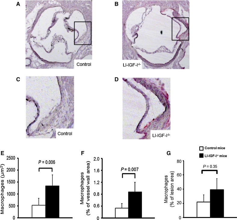
Fig. 2.
Increased macrophage content in aortic wall of female LI-IGF-I−/− mice. Female control (n = 13) and LI-IGF-I−/− (n = 9) mice received an atherogenic (modified Paigen) diet from 6 to 12 months of age. Macrophage area was quantified using immunostaining for Mac-2 in aortic root cryosections (40 µm after the aortic cusps). a–d Representative photomicrographs from female a, c control and b, d LI-IGF-I−/− mice are shown. Areas in the rectangles of a and b are enlarged in c and d, respectively. e, f Quantification of macrophage area in aortic wall of female mice given as e µm2 and as f % of aortic wall area. g Macrophage area was not significantly altered when normalized to the fatty streak area. Values in e–g are given as means (SEM). p values were calculated using unpaired t tests

