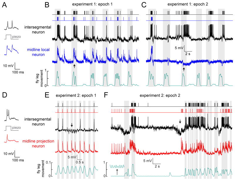
Figure 7. Parallel coding of complex stimuli in simultaneously-recorded central neuron pairs.
(A) Paired whole-cell recording from an intersegmental neuron and a midline local neuron. Example traces show the simultaneous responses of the two neurons to mechanical stimulation of a femur bristle. These responses confirm that this particular pair of neurons share input from some of the same bristles (Figure 3).
(B) During this epoch of the experiment, the fly makes large movements which cause both neurons to depolarize and fire correlated bursts of spikes (e.g., at the arrow). Fly movement is quantified in arbitrary units (see Supplemental Experimental Procedures). Movement bouts are shaded in gray. Spike times are represented with rasters above the raw voltage traces.
(C) During a later epoch of the same experiment, the midline local neuron stops being excited during movement bouts, and is instead inhibited by movement (e.g., at the arrow). This change corresponds to a switch between large movements of multiple legs, to smaller movements of the prothoracic leg.
(D) Same as (A) but for a simultaneously-recorded intersegmental and a midline projection neuron.
(E) The same pair of neurons as in (D), but now responding to spontaneous movement. Small periodic twitching movements of the fly’s leg (gray shading) evoke reliable responses in the midline projection neuron, but not in the intersegmental neuron. The periodic responses of the intersegmental neuron are interrupted by barrages of inhibitory postsynaptic potentials (e.g., at the arrow). Note the expanded vertical scale of the movement measurements.
(F) During a later epoch of the same experiment, experiment, the fly spontaneously switches from twitching to making larger movements of the entire leg. The midline projection neuron is depolarized during large movement bouts, while the intersegmental neuron responds with sequences of inhibition and excitation at movement onset (e.g., at the arrow). The inset in the movement trace (bottom) shows periodic movement on a 10-fold expanded vertical scale.

