Abstract
Acanthosis nigricans (AN), an entity recognized since the 19th century, is a dermatopathy associated with insulin-resistant conditions, endocrinopathies, drugs, chromosome abnormalities and neoplasia. The latter, also known as malignant AN, is mostly related to abdominal neoplasms. Malignant AN occurs frequently among elderly patients. In these cases, the onset is subtle, and spreading involves the flexural regions of the body, particularly the axillae, palms, soles, and mucosa. Gastric adenocarcinoma is the most frequent associated neoplasia, but many others have been reported. Renal cell carcinoma (RCC), although already reported, is rarely associated with malignant AN. The authors report the case of a woman who was being treated for depression but presented a long-standing and marked weight loss, followed by darkening of the neck and the axillary regions. Physical examination disclosed a tumoral mass in the left flank and symmetrical, pigmented, velvety, verrucous plaques on both axillae, which is classical for AN. The diagnostic work-up disclosed a huge renal mass, which was resected and further diagnosed as a RCC. The post-operative period was uneventful and the skin alteration was evanescent at the first follow-up consultation. The authors call attention to the association of AN with RCC.
Keywords : Acanthosis nigricans; Carcinoma, Renal Cell; Paraneoplastic Syndromes
CASE REPORT
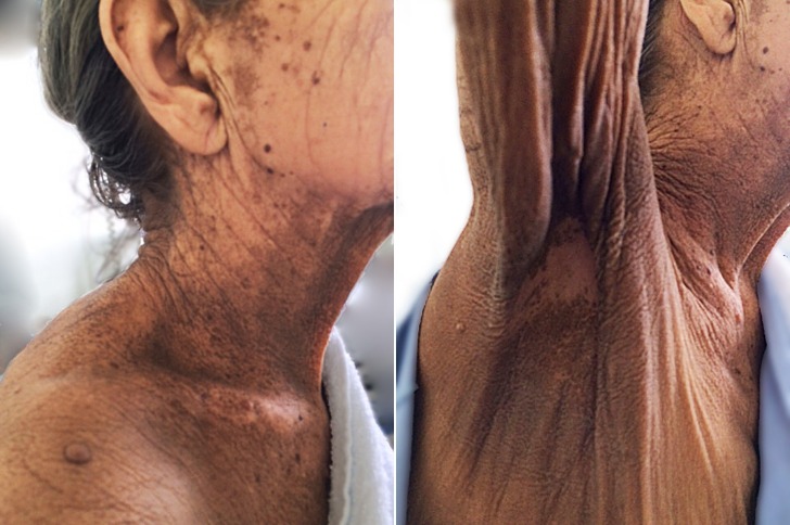
A 67-year-old woman sought medical attention complaining of progressive weight loss and loss of appetite over the last 10 months, which was treated with antidepressants because of the suspicion of a mood disorder. In the meantime, she noticed darkening of the skin, but her mucosa, palms, and soles were spared. At the time she was hospitalized, she had lost 21 kg and presenting daily fever (38°C) accompanied by rigor and marked weakness. She brought the results of a normal colonoscopy and an upper digestive endoscopy, which had recently been performed. The physical examination disclosed a pale and cachectic patient weighing 43 kg (BMI of 17) with normal hemodynamic and respiratory parameters. The skin of the neck, axillary, and inframammary regions was darkened and thickened with a velvety appearance, which is consistent with the clinical diagnosis of acanthosis nigricans (AN) (Figure 1).
Figure 1. Physical examination showing darkening and thickening of the skin. Note the darkening skin in the neck and the velvety appearance in the infra axillary region.
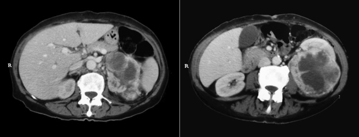
Peripheral lymphadenopathy was not found and physical examination of the lungs and heart was normal. However, although the abdomen was flat and flaccid, a hardened and painless mass was easily palpable in the left flank. The abdominal computed tomography confirmed the presence of a voluminous and heterogeneous mass, measuring 13.4 × 8.6 × 7.3 cm, predominantly hypoattenuating and with heterogeneous contrast enhancement. It anteriorly displaced the renal hilum, which was apparently without invasion (Figure 2).
Figure 2. Axial computed tomography of the abdomen after intravenous contrast medium injection showing a large, heterogeneous and mixed attenuating mass anteriorly displacing the left kidney.
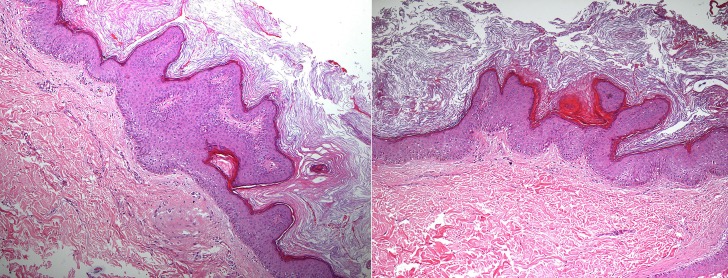
The laboratory work-up revealed microcytic hypochromic anemia, thrombocytosis, normal renal function, and moderate hyponatremia. The skin biopsy showed thickened epidermis by marked hyperkeratosis and papillomatosis. The papillomatosis resulted from finger-like projections of the dermal papillae to the surface, which was lined by thin epidermis. In between these projections, the epidermis was thicker than the epidermis overlying the papillae. There was no melanocytic proliferation or significant hyperpigmentation of the epidermal basal layer. The dermis had no significant inflammation. The pathological findings were consistent with AN (Figure 3).
Figure 3. Photomicrography of the skin showing epidermal thickening due to “finger-like” papillomatosis and hyperkeratosis without melanocytic proliferation.
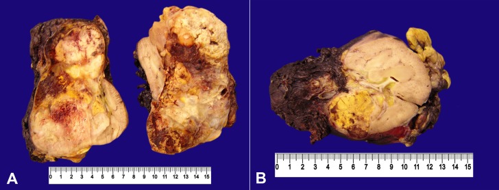
The patient was submitted to a laparoscopic left total nephrectomy followed by tumor removal through a Pfannenstiel incision. The surgical specimen comprised an irregular-contour monoblock weighing 597 g and measuring 14.5 × 13.0 × 8.3 cm, which, at the cut surface, showed a tumor mass occupying nearly 85% of the renal parenchyma, extending until the renal sinus but apparently sparing the vascular structures. The remaining renal tissue showed the corticomedullary limit, which was partially preserved in less than 10% of the kidney (Figure 4).
Figure 4. Gross findings of the formalin fixed surgical specimen. A and B – The huge extension of the tumor presentingthe golden color in some areas due to the intracellular lipid accumulation.
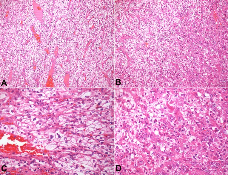
The microscopic examination showed that the morphology was consistent with renal clear-cell carcinoma (Figure 5), Fuhrman 4 nuclear grade with necrosis extending into nearly 40% of the neoplasia. The tumor was restricted to the kidney but invasion was present within the fat tissue of the renal sinus, as well as in the lymphatics and the venules within the tumor. The surgical margins were free of tumor.
Figure 5. Photomicrography of the tumor. A and B – nests and sheets of epithelial cells with clear cytoplasm and distinct cell membrane, interspersed by a rich network of thin-walled vascular tissue (H&E, 200X). C and D – Note areas of eosinophilic cytoplasm (often seen in high-grade neoplasm) and nuclei of pleomorphic size and shape with loose chromatin, and the presence of macronucleoli characterizing the Fuhrman 4 nuclear grading.
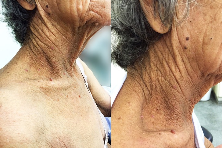
The post-operative period was uneventful and the patient was discharged on the seventh post-operative day with skin bleaching (Figure 6). She was referred to an oncological center for follow-up.
Figure 6. Skin examination of the neck region 50 days after tumor removal. Note the almost complete disappearance of the acanthosis nigricans in this area.
DISCUSSION
Renal cell carcinoma (RCC) accounts for 3% of all solid organ neoplasia, occurring mostly between the ages of 60 years and 70 years with a slight predominance among males.1 Although the typical presentation of RCC is represented by flank pain, palpable mass, and hematuria, only up to 20% of cases will present this triad, and it is common to diagnose large tumors in its origin before any previous accompanying symptoms. However, with the widespread use of imaging techniques, incidental diagnoses are becoming increasingly frequent, reaching almost 70% of cases.2 Nevertheless, the diagnosis is also frequently made by the metastatic presentation instead of being done through the detection of the primary tumor.3 In this setting the diagnosis is suspected or is held by manifestations on various areas and systems of the human body: (i) the skin; (ii) the musculoskeletal system; (iii) the nervous system; (iv) the blood system; (v) the vascular system; (vi) the lungs and pleura; (vii) the heart; (viii) the endocrine system; and (ix) the gastrointestinal system.3-18
Dermatological manifestations of RCC, although seldom observed, may be represented by nodules: (i) metastasis (presented in 3–6% of the RCC) mostly found in the scalp, the thorax, and the abdomen;1,19-22 or (ii) leukocytoclastic vasculitis, with or without ulceration.23 Even rarer, the skin manifestations may also be represented by paraneoplastic syndromes, such as AN, palmar fibromatosis, erythrodermia, and bullous pemphigoid.24,25
Paraneoplastic syndrome refers to conditions, signs, and symptoms secondary to the presence of a malignancy,26 which, in the case of RCC, may be present in up to 40% of the cases, and are also represented by fever, cachexia, Stauffer’s syndrome, polycytemia, hypercalcemia, hypertension, gynecomastia, galactorrhoea, and others, which subside after the tumor removal, and may upsurge with tumor relapse or metastatic disease.
Our patient’s situation represents one of those cases of pauci symptomatic RCC (only weight loss); however, the misinterpretation (mood disorder) of the cause of weight loss was very regrettable. Our patient was being followed by a mental health service where the clinical hypothesis was far from the correct diagnosis, giving an opportunity for the large growth of the tumor. The darkening of the skin was probably also neglected what added to the marked weight loss should have raised the suspicion for malignancy.
It is suspected that AN was first diagnosed in 1867 by Mitchel.27 However, it was only in 1890 that Pollitzer28 and Janvosky29 described the verrucous hyperpigmented dermatosis, which was named “acanthosis nigricans.” Since then, many descriptions proliferated in the literature associating AN with many clinical conditions mostly related to insulin-resistance entities30 such as: (i) obesity (up to 70% may present AN);31 (ii) type-2 diabetes; (iii) metabolic syndrome;32 and (iv) polycistic ovary syndrome,33 other endocrinopathies (acromegaly, Cushing syndrome, hypothyroidism), Down syndrome, drug use (e.g. nicotinic acid, fusidic acid, and hormones), and genetic syndromes.34 AN more often involves the neck, axilla, the region under the breast, the groin, and other skin folds. Less commonly, the back of the neck, face, periumbilical and anogenital areas, palms, soles, and mucosa can be involved. The lesions are often symmetrical and characterized by brownish, thickened, dry, and rough skin, with a velvet or leather appearance, sometimes showing verrucous or papillomatous excrescences. Patients with AN may complain that they have a kind of grime they cannot get rid of. Microscopically, the lesion is characterized by papillary hypertrophy (papillomatosis) and hyperkeratosis. Hyperpigmentation is due to hyperkeratosis rather than an increase in melanin pigment in the skin.
In 1893, Ferdinand-Jean Darier first described the association of AN with malignancy (two cases with the descriptive title of “Dystrophie papilaire et pigmentaire”,35 which, although clinically very similar, usually affects palms and soles, with rapid onset and spread, afflicting the elderly population.34,36 Among all malignancies, gastric adenocarcinoma is particularly associated with up to 61% of all malignant AN, followed by neoplasia of the ovary, lungs, and breast. Much less frequent, esophageal, intestinal, biliary bladder, pancreas, prostate, bladder, and other urothelial carcinoma have been reported in association with AN.36 Searching the PubMed database (irrespective of the period), using the keywords “acanthosis nigricans” crossed with “clear cell carcinoma,” “urothelial carcinoma,” “kidney,” “renal carcinoma,” and “paraneoplastic,” we found 168 citations related to malignant AN—among them the association with renal malignancy are described as follows. In the Riegel and Jacobs series,36 which comprised 277 cases of malignant AN, only 1 case of renal malignancy was reported; Moscardi et al.37 reported a case of renal malignancy without histological identification; and Lee et al.38 reported a case of renal urothelial carcinoma and AN. Therefore, we conclude that this association is very rare or at least under-reported. The pathogenesis of malignant AN, although not entirely understood, involves the action of an excess of transforming growth factor-α acting on the epidermal growth factor receptor inducing hyperstimulation of keratinocytes, which subside or disappear after tumor removal.39 Generally, in 61% of cases the skin involvement is concomitant with the neoplasia; however, in 18% of cases AN appears many years before the neoplasia.40
In our case, although delayed, AN was diagnosed concomitantly with the neoplasia since the skin alteration appeared when the patient had already started losing weight.
An evaluation of the patient during the early post-operative period (around the seventh day after surgery) showed a slight evanescence of the darkening color of the skin. On the second month after surgery, the AN was no longer detected on the neck, which showed its close association with the presence of the neoplasia (Figure 6).
The aim of this report was not only to show the association of RCC with AN, but also to call attention to this diagnosis in the elderly, most often when accompanied by weight loss without any other clinical feature that could raise the surveillance for neoplasia.
Footnotes
Campos FPF, Narvaez MRA, Reis PVS, et al. Acanthosis Nigricans associated with clear-cell renal cell carcinoma. Autopsy Case Rep [Internet]. 2016;6(1):33-40. http://dx.doi.org/10.4322/acr.2016.021/
REFERENCES
- 1.Smyth LG, Casey RG, Quinlan DM. Renal cell carcinoma presenting as an ominous matachronous scalp metastasis. Can Urol Assoc J. 2010;4(3):E64-6. PMid: [DOI] [PMC free article] [PubMed] [Google Scholar]
- 2.Sountoulides P, Metaxa L, Cindolo L. Atypical presentations and rare metastatic sites of renal cell carcinoma: a review of case reports. J Med Case Rep. 2011;5(1):429. http://dx.doi.org/10.1186/1752-1947-5-429. PMid: [DOI] [PMC free article] [PubMed] [Google Scholar]
- 3.Gharabaghi MA. Clinical Spectrum of Patients with Renal Cell Carcinoma. In: Amato R, editor. Emerging research and treatments in renal cell carcinoma. Rijeka: InTech; 2012 [cited 2016 Jan 5]. chap 10; p. 229-44. Available from: http://www.intechopen.com/books/emerging-research-and-treatments-in-renal-cellcarcinoma/clinical-spectrum-of-patients-with-renal-cell-carcinoma [Google Scholar]
- 4.Fottner A, Szalantzy M, Wirthmann L, et al. Bone metastases from renal cell carcinoma: patient survival after surgical treatment. BMC Musculoskelet Disord. 2010;11(1):145-50. http://dx.doi.org/10.1186/1471-2474-11-145. PMid: [DOI] [PMC free article] [PubMed] [Google Scholar]
- 5.Klausner AP, Ost MC, Waterhouse RL Jr, Savage SJ. Occult renal cell carcinoma in a patient with polymyositis. Urology. 2002;59(5):773-6. http://dx.doi.org/10.1016/S0090-4295(02)01533-9. PMid: [DOI] [PubMed] [Google Scholar]
- 6.Respicio G, Shwaiki W, Abeles MA. 58-year-old man with anti-Jo-1 syndrome and renal cell carcinoma: a case report and discussion. Conn Med. 2007;71(3):151-3. PMid: [PubMed] [Google Scholar]
- 7.Koukoulis A, Cimas I, Gómara S. Paraneoplastic opsoclonus associated with papillary renal cell carcinoma. J Neurol Neurosurg Psychiatry. 1998;64(1):137-8. http://dx.doi.org/10.1136/jnnp.64.1.137. PMid: [DOI] [PMC free article] [PubMed] [Google Scholar]
- 8.Roy MJ, May EF, Jabbari B. Life-threatening polyneuropathy heralding renal cell carcinoma. Mil Med. 2002;167(12):986-9. PMid: [PubMed] [Google Scholar]
- 9.Mohammed Ilyas MI, Turner GD, Cranston D. Human chorionic gonadotropin secreting clear cell renal cell carcinoma with paraneoplastic gynaecomastia. Scand J Urol Nephrol. 2008;42(6):555-7. http://dx.doi.org/10.1080/00365590802468834. PMid: [DOI] [PubMed] [Google Scholar]
- 10.Hameed A, Pahuja A, Thwaini A, Nambirajan T. Subclavian vein thrombosis: an unusual presentation of renal cell carcinoma. Can Urol Assoc J. 2011;5(2):E27-8. http://dx.doi.org/10.5489/cuaj.10061. PMid: [DOI] [PMC free article] [PubMed] [Google Scholar]
- 11.Wallach JB, McGarry T, Torres J. Lymphangitic metastasis of recurrent renal cell carcinoma to the contralateral lung causing lymphangitic carcinomatosis and respiratory symptoms. Curr Oncol. 2011;18(1):e35-7. http://dx.doi.org/10.3747/co.v18i1.647. PMid: [DOI] [PMC free article] [PubMed] [Google Scholar]
- 12.Suyama H, Igishi T, Makino H, et al. Bronchial artery embolization before interventional bronchoscopy to avoid uncontrollable bleeding: a case report of endobronchial metastasis of renal cell carcinoma. Intern Med. 2011;50(2):135-9. http://dx.doi.org/10.2169/internalmedicine.50.3818. PMid: [DOI] [PubMed] [Google Scholar]
- 13.Teresa P, Grazia ZM, Doriana M, Irene P, Michele S. Malignant effusion of chromophobe renal-cell carcinoma: Cytological and immunohistochemical findings. Diagn Cytopathol. 2012;40(1):56-61. http://dx.doi.org/10.1002/dc.21599. PMid: [DOI] [PubMed] [Google Scholar]
- 14.Okubo Y, Yonese J, Kawakami S, et al. Obstinate cough as a sole presenting symptom of non-metastatic renal cell carcinoma. Int J Urol. 2007;14(9):854-5. http://dx.doi.org/10.1111/j.1442-2042.2007.01832.x. PMid: [DOI] [PubMed] [Google Scholar]
- 15.Miyamoto MI, Picard MH. Left atrial mass caused by metastatic renal cell carcinoma: an unusual site of tumor involvement mimicking myxoma. J Am Soc Echocardiogr. 2002;15(8):847-8. http://dx.doi.org/10.1067/mje.2002.120508. PMid: [DOI] [PubMed] [Google Scholar]
- 16.Gopan T, Toms SA, Prayson RA, Suh JH, Hamrahian AH, Weil RJ. Symptomatic pituitary metastases from renal cell carcinoma. Pituitary. 2007;10(3):251-9. http://dx.doi.org/10.1007/s11102-007-0047-5. PMid: [DOI] [PubMed] [Google Scholar]
- 17.Cimino-Mathews A, Sharma R, Netto GJ. Diagnostic use of PAX8, CAIX, TTF-1, and TGB in metastatic renal cell carcinoma of the thyroid. Am J Surg Pathol. 2011;35(5):757-61. http://dx.doi.org/10.1097/PAS.0b013e3182147fa8. PMid: [DOI] [PMC free article] [PubMed] [Google Scholar]
- 18.Kranidiotis GP, Voidonikola PT, Dimopoulos MK, Anastasiou-Nana MI. Stauffer's syndrome as a prominent manifestation of renal cancer: a case report. Cases J. 2009;2(1):49-52. http://dx.doi.org/10.1186/1757-1626-2-49. PMid: [DOI] [PMC free article] [PubMed] [Google Scholar]
- 19.Mueller TJ, Wu H, Greenberg RE, et al. Cutaneous metastases from genitourinary malignancies. Urology. 2004;63(6):1021-6. http://dx.doi.org/10.1016/j.urology.2004.01.014. PMid: [DOI] [PubMed] [Google Scholar]
- 20.Srinivasan N, Pakala A, Al-Kali A, Rathi S, Ahmad W. Papillary renal cell carcinoma with cutaneous metastases. Am J Med Sci. 2010;339(5):458-61. http://dx.doi.org/10.1097/MAJ.0b013e3181d5678d. PMid: [DOI] [PubMed] [Google Scholar]
- 21.Arrabal-Polo MA, Arias-Santiago SA, Aneiros-Fernandez J, Burkhardt-Perez P, Arrabal-Martin M, Naranjo-Sintes R. Cutaneous metastases in renal cell carcinoma: a case report. Cases J. 2009;2(1):7948-51. http://dx.doi.org/10.4076/1757-1626-2-7948. PMid: [DOI] [PMC free article] [PubMed] [Google Scholar]
- 22.Dorairajan LN, Hemal AK, Aron M, et al. Cutaneous metastases in renal cell carcinoma. Urol Int. 1999;63(3):164-7. http://dx.doi.org/10.1159/000030440. PMid: [DOI] [PubMed] [Google Scholar]
- 23.Maynard JW, Christopher-Stine L, Gelber AC. Testicular pain followed by microscopic hematuria, a renal mass, palpable purpura, polyarthritis, and hematochezia. J Clin Rheumatol. 2010;16(8):388-91. http://dx.doi.org/10.1097/RHU.0b013e3181fe8bbe. PMid: [DOI] [PubMed] [Google Scholar]
- 24.Klein T, Rotterdam S, Noldus J, Hinkel A. Bullous pemphigoid is a rara paraneoplastic syndrome in patients with renal cell carcinoma. Scand J Urol Nephrol. 2009;43(4):334-6. http://dx.doi.org/10.1080/00365590902811545. PMid: [DOI] [PubMed] [Google Scholar]
- 25.Tebbe B, Schlippert U, Garbe C, Orfanos CE. Erythroderma "en nappes claires" as a marker of metastatic kidney cancer. Lasting, successful treatment with rIFN-alpha-2a. Hautarzt. 1991;42(5):324-7. PMid: [PubMed] [Google Scholar]
- 26.Palapattu GS, Kristo B, Rajfer J. Paraneoplastic syndromes in urologic malignancy: the many faces of renal cell carcinoma. Rev Urol. 2002;4(4):163-70. PMid: [PMC free article] [PubMed] [Google Scholar]
- 27.Mitchel SW. Extensive encephaloid cancer of the subserous tissues of the peritoneum and pleura: Intensely bronzed-cloured skin; supra-renal capsules healthy. Proc. Path. Soc. Philad. 1867;3:35. [Google Scholar]
- 28.Politzer S. Acanthosis nigricans. In: Unna PG, Morris M, Besnier E, Duhring LA, editors. International atlas of rare skin diseases. London: HK Lewis & Co. Ltda; 1890. Pt. IV, No. XI, p. 6. [Google Scholar]
- 29.Janvosky V. Acanthosis nigricans. In: Unna PG, Morris M, Besnier E, Duhring LA, editors. International atlas of rare skin diseases. London: HK Lewis & Co. Ltda; 1890. Pt. IV, No. XI, p. 1. [Google Scholar]
- 30.Rendon MI, Cruz PD Jr, Sontheimer RD, Berg-stresser PR. Acanthosis nigricans: a cutaneous marker of tissue resistance to insulin. J Am Acad Dermatol. 1989;21(3 pt 1):461-9. http://dx.doi.org/10.1016/S0190-9622(89)70208-5. PMid: [DOI] [PubMed] [Google Scholar]
- 31.Hud JA Jr, Cohen JB, Wagner JM, Cruz PD Jr. Prevalence and significance of acanthosis nigricans in an adult obese population. Arch Dermatol. 1992;128(7):941-4. http://dx.doi.org/10.1001/archderm.1992.01680170073009. PMid: [PubMed] [Google Scholar]
- 32.Kalus AA, Chien AJ, Olerud JE. Chapter 151: diabetes mellitus and other endocrine diseases In: Goldsmith LA, Katz SI, Gilchrest BA, et al., editors. Fitzpatrick’s dermatology in general medicine. 8th ed. New York: McGraw-Hill; 2012. [Google Scholar]
- 33.Stulberg DL, Clark N, Tovey D. Common hyperpigmantation disorders in adults: part II. Melanoma, seborreic keratoses, acanthosis nigricans, melasma, diabetic dermopathy, tinea versicolor and post inflammatory hyperpigmentation. Am Fam Physician. 2003;68(10):1963-8. PMid: [PubMed] [Google Scholar]
- 34.Kubicka-Wołkowska J, Dębska-Szmich S, Lisik-Habib M, Noweta M, Potemski P. Malignant acanthosis nigricans associated with prostate cancer: a case report. BMC Urol. 2014;14(1):88. http://dx.doi.org/10.1186/1471-2490-14-88. PMid: [DOI] [PMC free article] [PubMed] [Google Scholar]
- 35.Pollitzer S. Acanthosis nigricans: a symptom of a disoreder of the abdominal sympathetic. JAMA. 1909;LIII(17):1369-73. http://dx.doi.org/10.1001/jama.1909.92550170001001g. [Google Scholar]
- 36.Rigel DS, Jacobs MI. Malignant acanthosis nigricans: a review. J Dermatol Surg Oncol. 1980;6(11):923-7. http://dx.doi.org/10.1111/j.1524-4725.1980.tb01003.x. PMid: [DOI] [PubMed] [Google Scholar]
- 37.Moscardi JL, Macedo NA, Espasandin JA, Piñeyro MI. Malignant acanthosis nigricans associated with a renal tumor. Int J Dermatol. 1993;32(12):893-4. http://dx.doi.org/10.1111/j.1365-4362.1993.tb01410.x. PMid: [DOI] [PubMed] [Google Scholar]
- 38.Lee HC, Ker KJ, Chong WS. Oral malignant acanthosis nigricans and tripe palms associated with renal urothelial carcinoma. JAMA Dermatol. 2015;151(12):1381-3. http://dx.doi.org/10.1001/jamadermatol.2015.2139. PMid: [DOI] [PubMed] [Google Scholar]
- 39.Koyama S, Ikeda K, Sato M, et al. Transforming growth factor alpha (TGF- alpha)–producing gastric carcinoma with acanthosis nigricans: an endocrine effect of TGF- alpha in the pathogenesis of cutaneous paraneoplastic syndrome and epithelial hyperplasia of the esophagus. J Gastroenterol. 1997;32(1):71-7. http://dx.doi.org/10.1007/BF01213299. PMid: [DOI] [PubMed] [Google Scholar]
- 40.Krawczyk M, Mykała-Cieśla J, Kołodziej-Jaskuła A. Acanthosis nigricans as a paraneoplastic syndrome. Case reports and review of literature. Pol Arch Med Wewn. 2009;119(3):180-3. PMid: [PubMed] [Google Scholar]


