Abstract
Mucormycosis is a rare opportunistic fungal infection that occurs in certain immunocompromised patients. We present 2 cases of invasive mucormycosis due to Rhizopus spp. in patients with chronic granulomatous disease (CGD) and discuss their clinical presentation, management challenges, and outcomes.
Mucormycosis is an uncommon fungal infection caused by members of the order Mucorales. Populations at risk for this potentially life-threatening infection include hematopoietic stem-cell transplant (HSCT) recipients, patients with hematological malignancies, diabetes mellitus, ketoacidosis, burns, trauma, premature neonates, and patients receiving iron chelation. Rhizopus is the most commonly identified species, followed by Mucor spp. Common patterns of mucormycosis are cutaneous, gastrointestinal, rhinocerebral, and pulmonary. Amphotericin B is the antifungal drug of choice for treatment of mucormycosis. Oral posaconazole is the only triazole that has activity against these fungi and has been used successfully as salvage therapy.1 Combination polyene-caspofungin treatment was found to be associated with improved survival in patients with rhino-orbital-cerebral mucormycosis, compared to polyene monotherapy.2 Surgery is an important adjunctive therapy and was shown to decrease mortality in patients with mucormycosis.1 Here we describe rare presentations of pulmonary mucormycosis caused by Rhizopus spp. in 2 patients with CGD; with chest wall and spinal involvement in a child, and cardiovascular involvement in an adult patient.
Case Report
Case 1
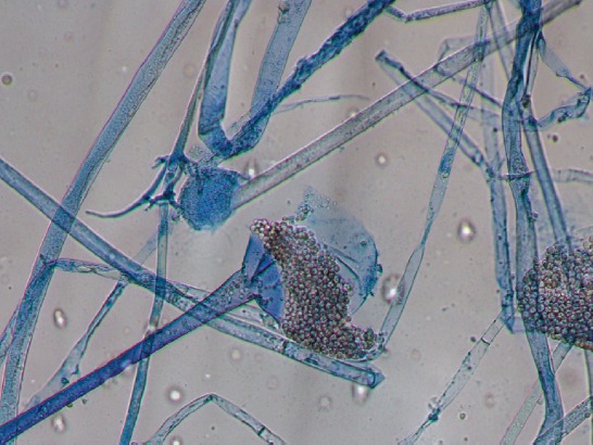
A 2-year-old girl with sickle cell anemia was transferred to King Fahad Medical City in Riyadh, Saudi Arabia with pneumonia and pleural effusion that failed to respond to prolonged courses of broad spectrum antibiotics and pleural drainage. Her past medical history was negative for recurrent infections, exposure to blood transfusions or immunosuppressive drugs. Examination revealed a febrile, malnourished child with enlarged liver and spleen. Chest examination showed a firm mass extending from the axial to the posterior aspect of the right chest wall. Initial laboratory results were: hemoglobin (77 g/L; normal range: 105-140 g/L), white blood cell count (29 x 109/L; normal range: 5-14.5 x 10⁹/L), erythrocyte sedimentation rate (80 mm/hour; normal range: 0-10 mm/hour), C-reactive protein (320 mg/L; normal range: 0-8 mg/L). Computed tomography scan showed consolidation involving right lower lobe, middle lobe, and posterior segment of the right upper lobe with pleural effusion. A right chest wall lesion with intraspinal extension was also noted. Magnetic resonance imaging of chest and spine showed an extradural extramedullary intensely enhancing mass extending from T2 through T7 and in continuity with a heterogeneously enhancing, necrotic, right sided chest wall mass. There were also destructive bony changes involving the costovertebral junction. Cultures of tissue obtained from surgical biopsy of the chest wall mass grew Rhizopus spp. (Figure 1).
Figure 1.

Rhizopus spp. grown from intraoperatively obtained lung tissue specimen.
Histopathology revealed acute and chronic necrotizing granulomatous inflammation with eosinophils. Fungal hyphae with pauciseptations were also evident on Gomori methenamine silver (GMS) stain. She was subsequently diagnosed to have CGD based on oxidative burst test result. Treatment with liposomal amphotericin B was initiated at a dose of 5 mg/kg/day then increased to 7 mg/kg/day. Caspofungin and interferon γ (IFN-γ) were added to treatment. She underwent surgical debulking of the chest wall mass and near-total pneumonectomy. Since she had no neurological symptoms from the intraspinal extension of the infection, conservative management with antifungal treatment and close follow up were planned. The intraspinal lesion continued to regress on further MRI studies. While on treatment, she developed a gaping wound at the surgical site due to poor healing and osteomyelitis of the ribs. Surgical debridement followed by excision of 2 ribs and primary closure of the wound were performed. She showed gradual improvement, but did not show complete recovery. She was then referred to a specialized center for hematopoietic stem cell transplantation (HSCT).
Case 2
A 22-year-old male known to have CGD was referred to King Faisal Specialist Hospital and Research Center (KFSHRC) in Riyadh, Saudi Arabia for surgical management of a large right lung abscess. He was diagnosed to have CGD at the age of 17 years and maintained solely on trimethoprim/sulfamethoxazole prophylaxis. His current illness started as fever and cough for 3 weeks for which he was admitted to a local hospital and started on broad spectrum antibiotics. Chest imaging with CT scan revealed a large abscess in the middle and lower lobes of the right lung and pleural effusion. Thoracotomy and drainage was performed at the referring hospital; however, he failed to show clinical improvement and no microbial etiology was identified. At KFSHRC, initial investigations were: white blood cell count (16 x 109/L; normal range: 4-11 x109/L), hemoglobin (85 g/L; normal range: 125-180 g/L), and procalcitonin (4.78 mcg/L; normal range: <0.5 mcg/L). Computed tomography scan of the chest and abdomen showed a right hemothorax mass lesion measuring 11 x 10 cm with invasion to the mediastinum, superior vena cava, right atrium, part of the inferior vena cava, diaphragm, part of the liver, and anterior chest wall. Computed tomography guided biopsy showed inflammatory cells with fungal elements. He was treated initially with voricaonazole and caspofungin in addition to antibacterial therapy. Tissue from open lung biopsy showed necrotizing granulomatous inflammation and cultures grew Rhizopus spp. Voricaonazole was changed to liposomal amphoterecin B and caspofungin continued. He underwent sternotomy with attempted mass resection. The procedure was aborted after identifying that the mass was unresectable and adherent to the right atrium and ventricle. Echocardiography showed a low ejection fraction of 35% with severely reduced right ventricular function. Unfortunately, he died several days later due to severe hypotension with bradycardia and pulseless electrical activity.
Discussion
Chronic granulomatous disease (CGD) is a genetic disorder that results from mutations in genes encoding the phagocyte nicotinamide adenine dinucleotide phosphate oxidase leading to defective generation of reactive oxygen species. This predisposes CGD patients to recurrent life-threatening bacterial and invasive fungal infections (IFIs) and multiple granuloma formations. Lungs and chest wall are the most frequent sites of infection in these patients. Aspergillus spp. is the most commonly isolated pathogen. Other sites of infection include bone, brain, and liver. Extension of infection to the vertebrae, with or without spinal cord involvement, has been reported more with invasive aspergillosis.3 An emergency presentation of CGD is mulch pneumonitis, a syndrome similar to hypersensitivity pneumonitis which presents with fever, dyspnea, and diffuse interstitial infiltrates following exposure to aerosolized mulch or organic material. In patients with mulch pneumonitis, bronchoscopy and biopsy may yield Aspergillus and other fungi including Rhizopus spp.4 In patients with CGD, invasive mucormycosis is rare and was proposed to occur mainly in patients receiving significant immunosuppression for treatment of CGD autoinflammatory manifestations.5 Disseminated Absidia corymbifera was reported in a CGD patient treated with high dose steroids for mulch pneumonitis.6 However, gastrointestinal and cutaneous mucormycosis due to Rhizopus spp. were reported as the initial presentation of CGD without iatrogenic immunosuppression.7,8 A recent systematic review showed that the most prevalent non-Aspergillus fungal infections in CGD were associated with Rhizopus spp. and Trichosporon spp. with the lungs being the most commonly affected organs. The lower respiratory tract was involved as a single site or as one of multiple organ involvement. Following Rhizopus spp., the most prevalent isolated non-Aspergillus molds were Scedosporium spp., Paecilomyces spp., and Geosmithia spp.9 Management of IFIs in CGD is complex and challenging. Opportunistic filamentous fungi other than Aspergillus are being increasingly recovered particularly from patients receiving itraconazole prophylaxis. Therefore, accurate fungal identification and susceptibility testing, along with monitoring triazoles drug levels for susceptible molds, are important factors for optimal therapy.3,8 In addition to single agent or combination antifungal therapy, surgical intervention may be required. Use of IFN-γ as a prophylaxis, and granulocyte transfusion for severe infections in CGD were described. Allogeneic HSCT is considered the definitive cure for CGD and can be performed in the setting of refractory infection.10
In conclusion, infection due to Rhizopus spp. in CGD patients can affect many systems including the respiratory, cutaneous, gastrointestinal, central nervous system, and deep organs. Pulmonary manifestations are variable and include mulch pneumonitis, consolidation with plural effusion, lung abscess, and invasive parenchymal disease which can extend to cause chest wall, vertebral, or cardiovascular involvement. As it is a potentially life-threatening infection, early diagnosis and aggressive medical and surgical management of Rhizopus spp. infections in CGD are crucial factors in improving the outcome.
Footnotes
References
- 1.Zaoutis TE, Roilides E, Chiou CC, Buchanan WL, Knudsen TA, Sarkisova TA, et al. Zygomycosis in children: a systematic review and analysis of reported cases. Pediatr Infect Dis J. 2007;26:723–727. doi: 10.1097/INF.0b013e318062115c. [DOI] [PubMed] [Google Scholar]
- 2.Reed C, Bryant R, Ibrahim AS, Edwards J, Jr, Filler SG, Goldberg R, et al. Combination polyene-caspofungin treatment of rhino-orbital-cerebral mucormycosis. Clin Infect Dis. 2008;47:364–371. doi: 10.1086/589857. [DOI] [PMC free article] [PubMed] [Google Scholar]
- 3.Blumental S, Mouy R, Mahlaoui N, Bougnoux ME, Debré M, Beauté J, et al. Invasive mold infections in chronic granulomatous disease: a 25-year retrospective survey. Clin Infect Dis. 2011;53:e159–e169. doi: 10.1093/cid/cir731. [DOI] [PubMed] [Google Scholar]
- 4.Siddiqui S, Anderson VL, Hilligoss DM, Abinun M, Kuijpers TW, Masur H, et al. Fulminant mulch pneumonitis: an emergency presentation of chronic granulomatous disease. Clin Infect Dis. 2007;45:673–681. doi: 10.1086/520985. [DOI] [PubMed] [Google Scholar]
- 5.Vinh DC, Freeman AF, Shea YR, Malech HL, Abinun M, Weinberg GA, et al. Mucormycosis in chronic granulomatous disease: association with iatrogenic immunosuppression. J Allergy Clin Immunol. 2009;123:1411–1413. doi: 10.1016/j.jaci.2009.02.020. [DOI] [PMC free article] [PubMed] [Google Scholar]
- 6.Abinun M, Wright C, Gould K, Flood TJ, Cassidy J. Absidia corymbifera in a patient with chronic granulomatous disease. Pediatr Infect Dis J. 2007;26:1167–1168. doi: 10.1097/INF.0b013e31815a0a2c. [DOI] [PubMed] [Google Scholar]
- 7.Dekkers R, Verweij PE, Weemaes CM, Severijnen RS, Van Krieken JH, Warris A. Gastrointestinal zygomycosis due to Rhizopus microsporus var. rhizopodiformis as a manifestation of chronic granulomatous disease. Med Mycol. 2008;46:491–494. doi: 10.1080/13693780801946577. [DOI] [PubMed] [Google Scholar]
- 8.Wildenbeest JG, Oomen MW, Brüggemann RJ, de Boer M, Bijleveld Y, van den Berg JM, et al. Rhizopus oryzae skin infection treated with posaconazole in a boy with chronic granulomatous disease. Pediatr Infect Dis J. 2010;29:578. doi: 10.1097/INF.0b013e3181dc8352. [DOI] [PubMed] [Google Scholar]
- 9.Dotis J, Pana ZD, Roilides E. Non-Aspergillus fungal infections in chronic granulomatous disease. Mycoses. 2013;56:449–462. doi: 10.1111/myc.12049. [DOI] [PubMed] [Google Scholar]
- 10.Falcone EL, Holland SM. Invasive fungal infection in chronic granulomatous disease: insights into pathogenesis and management. Curr Opin Infect Dis. 2012;25:658–669. doi: 10.1097/QCO.0b013e328358b0a4. [DOI] [PubMed] [Google Scholar]


