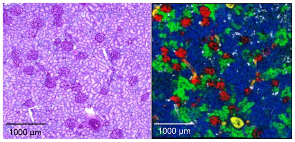
Figure 2.
Molecular images (right panel) of specific peptides and proteins acquired by MALDI IMS at 10 μm spatial resolution from a section of human kidney and the corresponding microscopy image (left panel) corresponding periodic acid Schiff stain of the same section after the MS image was taken. The glomeruli are shown in red and the tubules in green and blue on the MS image. See text for details. (Images courtesy: Kerri Grove, National Resource for Imaging Mass Spectrometry, Vanderbilt University.)

