

Abstract
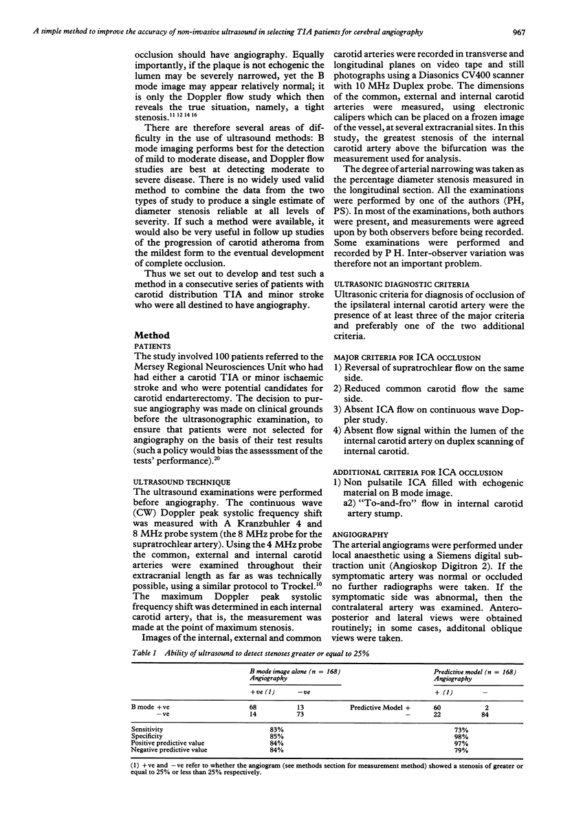
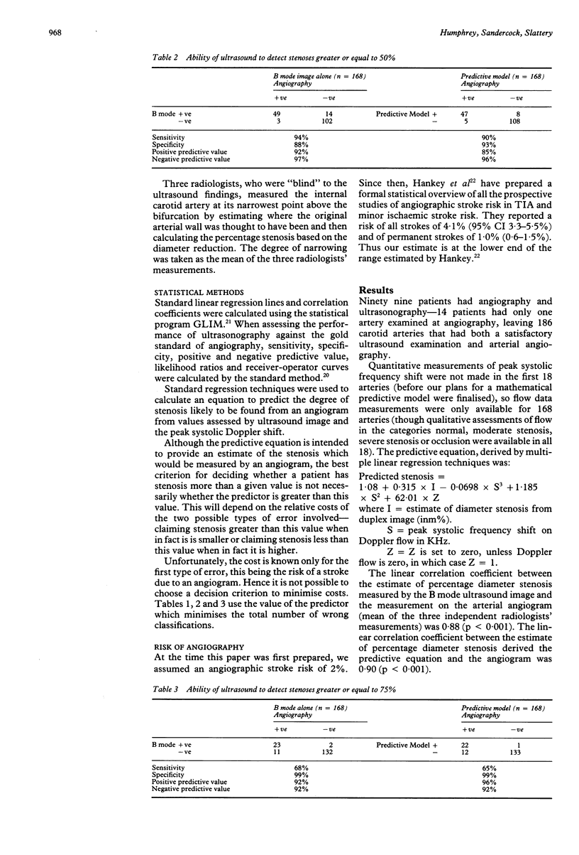
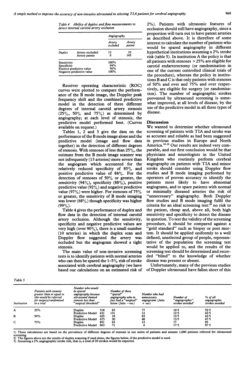
A prospective study is reported of the ability of B mode ultrasound imaging and continuous wave Doppler flow studies to detect different degrees of stenosis of the extracranial internal carotid artery (ICA) in 186 arteries in 99 patients with transient ischaemic attacks (TIA) and minor ischaemic stroke. A simple mathematical equation has been developed which combines the image and flow data to provide a single predictor of the degree of angiographic stenosis which has advantages over either ultrasonic modality used alone. The sensitivity and specificity of the predictive model in the detection of stenosis greater than or equal to 25% was 73% and 98%, of stenosis greater than or equal to 50% was 90% and 93%, of stenosis greater than or equal to 75%, 65% and 99% and occlusion 100% and 94% respectively. The principal clinical value of ultrasound screening is to spare patients with "non-significant" stenosis the risk of unnecessary angiography. Thus a simple measure of the Duplex screening tests' performance is the proportion of all strokes occurring as a complication of angiography that are avoided by changing the investigation policy from "angiograms for all carotid TIA and minor ischaemic stroke patients" to "angiograms for all patients with abnormal ultrasound results". If Duplex scanning were used to select patients most likely to have a significant abnormality on angiography, depending on the degree of stenosis to be detected, 52-85% of angiographic strokes might be avoided. If the predictive equation were used 62-88% of angiographic strokes might be avoided.
Full text
PDF
Selected References
These references are in PubMed. This may not be the complete list of references from this article.
- Dion J. E., Gates P. C., Fox A. J., Barnett H. J., Blom R. J. Clinical events following neuroangiography: a prospective study. Stroke. 1987 Nov-Dec;18(6):997–1004. doi: 10.1161/01.str.18.6.997. [DOI] [PubMed] [Google Scholar]
- Hankey G. J., Warlow C. P., Sellar R. J. Cerebral angiographic risk in mild cerebrovascular disease. Stroke. 1990 Feb;21(2):209–222. doi: 10.1161/01.str.21.2.209. [DOI] [PubMed] [Google Scholar]
- Harward T. R., Bernstein E. F., Fronek A. Range-gated pulsed Doppler power frequency spectrum analysis for the diagnosis of carotid arterial occlusive disease. Stroke. 1986 Sep-Oct;17(5):924–928. doi: 10.1161/01.str.17.5.924. [DOI] [PubMed] [Google Scholar]
- Hennerici M., Reifschneider G., Trockel U., Aulich A. Detection of early atherosclerotic lesions by duplex scanning of the carotid artery. J Clin Ultrasound. 1984 Oct;12(8):455–463. doi: 10.1002/jcu.1870120802. [DOI] [PubMed] [Google Scholar]
- Jacobs N. M., Grant E. G., Schellinger D., Byrd M. C., Richardson J. D., Cohan S. L. Duplex carotid sonography: criteria for stenosis, accuracy, and pitfalls. Radiology. 1985 Feb;154(2):385–391. doi: 10.1148/radiology.154.2.3880910. [DOI] [PubMed] [Google Scholar]
- Levien L. J., Voll C. L., Lithgow-Jolly P., Fritz V. U. The value of noninvasive investigation in the diagnosis of total occlusion of the internal carotid artery. Stroke. 1985 Nov-Dec;16(6):945–949. doi: 10.1161/01.str.16.6.945. [DOI] [PubMed] [Google Scholar]
- Pessin M. S., Duncan G. W., Mohr J. P., Poskanzer D. C. Clinical and angiographic features of carotid transient ischemic attacks. N Engl J Med. 1977 Feb 17;296(7):358–362. doi: 10.1056/NEJM197702172960703. [DOI] [PubMed] [Google Scholar]
- Toole J. F., Janeway R., Choi K., Cordell R., Davis C., Johnston F., Miller H. S. Transient ischemic attacks due to atherosclerosis. A prospective study of 160 patients. Arch Neurol. 1975 Jan;32(1):5–12. doi: 10.1001/archneur.1975.00490430027003. [DOI] [PubMed] [Google Scholar]
- Trockel U., Hennerici M., Aulich A., Sandmann W. The superiority of combined continuous wave Doppler examination over periorbital Doppler for the detection of extracranial carotid disease. J Neurol Neurosurg Psychiatry. 1984 Jan;47(1):43–50. doi: 10.1136/jnnp.47.1.43. [DOI] [PMC free article] [PubMed] [Google Scholar]
- Walsh J., Markowitz I., Kerstein M. D. Carotid endarterectomy for amaurosis fugax without angiography. Am J Surg. 1986 Aug;152(2):172–174. doi: 10.1016/0002-9610(86)90236-9. [DOI] [PubMed] [Google Scholar]
- Wolverson M. K., Heiberg E., Sundaram M., Tantanasirviongse S., Shields J. B. Carotid atherosclerosis: high-resolution real-time sonography correlated with angiography. AJR Am J Roentgenol. 1983 Feb;140(2):355–361. doi: 10.2214/ajr.140.2.355. [DOI] [PubMed] [Google Scholar]
- Zwiebel W. J., Austin C. W., Sackett J. F., Strother C. M. Correlation of high-resolution, B-mode and continuous-wave Doppler sonography with arteriography in the diagnosis of carotid stenosis. Radiology. 1983 Nov;149(2):523–532. doi: 10.1148/radiology.149.2.6622699. [DOI] [PubMed] [Google Scholar]