Abstract
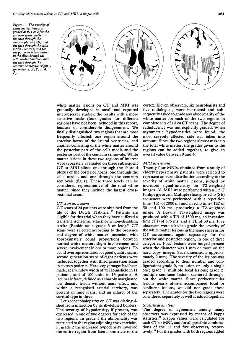
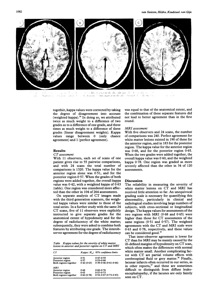
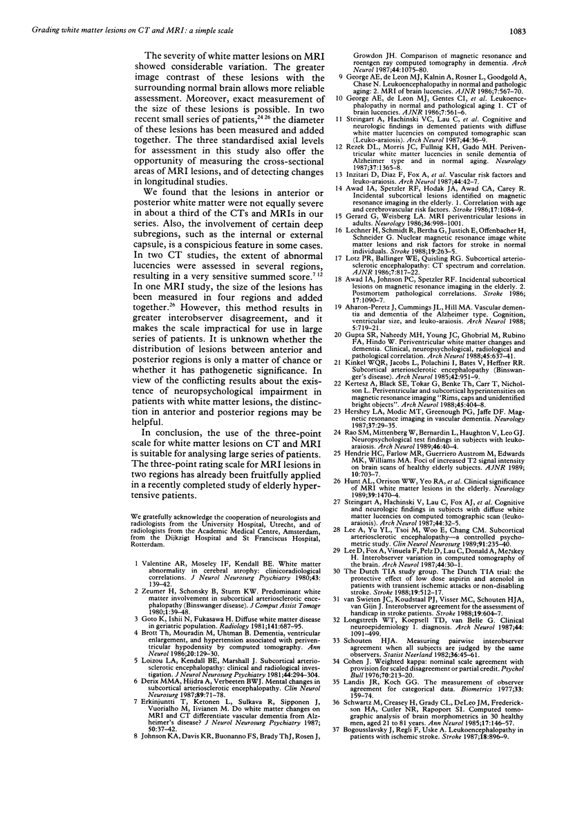
We developed and tested a simple three-point scale for grading white matter lesions in anterior and posterior regions of the brain. Twenty four CT scans and 24 MRI scans were separately judged by 11 and five observers, respectively, on the presence and severity of white matter lesions. The observers were radiologists and neurologists. For CT scans, these periventricular changes were graded according to their extent as absent, or partly involving the white matter, or extending up to the subcortical region. The MRI lesions were graded as no lesion or only a single one, multiple focal lesions, and multiple confluent lesions. The pairwise agreements of all possible combinations of observers for each scan were corrected for chance (kappa statistics; maximal agreement 1.0). The weighted kappa value, for anterior and posterior regions combined, was 0.63 for CT scans, and 0.78 for MRI scans. This three-point scale for two separate regions seems suitable as a basis for cross-sectional or longitudinal studies of large series of patients.
Full text
PDF


Images in this article
Selected References
These references are in PubMed. This may not be the complete list of references from this article.
- Aharon-Peretz J., Cummings J. L., Hill M. A. Vascular dementia and dementia of the Alzheimer type. Cognition, ventricular size, and leuko-araiosis. Arch Neurol. 1988 Jul;45(7):719–721. doi: 10.1001/archneur.1988.00520310025011. [DOI] [PubMed] [Google Scholar]
- Awad I. A., Johnson P. C., Spetzler R. F., Hodak J. A. Incidental subcortical lesions identified on magnetic resonance imaging in the elderly. II. Postmortem pathological correlations. Stroke. 1986 Nov-Dec;17(6):1090–1097. doi: 10.1161/01.str.17.6.1090. [DOI] [PubMed] [Google Scholar]
- Awad I. A., Spetzler R. F., Hodak J. A., Awad C. A., Carey R. Incidental subcortical lesions identified on magnetic resonance imaging in the elderly. I. Correlation with age and cerebrovascular risk factors. Stroke. 1986 Nov-Dec;17(6):1084–1089. doi: 10.1161/01.str.17.6.1084. [DOI] [PubMed] [Google Scholar]
- Bogousslavsky J., Regli F., Uske A. Leukoencephalopathy in patients with ischemic stroke. Stroke. 1987 Sep-Oct;18(5):896–899. doi: 10.1161/01.str.18.5.896. [DOI] [PubMed] [Google Scholar]
- Derix M. M., Hijdra A., Verbeeten B. W., Jr Mental changes in subcortical arteriosclerotic encephalopathy. Clin Neurol Neurosurg. 1987;89(2):71–78. doi: 10.1016/0303-8467(87)90179-x. [DOI] [PubMed] [Google Scholar]
- Erkinjuntti T., Ketonen L., Sulkava R., Sipponen J., Vuorialho M., Iivanainen M. Do white matter changes on MRI and CT differentiate vascular dementia from Alzheimer's disease? J Neurol Neurosurg Psychiatry. 1987 Jan;50(1):37–42. doi: 10.1136/jnnp.50.1.37. [DOI] [PMC free article] [PubMed] [Google Scholar]
- George A. E., de Leon M. J., Gentes C. I., Miller J., London E., Budzilovich G. N., Ferris S., Chase N. Leukoencephalopathy in normal and pathologic aging: 1. CT of brain lucencies. AJNR Am J Neuroradiol. 1986 Jul-Aug;7(4):561–566. [PMC free article] [PubMed] [Google Scholar]
- George A. E., de Leon M. J., Kalnin A., Rosner L., Goodgold A., Chase N. Leukoencephalopathy in normal and pathologic aging: 2. MRI of brain lucencies. AJNR Am J Neuroradiol. 1986 Jul-Aug;7(4):567–570. [PMC free article] [PubMed] [Google Scholar]
- Gerard G., Weisberg L. A. MRI periventricular lesions in adults. Neurology. 1986 Jul;36(7):998–1001. doi: 10.1212/wnl.36.7.998. [DOI] [PubMed] [Google Scholar]
- Goto K., Ishii N., Fukasawa H. Diffuse white-matter disease in the geriatric population. A clinical, neuropathological, and CT study. Radiology. 1981 Dec;141(3):687–695. doi: 10.1148/radiology.141.3.7302224. [DOI] [PubMed] [Google Scholar]
- Gupta S. R., Naheedy M. H., Young J. C., Ghobrial M., Rubino F. A., Hindo W. Periventricular white matter changes and dementia. Clinical, neuropsychological, radiological, and pathological correlation. Arch Neurol. 1988 Jun;45(6):637–641. doi: 10.1001/archneur.1988.00520300057019. [DOI] [PubMed] [Google Scholar]
- Hendrie H. C., Farlow M. R., Austrom M. G., Edwards M. K., Williams M. A. Foci of increased T2 signal intensity on brain MR scans of healthy elderly subjects. AJNR Am J Neuroradiol. 1989 Jul-Aug;10(4):703–707. [PMC free article] [PubMed] [Google Scholar]
- Hershey L. A., Modic M. T., Greenough P. G., Jaffe D. F. Magnetic resonance imaging in vascular dementia. Neurology. 1987 Jan;37(1):29–36. doi: 10.1212/wnl.37.1.29. [DOI] [PubMed] [Google Scholar]
- Hunt A. L., Orrison W. W., Yeo R. A., Haaland K. Y., Rhyne R. L., Garry P. J., Rosenberg G. A. Clinical significance of MRI white matter lesions in the elderly. Neurology. 1989 Nov;39(11):1470–1474. doi: 10.1212/wnl.39.11.1470. [DOI] [PubMed] [Google Scholar]
- Inzitari D., Diaz F., Fox A., Hachinski V. C., Steingart A., Lau C., Donald A., Wade J., Mulic H., Merskey H. Vascular risk factors and leuko-araiosis. Arch Neurol. 1987 Jan;44(1):42–47. doi: 10.1001/archneur.1987.00520130034014. [DOI] [PubMed] [Google Scholar]
- Johnson K. A., Davis K. R., Buonanno F. S., Brady T. J., Rosen T. J., Growdon J. H. Comparison of magnetic resonance and roentgen ray computed tomography in dementia. Arch Neurol. 1987 Oct;44(10):1075–1080. doi: 10.1001/archneur.1987.00520220071020. [DOI] [PubMed] [Google Scholar]
- Kertesz A., Black S. E., Tokar G., Benke T., Carr T., Nicholson L. Periventricular and subcortical hyperintensities on magnetic resonance imaging. 'Rims, caps, and unidentified bright objects'. Arch Neurol. 1988 Apr;45(4):404–408. doi: 10.1001/archneur.1988.00520280050015. [DOI] [PubMed] [Google Scholar]
- Kinkel W. R., Jacobs L., Polachini I., Bates V., Heffner R. R., Jr Subcortical arteriosclerotic encephalopathy (Binswanger's disease). Computed tomographic, nuclear magnetic resonance, and clinical correlations. Arch Neurol. 1985 Oct;42(10):951–959. doi: 10.1001/archneur.1985.04060090033010. [DOI] [PubMed] [Google Scholar]
- Landis J. R., Koch G. G. The measurement of observer agreement for categorical data. Biometrics. 1977 Mar;33(1):159–174. [PubMed] [Google Scholar]
- Lechner H., Schmidt R., Bertha G., Justich E., Offenbacher H., Schneider G. Nuclear magnetic resonance image white matter lesions and risk factors for stroke in normal individuals. Stroke. 1988 Feb;19(2):263–265. doi: 10.1161/01.str.19.2.263. [DOI] [PubMed] [Google Scholar]
- Lee A., Yu Y. L., Tsoi M., Woo E., Chang C. M. Subcortical arteriosclerotic encephalopathy--a controlled psychometric study. Clin Neurol Neurosurg. 1989;91(3):235–241. doi: 10.1016/0303-8467(89)90117-0. [DOI] [PubMed] [Google Scholar]
- Loizou L. A., Kendall B. E., Marshall J. Subcortical arteriosclerotic encephalopathy: a clinical and radiological investigation. J Neurol Neurosurg Psychiatry. 1981 Apr;44(4):294–304. doi: 10.1136/jnnp.44.4.294. [DOI] [PMC free article] [PubMed] [Google Scholar]
- Longstreth W. T., Jr, Koepsell T. D., van Belle G. Clinical neuroepidemiology. I. Diagnosis. Arch Neurol. 1987 Oct;44(10):1091–1099. doi: 10.1001/archneur.1987.00520220087023. [DOI] [PubMed] [Google Scholar]
- Rao S. M., Mittenberg W., Bernardin L., Haughton V., Leo G. J. Neuropsychological test findings in subjects with leukoaraiosis. Arch Neurol. 1989 Jan;46(1):40–44. doi: 10.1001/archneur.1989.00520370042017. [DOI] [PubMed] [Google Scholar]
- Rezek D. L., Morris J. C., Fulling K. H., Gado M. H. Periventricular white matter lucencies in senile dementia of the Alzheimer type and in normal aging. Neurology. 1987 Aug;37(8):1365–1368. doi: 10.1212/wnl.37.8.1365. [DOI] [PubMed] [Google Scholar]
- Schwartz M., Creasey H., Grady C. L., DeLeo J. M., Frederickson H. A., Cutler N. R., Rapoport S. I. Computed tomographic analysis of brain morphometrics in 30 healthy men, aged 21 to 81 years. Ann Neurol. 1985 Feb;17(2):146–157. doi: 10.1002/ana.410170208. [DOI] [PubMed] [Google Scholar]
- Steingart A., Hachinski V. C., Lau C., Fox A. J., Diaz F., Cape R., Lee D., Inzitari D., Merskey H. Cognitive and neurologic findings in subjects with diffuse white matter lucencies on computed tomographic scan (leuko-araiosis). Arch Neurol. 1987 Jan;44(1):32–35. doi: 10.1001/archneur.1987.00520130024012. [DOI] [PubMed] [Google Scholar]
- Steingart A., Hachinski V. C., Lau C., Fox A. J., Fox H., Lee D., Inzitari D., Merskey H. Cognitive and neurologic findings in demented patients with diffuse white matter lucencies on computed tomographic scan (leuko-araiosis). Arch Neurol. 1987 Jan;44(1):36–39. doi: 10.1001/archneur.1987.00520130028013. [DOI] [PubMed] [Google Scholar]
- Valentine A. R., Moseley I. F., Kendall B. E. White matter abnormality in cerebral atrophy: clinicoradiological correlations. J Neurol Neurosurg Psychiatry. 1980 Feb;43(2):139–142. doi: 10.1136/jnnp.43.2.139. [DOI] [PMC free article] [PubMed] [Google Scholar]
- van Swieten J. C., Koudstaal P. J., Visser M. C., Schouten H. J., van Gijn J. Interobserver agreement for the assessment of handicap in stroke patients. Stroke. 1988 May;19(5):604–607. doi: 10.1161/01.str.19.5.604. [DOI] [PubMed] [Google Scholar]


