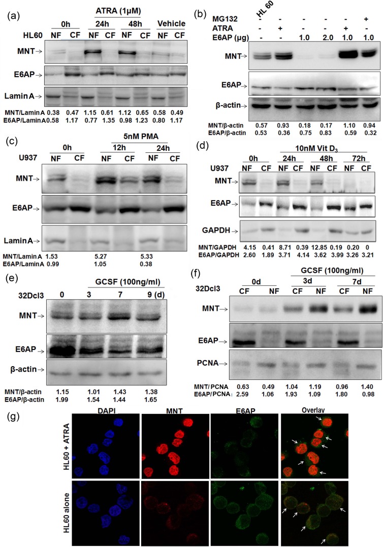
Figure 3. MNT expression is upregulated during myeloid differentiation.
a. HL60 cells were treated with 1μM ATRA. Post-ATRA treatment for 0, 24 and 48h lysates from nuclear and cytoplasmic fractions were prepared, resolved on 10% SDS-PAGE and immunoblotted with anti-MNT and anti-E6AP antibodies. Lamin A was used as loading control for the quality of nuclear fractions. DMSO was used as vehicle. b. HL60 cells were transfected with increasing amounts of E6AP (1.0 and 2.0μg), treated with 1μM ATRA for 24h in control cells and 24h post transfection for next 24h in 1.0μg E6AP transfected cells. As indicated 10μM MG132 treatment was given 6h prior to lysate preparation. Lysates resolved on 10% SDS-PAGE were immunoblotted with anti-MNT, E6AP and β-actin antibodies. c. U937 cells were treated with 5nM PMA. Post-PMA treatment for 0, 12 and 24h lysates from nuclear and cytoplasmic fractions were prepared, resolved on 10% SDS-PAGE and immunoblotted with anti-MNT followed by anti-E6AP antibodies. Lamin A was used as loading control. d. U937 cells were treated with 10nM Vitamin D3, post-Vitamin D3 treatment for 0, 24, 48 and 72h, lysates from nuclear and cytoplasmic fractions were prepared, resolved on 10% SDS-PAGE and immunoblotted with anti-MNT followed by anti-E6AP and anti-GAPDH antibodies. e. 32Dcl3 cells were treated with 100ng/ml G-CSF for 0, 3, 7 and 9 days as indicated. Post-induction, WCEs were prepared, resolved on 10% SDS-PAGE and immunoblotted with anti-MNT and anti-E6AP antibodies. β-actin was used as loading control. f. 32Dcl3 cells were treated with 100ng/ml G-CSF as indicated. Post-induction, nuclear/cytoplasmic fractions were prepared, resolved on 10% SDS-PAGE followed by immunoblotting with anti-MNT and anti-E6AP antibodies. PCNA was used a loading control for the quality of nuclear fractions. g. Indirect immunofluorescence staining for MNT and E6AP was performed using respective primary and conjugated secondary antibodies. Stained cells were visualized under fluorescence microscope (63X) to assess their localization. Results are representative of three minimum independent experiments.

