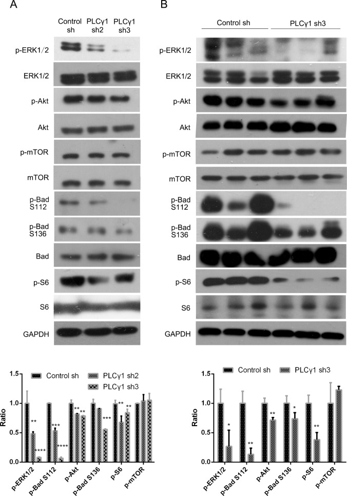
Figure 4. Effect of PLCγ1 inhibition on signal molecules associated with tumor growth and metastasis of human gastric adenocarcinoma.
(A) Cells were transduced with lentivirus-mediated PLCγ1shRNA2/3 vectors. The protein levels of Akt, p-Akt, ERK1/2, p-ERK1/2, mTOR, p-mTOR, S6, p-S6(Ser235/236), Bad, p-Bad (Ser112), p-Bad (Ser136), PLCγ1, and GAPDH were detected with Western blotting analysis as described in Materials and Methods. (B) A nude mouse model harboring tumor xenografts derived from BGC-823 cells transduced with PLCγ1-shRNA3 vector was constructed. The protein levels of Akt, p-Akt, ERK1/2, p-ERK1/2, mTOR, p-mTOR, S6, p-S6(Ser235/236), Bad, p-Bad (Ser112), p-Bad (Ser136), PLCγ1, and GAPDH in the tumor samples were detected by Western blotting analysis as described in Materials and Methods. Data are reported as means ± S.D. of three independent experiments (*P < 0.05, **P < 0.01, ***P < 0.001, ****P < 0.0001, vs respective control).

