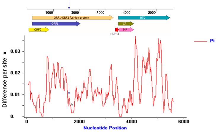
Figure 7.
Distribution of MaYMV genetic variation estimated by nucleotide diversity (π). The sliding window was 100 sites wide with slide set at 25-site intervals. The relative positions of the seven ORFs of the MaYMV genome are illustrated by lines above the plot. The positions of conserved frameshifting sequence and pseudoknot sequences are shown as blue arrow and black asterisks, respectively.

