

Abstract
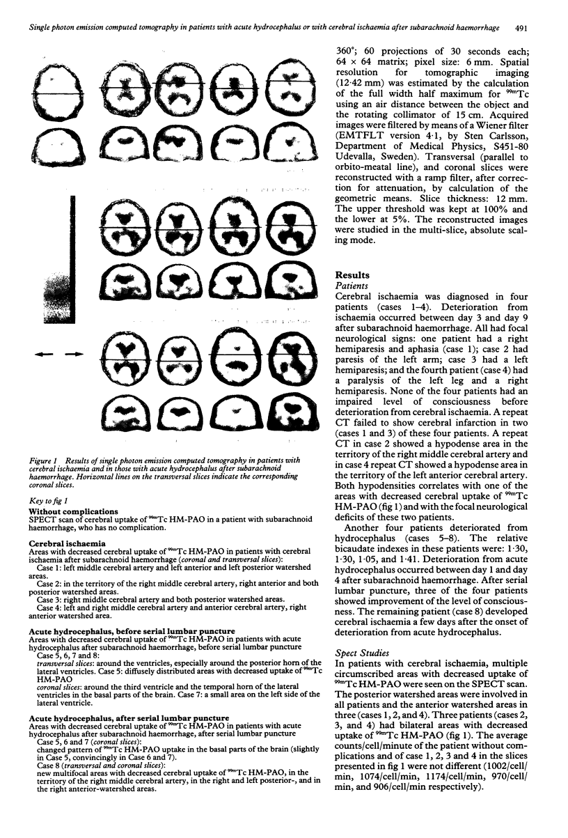
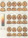
Using single photon emission computed tomography (SPECT), cerebral blood flow was studied in eight patients with gradual deterioration in the level of consciousness after subarachnoid haemorrhage. Four had cerebral ischaemia and four had acute hydrocephalus. In patients with cerebral ischaemia, single photon emission computed tomography scanning showed multiple regions with decreased uptake of technetium-99M labelled d,l-hexamethyl-propylene amine oxime (99mTcHM-PAO) mainly in watershed areas. In patients with acute hydrocephalus, decreased uptake was seen mainly in the basal parts of the brain: around the third ventricle, around the temporal horns of the lateral ventricles, and in the basal part of the frontal lobe. After serial lumbar puncture, there was improvement of the uptake of 99mTc HM-PAO in these basal areas in three (convincingly in two and slightly in the other) of the four patients accompanied by clinical improvement in these three patients. These results suggest that patients with acute hydrocephalus and impaired consciousness after SAH, in contrast to patients with cerebral ischaemia, have decreased cerebral blood flow predominantly in the basal parts of the brain.
Full text
PDF
Images in this article
Selected References
These references are in PubMed. This may not be the complete list of references from this article.
- Brooks D. J., Beaney R. P., Powell M., Leenders K. L., Crockard H. A., Thomas D. G., Marshall J., Jones T. Studies on cerebral oxygen metabolism, blood flow, and blood volume, in patients with hydrocephalus before and after surgical decompression, using positron emission tomography. Brain. 1986 Aug;109(Pt 4):613–628. doi: 10.1093/brain/109.4.613. [DOI] [PubMed] [Google Scholar]
- Davis S., Andrews J., Lichtenstein M., Kaye A., Tress B., Rossiter S., Salehi N., Binns D. A single-photon emission computed tomography study of hypoperfusion after subarachnoid hemorrhage. Stroke. 1990 Feb;21(2):252–259. doi: 10.1161/01.str.21.2.252. [DOI] [PubMed] [Google Scholar]
- Graff-Radford N. R., Rezai K., Godersky J. C., Eslinger P., Damasio H., Kirchner P. T. Regional cerebral blood flow in normal pressure hydrocephalus. J Neurol Neurosurg Psychiatry. 1987 Dec;50(12):1589–1596. doi: 10.1136/jnnp.50.12.1589. [DOI] [PMC free article] [PubMed] [Google Scholar]
- Géraud G., Tremoulet M., Guell A., Bes A. The prognostic value of noninvasive CBF measurement in subarachnoid hemorrhage. Stroke. 1984 Mar-Apr;15(2):301–305. doi: 10.1161/01.str.15.2.301. [DOI] [PubMed] [Google Scholar]
- Hasan D., Vermeulen M., Wijdicks E. F., Hijdra A., van Gijn J. Management problems in acute hydrocephalus after subarachnoid hemorrhage. Stroke. 1989 Jun;20(6):747–753. doi: 10.1161/01.str.20.6.747. [DOI] [PubMed] [Google Scholar]
- Hayashi M., Kobayashi H., Kawano H., Yamamoto S., Maeda T. Cerebral blood flow and ICP patterns in patients with communicating hydrocephalus after aneurysm rupture. J Neurosurg. 1984 Jul;61(1):30–36. doi: 10.3171/jns.1984.61.1.0030. [DOI] [PubMed] [Google Scholar]
- Hijdra A., Van Gijn J., Stefanko S., Van Dongen K. J., Vermeulen M., Van Crevel H. Delayed cerebral ischemia after aneurysmal subarachnoid hemorrhage: clinicoanatomic correlations. Neurology. 1986 Mar;36(3):329–333. doi: 10.1212/wnl.36.3.329. [DOI] [PubMed] [Google Scholar]
- Lassen N. A. Cerebral blood flow measured by xenon-133. Nucl Med Commun. 1987 Jul;8(7):535–547. [PubMed] [Google Scholar]
- Lear J. L. Quantitative local cerebral blood flow measurements with technetium-99m HM-PAO: evaluation using multiple radionuclide digital quantitative autoradiography. J Nucl Med. 1988 Aug;29(8):1387–1392. [PubMed] [Google Scholar]
- Lindsay K. W., Teasdale G. M., Knill-Jones R. P. Observer variability in assessing the clinical features of subarachnoid hemorrhage. J Neurosurg. 1983 Jan;58(1):57–62. doi: 10.3171/jns.1983.58.1.0057. [DOI] [PubMed] [Google Scholar]
- Mamo H. L., Meric P. C., Ponsin J. C., Rey A. C., Luft A. G., Seylaz J. A. Cerebral blood flow in normal pressure hydrocephalus. Stroke. 1987 Nov-Dec;18(6):1074–1080. doi: 10.1161/01.str.18.6.1074. [DOI] [PubMed] [Google Scholar]
- Meese W., Kluge W., Grumme T., Hopfenmüller W. CT evaluation of the CSF spaces of healthy persons. Neuroradiology. 1980 Apr;19(3):131–136. doi: 10.1007/BF00342387. [DOI] [PubMed] [Google Scholar]
- Menon D., Weir B., Overton T. Ventricular size and cerebral blood flow following subarachnoid hemorrhage. J Comput Assist Tomogr. 1981 Jun;5(3):328–333. doi: 10.1097/00004728-198106000-00002. [DOI] [PubMed] [Google Scholar]
- Neirinckx R. D., Canning L. R., Piper I. M., Nowotnik D. P., Pickett R. D., Holmes R. A., Volkert W. A., Forster A. M., Weisner P. S., Marriott J. A. Technetium-99m d,l-HM-PAO: a new radiopharmaceutical for SPECT imaging of regional cerebral blood perfusion. J Nucl Med. 1987 Feb;28(2):191–202. [PubMed] [Google Scholar]
- Powers W. J., Grubb R. L., Jr, Baker R. P., Mintun M. A., Raichle M. E. Regional cerebral blood flow and metabolism in reversible ischemia due to vasospasm. Determination by positron emission tomography. J Neurosurg. 1985 Apr;62(4):539–546. doi: 10.3171/jns.1985.62.4.0539. [DOI] [PubMed] [Google Scholar]
- Shoulson I., Fahn S. Huntington disease: clinical care and evaluation. Neurology. 1979 Jan;29(1):1–3. doi: 10.1212/wnl.29.1.1. [DOI] [PubMed] [Google Scholar]
- Tamaki N., Kusunoki T., Wakabayashi T., Matsumoto S. Cerebral hemodynamics in normal-pressure hydrocephalus. Evaluation by 133Xe inhalation method and dynamic CT study. J Neurosurg. 1984 Sep;61(3):510–514. doi: 10.3171/jns.1984.61.3.0510. [DOI] [PubMed] [Google Scholar]
- Teasdale G., Jennett B. Assessment of coma and impaired consciousness. A practical scale. Lancet. 1974 Jul 13;2(7872):81–84. doi: 10.1016/s0140-6736(74)91639-0. [DOI] [PubMed] [Google Scholar]
- Vorstrup S., Christensen J., Gjerris F., Sørensen P. S., Thomsen A. M., Paulson O. B. Cerebral blood flow in patients with normal-pressure hydrocephalus before and after shunting. J Neurosurg. 1987 Mar;66(3):379–387. doi: 10.3171/jns.1987.66.3.0379. [DOI] [PubMed] [Google Scholar]
- Yonekura Y., Nishizawa S., Mukai T., Fujita T., Fukuyama H., Ishikawa M., Kikuchi H., Konishi J., Andersen A. R., Lassen N. A. SPECT with [99mTc]-d,l-hexamethyl-propylene amine oxime (HM-PAO) compared with regional cerebral blood flow measured by PET: effects of linearization. J Cereb Blood Flow Metab. 1988 Dec;8(6):S82–S89. doi: 10.1038/jcbfm.1988.36. [DOI] [PubMed] [Google Scholar]
- van Gijn J., Hijdra A., Wijdicks E. F., Vermeulen M., van Crevel H. Acute hydrocephalus after aneurysmal subarachnoid hemorrhage. J Neurosurg. 1985 Sep;63(3):355–362. doi: 10.3171/jns.1985.63.3.0355. [DOI] [PubMed] [Google Scholar]
- van Gijn J., van Dongen K. J. Computerized tomography in subarachnoid hemorrhage: difference between patients with and without an aneurysm on angiography. Neurology. 1980 May;30(5):538–539. doi: 10.1212/wnl.30.5.538. [DOI] [PubMed] [Google Scholar]