Abstract
Reconstruction of chest wall tumor is very important link of chest wall tumor resection. Many implants have been reported to be used to reconstruct the chest wall, such as steelwire, titanium mesh and polypropylene mesh. It is really hard for clinicians to decide which implant is the best one to replace the chest wall. We herein report a 68-year-old man who had underwent a chest wall reconstruction with a hernia repair piece and a Dacron hernia repair piece. The patient has maintained an excellent cosmetic and functional outcome since surgery, which proves that the hernia piece still has its place in reconstruction of chest wall.
Keywords: Chest wall tumor, chest defect, chest wall reconstruction, hernia repair piece
Introduction
Resection of a giant rib tumor is a challenge for clinicians. When a large portion of the sternum or rib is invaded by a tumor, careful reconstruction is required to avoid serious secondary complications such as paradoxical respiration. The requirements of an ideal replacement for reconstruction of the chest wall are its availability, durability, non-reactivity, and resistance to infection. Traditionally, steelwire, titanium meshes, or polypropylene meshes are usually used for skeletal reconstruction. As new technologies have been developed, such as high-resolution three-dimensional printing technology, and autogenous implants have been used to provide better chest wall reconstruction. However, there is no consensus of which is the best material. In this article, we report our experience of using hernia repair mesh and provide a brief introduction to this material.
Case presentation
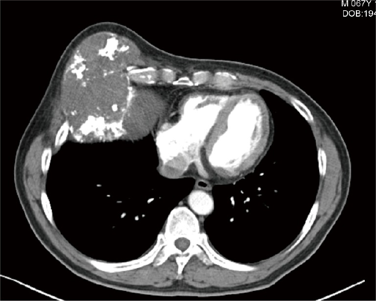
A 68-year-old man who had a mass in his chest wall for 12 years was admitted to our hospital. He showed no significant symptoms and the patient had no mental disease, general medical disease, chronic pain or psychoactive addiction in the past or at present. During his physical examination, we observed a huge mass measuring 10 cm × 8 cm × 8 cm in the right anterior side of his chest wall. A computed tomography (CT) scan showed a large tumor (15.7 cm × 15 cm) located in the right chest wall, which had destroyed the 5th rid completely and compressed the 4th and 6th rib seriously (Figure 1). The patient underwent a tumor radical resection and chest wall reconstruction with hernia repair pieces, and the postoperative pathology confirmed the chondrosarcoma. There are no postoperative complications such as paradoxical respire after the surgery and during our follow-up; furthermore, the patient has maintained good health since the surgery.
Figure 1.

The tumor destroyed the 5th rid completely and compressed the 4th and 6th rib seriously.
Operative techniques
The tumor was finally resected and a careful reconstruction was given to repair the defect. The operation procedure is shown in the Figure 2.
Figure 2.

Chest wall reconstruction after resection using hernia repair piece (1). Available online: http://www.asvide.com/articles/984
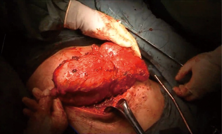
After general anesthesia and inserting a single-lumen endotracheal tube, the patient was in a horizontal position. A 30 cm incision was made on the right anterior side of the chest wall. The skin and muscles were separated to expose the tumor (Figure 3). We found the tumor had completely invaded the 5th rib as well as partly invaded the 4th rib. Then the surrounding tissue and tumor were separated carefully, the ribs and cartilago costalis were cut using a rib clamp under the premise of ensuring the negative margin (3 cm). The tumor, which measured 22 cm × 12 cm × 12 cm and weighed 1,335 g, was then completely resected. The intraoperative pathology confirmed the chondrosarcoma.
Figure 3.

the tumor was resected under the premise of ensuring the negative margin, which measured 22 cm × 12 cm × 12 cm and weighed 1,335 g.
Next, the patient received a chest wall reconstruction. First, we measured the size of the defect and adjusted the shape of a hernia repair piece (Johnson & Johnson, USA). The patch was overlaid on the defect, continuously monofilament nonabsorbable suture was used to fix the patch to the edge of the defect. And another Dacron patch (Johnson & Johnson, USA) was sutured on the first patch to increase its structural strength. After placement of drainage tube, IHE chest wall was then closed in layers in the usual manner. The total amount of blood lost was about 300 mL.
Comments
Chest wall reconstruction is a very crucial step in the resection of chest wall tumor. While implants play very important roles in the chest wall reconstruction. An appropriate implant can provide an excellent cosmetic and functional outcome and avoid many postoperative complications. Steelwire, titanium mesh or polypropylene mesh was once common used as the material of the skeleton reconstruction. With the development of new technology, more and more new implants have been used to repair chest wall defect recently, such as 3D printing model (2), biomaterial artificial rib (3), autogenous rib graft (4) and so on. However, invention of new materials didn’t mean traditional implants have been out of time. Each material has its own advantages and there is no consensus that which is the best one.
While the hernia repair piece, as a kind of very traditional material, still has a very wonderful performance in chest wall reconstruction. In our follow-up, this patient who underwent reconstruction using hernia repair piece had an excellent outcome and no postoperative complications. What’s more, hernia piece also has many own irreplaceable advantages. Firstly, hernia repair piece make the reconstruction much easier and more convenient. The shape of the patch can be adjusted flexibly according to the sharp of the defect. What the performer needs to do is just fixing the patch to the edge of the defect. Of course, the using method can also be adjusted according to the requests of the performer. In fact, in our surgery, we added one more Dacron patch to enhance the structural strength of the reconstruction. Secondly, the hernia repair piece is much cheaper than high-tech implant. For poor patient, reconstruction with hernia piece is obvious more acceptable. And what’s more, hernia repair piece is so common that it can be found everywhere, make the reconstruction avoid anywhere and anytime.
As we have mentioned above, hernia repair piece proves it still has its place in reconstruction of chest wall. However, every material has its own optimum situation. From our experience, we recommend that hernia pieces should be used to reconstruct the defect when the defect only involved two or three ribs. In this condition, reconstruction of defect is undoubtedly needed to maintain the function of chest wall and the hernia patch has just enough firmness to replace the chest wall. Thus, the procedure described is safe and technically feasible and appears to be promising as an alternative approach for the reconstruction of chest wall after resection.
Acknowledgements
None.
Footnotes
Conflicts of Interest: The authors have no conflicts of interest to declare.
References
- 1.Wu Y, Zhang G, Zhu Z, et al. Chest wall reconstruction after resection using hernia repair piece. Asvide 2016;3:225. Available online: http://www.asvide.com/articles/984 [DOI] [PMC free article] [PubMed]
- 2.Butscher A, Bohner M, Hofmann S, et al. Structural and material approaches to bone tissue engineering in powder-based three-dimensional printing. Acta Biomater 2011;7:907-20. 10.1016/j.actbio.2010.09.039 [DOI] [PubMed] [Google Scholar]
- 3.Zhang LJ, Wang WP, Li WY, et al. A new alternative for bony chest wall reconstruction using biomaterial artificial rib and pleura: animal experiment and clinical application. Eur J Cardiothorac Surg 2011;40:939-47. [DOI] [PubMed] [Google Scholar]
- 4.Li W, Zhang G, Ye C, et al. Autogenous rib graft for reconstruction of sternal defects. J Thorac Dis 2014;6:1851-2. [DOI] [PMC free article] [PubMed] [Google Scholar]


