Abstract
The age of liver donors has been increasing in the past several years because of a donor shortage. In the United States, 33% of donors are age 50 years or older, as are more than 50% in some European countries. The impact of donor age on liver transplantation (LT) has been analyzed in several studies with contradictory conclusions. Nevertheless, recent analyses of the largest databases demonstrate that having an older donor is a risk factor for graft failure. Donor age is included as a risk factor in the more relevant graft survival scores, such as the Donor Risk Index, donor age and Model for End-stage Liver Disease, Survival Outcomes Following Liver Transplantation, and the Balance of Risk. The use of old donors is related to an increased rate of biliary complications and hepatitis C virus-related graft failure. Although liver function does not seem to be significantly affected by age, the incidence of several liver diseases increases with age, and the capacity of the liver to manage or overcome liver diseases or external injuries decreases. In this paper, the importance of age in LT outcomes, the role of donor age as a risk factor, and the influence of aging on liver regeneration are reviewed.
Keywords: Liver transplantation, Liver regeneration, Graft survival, Old donor, Aging
Core tip: Because of a donor shortage, the use of grafts from old donors has become widespread. Donor age is related to worse outcomes after liver transplantation, higher rates of graft failure, biliary complications and a worse graft survival. In recipients with hepatitis C, the impact of donor age is even more evident. Aging-related changes at the hepatocellular level may contribute to a decreased capacity of the liver to manage or overcome liver diseases and injuries. This review summarizes the evidence regarding the impact of donor age on liver transplantation outcomes.
INTRODUCTION
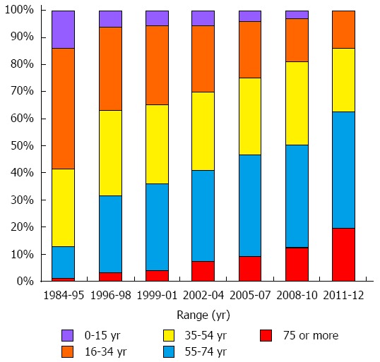
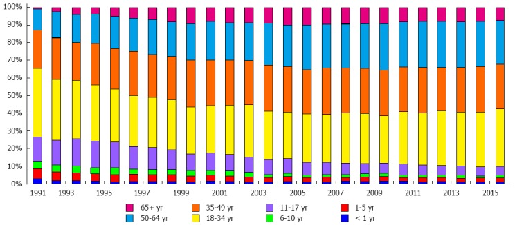
In the recent years a considerable change in donor age distribution of liver transplantation (LT) has been observed, as shown in the figures from the European Liver Transplant Registry (ELTR), with a rising percentage of livers proceeding from donors older than 60 years. In 1989, only 1% of livers proceeded from donors over 60 years of age. This rate escalates to 15% in 1999, 20% in 2001, and 29% in 2009[1,2]. In Spain, between 1984 and 1995, only 11.5% of donors were age 55 years or older, while between 2011 and 2012, 61.8% of donors were 55 years or older (Figure 1)[3]. The United Network for Organ Sharing (UNOS), in the United States, reports that in 1989, 2.4% of donors were age 50 years or older, but this rate increased to 29% in 1999 and to 33% in 2013 (Figure 2).
Figure 1.

Change in distribution of donor age in recent years. Source: Spanish Liver Transplant Registry.
Figure 2.

Change in distribution of donor age in recent years. Source: United Network for Organ Sharing reports.
The impact of donor age on LT has been evaluated in different studies with contradictory results. Many studies did not observe differences in graft survival according to donor age[4,5], on the contrary others report an increases of complication rates and poorer survival following transplantation from older donors[6,7]. Furthermore, a relationship has been described between allografts obtained from older donors and a faster progression of fibrosis after LT in recipients infected with hepatitis C virus (HCV)[8,9].
IMPACT OF DONOR AGE ON LIVER TRANSPLANT OUTCOMES
Deceased donor liver transplant
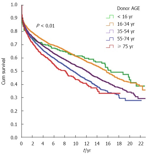
Studies based on institutional registries have evaluated the effects of donor age on patient and graft survival in the largest patient series[1-3,6,7]. In the ELTR, the 1-year survival of patients who received transplants between 1998 and 2001 was similar for all donor age groups[2]. In a recent analysis of the same ELTR database, graft survival was significantly higher if the organs proceed from donors younger than 55 years vs donors older than 65 years (65% vs 57%, P < 0.0001)[1]. An analysis of the data collected by the Spanish Registry for Liver Transplantation between 1991 and 2013 shows that donor age influences LT outcome (Figure 3). LT performed with deceased donors over age 55 years had a slight but significant worsening in actuarial graft survival one year after LT compared with those realized with graft from donors younger than 55 years. The difference in graft survival between the two groups was more evident at 5 years after LT[3]. Feng et al[10] recently analyzed donor risk factors in LTs finding that donor age over 60 years was the strongest risk factor for graft failure. In this analysis of the data collected from the Scientific Registry of Transplant Recipients, donor age over 40 years and especially over 60 years, donation after cardiac death, and split/partial grafts were strongly associated with graft failure. In a retrospective analysis performed using data obtained from the UNOS, Reese et al[11] found that performing LTs with donors who were ≥ 45 years old increased the risk of graft failure at 90 d after transplantation. Moreover, these authors found that a combination of prolonged cold ischemia time and older donor age were associated with a decrease in graft survival after LT. We performed a prospective analysis to establish if donor age over 60 years could be a risk factor for higher incidence of complications or graft failure[12]. We did not observe differences in the initial graft function between groups. Moreover in the older donor group we did not observe any case of primary non-function and patient survival was not affected. Nevertheless, graft survival at 12 mo was decreased by about 15% in the older donor group, although patient survival was not affected.
Figure 3.

Graft survival depending on donor age. Source: Spanish Liver Transplant Registry.
Other studies show different results. Anderson et al[13] analyzed 741 LTs performed between 1990 and 2007 and did not found significant difference in overall graft and patient survival with donors younger than 60 years compared to those aged 60 or older. However, when cases with donors ≥ 60 years were compared with each other from different time period, the authors observed that the LT performed after 2001 had a better patient and graft survival. LT performed before 2001 had significantly longer cold ischemic times compared with those performed after 2001. From this study, these authors concluded that donor age per se is not a disadvantage for graft or patient survival, but that there was a possible interaction between donor age and other factors such as ischemia time.
Alamo et al[14] conducted a case-control single-center study and examined the outcomes of 129 livers transplanted from donors older than age 70 years. The authors observed no differences in survival but did identify a greater incidence of ascites and primary dysfunction, probably secondary to a delayed start in graft function. They recognized that recipient Model for End-Stage Liver Disease (MELD) score and cold ischemia time were parameters associated with a poor prognosis. In addition the authors found that some donor factors were associated with a poor prognosis: diabetes, hypertension, and weight greater than 90 kg. With these results, this group concluded that LT with liver grafts from elderly donors is safe but that the selection of donors and recipients must be done with care. Kim et al[15] retrospectively analyzed outcomes of LT using livers from donors age 65 years and older and tried to identify those factors that affected graft survival. The results indicated that these factors were hepatitis C as the etiology of liver disease, MELD score higher than 20, donor serum glucose level higher than 200 mg/dL at the time of liver recovery, and skin incision to aortic cross-clamp time longer than 40 min in the donor surgery. In the analysis, the authors observed that the 5-year cumulative graft survival rate of none, one, two, three, and four unfavorable characteristics was 100%, 82%, 81.7%, 39.3%, and 25%, respectively (P < 0.05). The authors suggested that the grafts from older donors should not be considerate useless based only on age and that in selected cases, they can result in good graft survival. All these studies are summarized in the Table 1.
Table 1.
Studies that analyze impact of donor age on liver transplant outcomes
| Ref. | Type of donor | Cut-off age | No. of patients | Outcomes |
| Adam et al[1] | Deceased donor | < 55 yr vs > 65 yr | 80347 | Higher graft survival with donors younger than 55 yr |
| Adam et al[2] | Deceased donor | Multiple age groups | 41522 | No differences in one-year survival |
| Cuervas-Mons et al[3] | Deceased donor | 55 yr | 18568 | Lower graft 5-yr survival rate with older donors |
| Feng et al[10] | Deceased donor | 60 yr | 20023 | Higher rate of graft failure with older donors |
| Reese et al[11] | Deceased donor | 45 yr | 14756 | Higher rate of graft failure at 90 d after LT with older donors |
| Serrano et al[12] | Deceased donor | 60 yr | 149 | Lower graft survival rate with older donors |
| Anderson et al[13] | Deceased donor | 60 yr | 741 | No differences were observed |
| Alamo et al[14] | Deceased donor | 70 yr | 129 | No differences were observed in selected recipients (non HCV, low MELD, younger than 60 yr) |
| Kim et al[15] | Deceased donor | 65 yr | 100 | Donor age should not be an absolute contraindication |
| Han et al[16] | Living donor | 55 yr | 604 | Higher mortality rate with older donors |
| Dayangac et al[17] | Living donor | 50 yr | 150 | Higher rate of major complication with older donors |
| Ikegami et al[18] | Living donor | < 30 yr vs > 50 yr | 34 | Better graft function and regeneration rates with donors < 30 yr |
| Ikegami et al[19] | Living donor | 50 yr | 232 | Higher rate of small for size syndrome with older donors |
| Iwamoto et al[20] | Living donor | 50 yr | 232 | Worse survival and high bilirubin levels with older donors |
| Ono et al[21] | Living donor | < 30 yr vs > 50 yr | 15 | Lower regeneration rate a week after LT with older donors |
| Uchiyama et al[22] | Living donor | 48 yr | 321 | Higher rate of small for size syndrome with older donors |
| Li et al[23] | Living donor | 70 yr | 129 | No differences in recipient survival rate at 1, 3 and 5 yr |
| Wang et al[24] | Living donor | 50 yr | 159 | No differences in recipient survival rate at 1, 3 and 5 yr |
LT: Liver transplantations; HCV: Hepatitis C virus; MELD: Model for End-stage Liver Disease.
Living donor liver transplant
Han et al[16] recently demonstrated that living donor LT (LDLT) using elderly donors, defined as those ≥ 55 years of age, could be related with more serious complications and higher mortality rates. In that retrospective analysis including 604 LDLTs, the mortality rate was significantly higher in the elderly vs the younger donor group. The 5-year survival rate was 44.6% in the elderly group and 80.7% in the younger group, and the median overall survival was significantly shorter in the elderly group (31.2 ± 31.3 mo vs 51.4 ± 40.8 mo, P = 0.014). Biliary (41.7%) and arterial complications (16.7%) were the more frequent causes of death in the elderly group, which were both significantly higher than in the younger group. This study was limited because of its retrospective analysis that included a small number of patients in the elderly group; nevertheless, the results suggest that donor age directly affects overall survival and complication rate in LDLT.
Another recent study[17] demonstrated a significant association between surgical technique aspects and the rate of major complications when grafts from donors aged ≥ 50 years are used. In LDLT, enlarging the limits of surgery is associated with more complications in elderly donors. With donors who are ≥ 50 years old, these authors recommend avoiding right hepatectomy with middle hepatic vein harvesting or resulting in an estimated remnant liver volume less than 35%. Other reports suggest that donor age might have a major effect on recipient outcome in adult LDLT. Ikegami et al[18] demonstrated that LT performed with living donors ≤ 30 years old resulted in better function and regeneration rates within the first month than those performed with donors > 50 years of age. However, the outcome was not affected by the age of the liver graft. In a further study[19], the same authors demonstrated a greater incidence of small-for-size syndrome in recipients from living donors older than 50 years compared to those transplanted with livers from donors ≤ 50 years old. In addition, Iwamoto et al[20] reported significantly higher bilirubin levels and worse survival following transplantations using donors age 50 years or older. Recently, Ono et al[21] analyzed hepatic regeneration in living donors and observed that the regeneration rate a week after hepatectomy was significantly higher in donors who were ≤ 30 years old than in those ≥ 50 years old; however, the differences disappeared within a month after LT.
These results are consistent with the more recent work of Uchiyama et al[22], who retrospectively analyzed 321 consecutive LDLTs performed between 2004 and 2014 and found that donor age was a significant risk factor for small-for-size graft syndrome. In the conclusions, the authors suggest that the use of hepatic grafts from older donors should be avoided if possible to minimize post-transplant complications[22].
On the contrary Li et al[23], in a retrospective analysis, found no differences in complication rates and recipient survival at 1, 3, and 5 years. These data suggest that LDLT using older donors had no negative influence on the outcomes of both donors and recipients.
These results are consistent with other recent studies. Wang et al[24] analyzed the outcome of 159 LDLTs divided by donor age into older or younger than 50 years and found no significant difference in graft or recipient survival at 1, 3, and 5 years. However, the volume of red blood cells transfused during the surgical procedure was greater in the older donor group (1.900 mL vs 1.200 mL, P = 0.023). From these results, the authors suggested that LDLT with donors older than 50 years old is safe and that there are not significant adverse effects in terms of graft function and long-term donor and patient survival. All these studies are summarized in the Table 1.
DONOR AGE AS RISK FACTOR IN PROGNOSTIC SCORES
In the past several years, donor quality has been decreasing. Some studies have tried to detect the most important risk factors and to develop several mathematical formulas designed to predict graft outcome. All of them include donor age as a risk factor (Table 2). Feng et al[10] performed one of the most relevant studies; this group used the UNOS database to identify eight donor factors predicting graft failure after transplantation (donor age, donor height, donation after cardiac death, split liver donor, black race, vascular accident as cause of death, regional sharing, and cold ischemia time). A donor risk index (DRI) was developed, using these risk factors, to predict the isolated and cumulative effects of these variables on graft survival. Recipients of grafts with a DRI < 1.2 had a graft survival higher than 80% per year vs 71.4% in those transplanted with organs with a DRI > 2. In that study, donor age over 60 years was the strongest risk factor for graft failure (relative risk = 1.53 with a donor > 60; 1.65 if > 70). However, this index is not easily applicable in every country. In Europe, Eurotrasplant region database analysis showed that donor age (P < 0.0001), donation after cardiac death (P = 0.001), split/partial liver (P < 0.0001), latest serum GGT gamma-glutamyl transpeptidase (P = 0.006), allocation (P < 0.0001), and rescue allocation (P = 0.005) were significantly associated with an increased risk of graft failure. These six factors were used to construct a “new theoretical Eurotransplant risk index”[25].
Table 2.
Variables included in the most relevant survival scores
| Model | Variables included | Ref. |
| DRI | D-age, donor height, DCD, split, race, COD, allocation, CIT | Feng et al[10] |
| ET-DRI | D-age, DCD, Partial/Split, GGT, allocation, rescue allocation | Braat et al[25] |
| SOFT | D-age, COD, donor creatinine, R-age, R-BMI, previous OLT, previous abdominal surgery, R-albumin, dialysis, UNOS status, MELD score, encephalopathy, PVT, ascites, portal bleed, life support, allocation, CIT | Rana et al[26] |
| D-MELD | D-age, MELD score | Halldorson et al[27] |
| BAR | MELD score, CIT, R-age, D-age, previous OLT, life support | Dutkowski et al[28] |
COD: Cause of death; CIT: Cold ischemia time; DCD: Donation after cardiac death; DRI: Donor risk index; D-age: Donor age; ET-DRI: Eurotrasplant donor risk index; SOFT: Survival outcomes following liver transplantation; R-age: Recipient age; OLT: Orthotopic liver transplant; PVT: Portal vein thrombosis; D-MELD: Donor age Model for End-stage Liver Disease; BAR: Balance of risk.
Because post-transplantation patient survival depends on both the preoperative medical condition and donor quality, physicians often face the difficult decision of whether to accept high-risk donor liver offers for high-risk patients. Thus, in contrast with DRI, the Survival Outcomes Following Liver Transplantation (SOFT) score includes donor and recipient factors and also ischemia times[26]. The overall result of the score could guide the clinician to either accept or reject the offered allograft, based on the projected risk calculation. Authors proposed that cold ischemia time might be estimated when the offer is performed. Donor age > 70 is the donor variable that has a greater weight in the SOFT score[26]. Halldorson et al[27] tried to identify poor donor/recipient matches that could help to direct allocation of organs to recipients in which the survival is greatest, maximizing the benefit of donor livers. They created the D-MELD score, which was calculated as the product of the MELD score and donor age and was demonstrated to be highly predictive of post-LT survival. A D-MELD cut-off of 1600 identified donor/recipient combinations with significantly poorer survival. This score could predict excessive donor/recipient match risk and improve resource use. Another risk score described by Dutkowski et al[28] is the balance of risk system, which detects unfavorable combinations of donor and recipient factors. It analyzes six factors including donor age. In summary, donor age is a variable included in all main scores that analyze the risk of death and graft loss after LT and is one of the factors that weighs the most in these models.
LIVER AGE AS RISK FACTOR IN LIVER TRANSPLANT COMPLICATIONS
Donor age also has been described as a risk factor in development of some specific complications such as biliary and aggressive recurrence of HCV. Here we describe the studies that support these data.
Biliary complications
In recent years, numerous studies have shown that donor age may be related to a higher prevalence of biliary strictures. Thorsen et al[29] found that LTs performed with donors older than 75 years presented more biliary complications when compared with those patients who received a graft from donors aged 20 to 49 years (29.6% vs 13%). However, survival did not differ between groups. Verdonk et al[30] found that the incidence of anastomotic strictures (AS) increased from 5.3% before 1995 to 16.7% after 1995, possibly related to an increase in the use of grafts from donors with extended criteria. Similarly, Sundaram et al[31] found that biliary AS rate increases after the introduction of MELD for graft allocation (6.4% in the pre-MELD era vs 15.4% in the post-MELD era). Transplantation in the post-MELD era was an independent risk factor for biliary AS (OR = 2.30; 95%CI: 1.60-3.32, P = 0.001). Other risk factors were donor age (OR = 1.01; 95%CI: 1.00-1.02, P = 0.015), a prior bile leak (OR = 2.24; 95%CI: 1.32-3.76, P = 0.003), and a choledochocholedochostomy (OR = 2.22; 95%CI: 1.23-4.06, P = 0.008). Nevertheless, in most studies, age was not a risk factor for AS, but it is in non-AS. Lüthold et al[32] in a recent study in a pediatric population showed that risk factors for intrahepatic biliary strictures were donor age over 48 years (increase 1.09 fold) and MELD score higher than 30 (increase 1.2 fold). Heidenhain et al[33] analyzed nearly 2000 patients retrospectively and found that donor age (P = 0.028) and cold ischemia time (P = 0.002) were significant risk factors for the development of ischemic-type biliary lesions after liver transplant.
In the study performed by our group and mentioned above, we detected that non-anastomotic biliary strictures (NAS) were four times more frequent in the older donor group. In multivariate analysis (stepwise multiple logistic regression was performed) receiving a graft from a donor 60 years or older (OR = 4.2; 95%CI: 1.24-13.35, P < 0.01) and arterial complications (AC) (OR = 67; 95%CI: 11.39-394, P < 0.0001) were both independent risk factors associated with NAS. Almost one half of the LT patients with NAS did not have arterial thrombosis. In the logistic regression analysis donor age ≥ 60 years, emerge as an independent risk factor for intrahepatic non-ischemic strictures (OR = 15.4; 95%CI: 1.42-168.1, P = 0.024)[12]. NAS development in these cases could be related to ischemia-reperfusion injury. Despite there were no differences in ischemia time between the two groups it is possible that grafts from older donors were less tolerant to ischemic reperfusion injury. Similar complications have been described with non-beating-heart liver donors; the incidences of both NAS and ischemia-reperfusion injury is higher than with beating-heart donors[34]. Experimental data demonstrated that ischemia-reperfusion injury significantly affects the biliary tree. In vitro studies performed on human samples have demonstrated histological and molecular changes in the bile duct that are related to ischemic injury and indicate that biliary tract is the most sensitive structure to this type of injury[35]. Cells from bile duct are more exposed to re-oxygenation damage because they express lower levels of glutathione than hepatocytes[36].
In a recent work, Ghinolfi et al[37] demonstrated than LT with liver of donors older than 80 years of age is associated with a higher rate of NAS. Nevertheless the authors suggest that, with appropriate donor/recipient selection, suitable outcomes can be achieved. A higher MELD recipient and donor hemodynamic instability were associated with NAS and poorer graft survival[37].
HCV reinfection
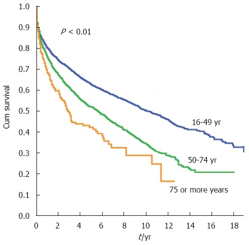
The deleterious effect of donor age on the recurrence of HCV infection has been fully demonstrated. Berenguer et al[8] reported that the survival of transplant patients with HCV infection is decreasing, and aging donors is one of the main factors. Donor age is an independent factor associated with the risk of developing cirrhosis and decreased survival. Lake et al[38], using data from the American Scientific Registry of Transplant Recipients, analyzed the impact of donor age on the survival of 778 hepatitis B, 3463 hepatitis C, and 7429 non-viral recipients. In HCV-infected recipients, the strongest predictor of graft loss was donor age. Transplantation with organs from donors between ages 41 and 50, 51 and 60, and > 60 years old was associated with a linear increase in the risk of graft loss. Subsequent single or multicenter studies confirmed these findings[39-43]. Analysis of the Spanish Registry for Liver Transplantation presented a lower graft survival in HCV-infected patients when organs were procured from donors older than 50 years[43] (Figure 4). Ghinolfi et al[39] analyzed the use of octogenarian donors for LT. In those ≥ 80 years old, the 5-year graft survival was lower for HCV-positive vs HCV-negative recipients (62.4% vs 85.6%, P = 0.034).
Figure 4.

Graft survival in hepatitis C virus-infected patients depending on donor age. Source: Spanish Liver Transplant Registry.
A correlation between accelerated fibrosis and worse outcome in grafts from older donors has been demonstrated[9,44]. Machicao et al[9] and Wali et al[44] reported that donors age 50 years or more had a median fibrosis progression rate of 2.7 units/year and time to cirrhosis of 2.2 years post-transplant. Donor age was also a strong factor in determining the likelihood of antiviral treatment success[45,46].
The impact on the LT outcomes of new direct-acting antiviral agents (DAA) against HCV has not been well established. These new drugs allow more simple treatment regimens and minimal toxicity, and when used in combination, achieve viral eradication in most HCV patients who undergo treatment[47,48]. The high cost of DAA still limits treatment on a large scale in most countries. In the next decades, DAA may lead to a significant reduction in patients needing a liver transplant for HCV and improve graft survival rate by decreasing the reinfection rate after LT[49]. In HCV-positive recipients, the impact of donor age on LT outcomes may someday be the same as that if HCV-negative recipients.
LIVER REGENERATION AND AGING
Morphological and structural changes occur in the liver with aging. At the macroscopic level, the liver suffers a reduction in size and a decline in blood flow[50,51]. At the hepatocellular level, changes include a loss of the smooth endoplasmic reticulum, a loss in the number of mitochondria accompanied by an increase in their volume, an increase in the volume of the dense body compartment (secondary lysosomes, residual bodies, lipofuscin), and an increase in hepatocyte polyploidy[52]. Despite morphological changes, the performed clinical studies do not allow for the identification of important age-associated deficits in liver function, and it is generally assumed that the majority of liver functions are relatively well maintained with age[53].
Although age does not seem to significantly affect liver function, the incidence of several liver diseases increases with age whereas the capacity of the liver to manage or overcome liver diseases or external injuries decreases. In fact, the most dramatic and well-documented effect of aging in the liver is the impairment of liver regeneration. Hepatocytes are normally quiescent cells, but in response to liver injury, they can undergo extensive replication to restore the liver. This cellular transition from quiescence to proliferation requires activation of S-phase and mitotic-specific genes. However, fewer hepatocytes in elderly humans enter S-phase in comparison to younger people, and those that do are slower in doing so, compromising the rate of liver regeneration[53].
The loss of liver regenerative capacity is expressed by the decrease in cell cycle and the increase in autophagy and apoptosis[54]. However, despite these phenomena, reported over 50 years ago, the cellular and molecular basis for the loss of an aged liver’s regenerative capacity has not been fully elucidated.
Different mechanisms have been suggested as implicated in the loss of this capacity with aging. Reduction in hepatocyte telomere length is one of these suggested mechanisms because it diminishes cell mitosis and apoptosis and thus produces a decline in cell proliferation. Takubo et al[55], after studying liver specimens from 94 individuals aged 0-101 years, found significant telomere shortening with age. Similar results were also observed in studies by Aikata et al[56] and Aini et al[57]. Hepatocytes presenting telomere shortening and karyotypic alterations were found in long-term transplanted human allografts. It appears that telomere shortening in liver cells is more significant in the early years, before the age of 40, when tissue turnover and growth are elevated[55,58]. This timing should be taken into consideration when comparing studies with controversial results because different donor age ranges were used.
Despite the clear connection between telomere shortening and reduction in cell proliferation, this association has not always implied impairment in liver regeneration. Experiments in a telomere restriction fragment-deficient mouse model demonstrated that liver regeneration after partial hepatectomy was not compromised by the loss of telomere integrity[59]. Post-hepatectomy regeneration was accomplished, increasing cell growth and yielding polyploid cells, indicating a switch from a proliferative to a cell growth pathway.
Another factor that suggests involvement of the decline in liver regeneration with aging is the inhibition of regeneration at an epigenetic level. Studies by Timchenko’s group[60] indicate that the reduced proliferative response of aged livers is likely to be related to alterations in signal-transduction pathways (at the translational and/or post-translational levels). The decline in the regenerative capacity of old livers seems to be related to epigenetic silencing of E2F-regulated genes as a result of several age-dependent signal-transduction pathways. A decline in growth hormone with age leads to higher cyclin D3 levels that activate cdk4. Activated cdk4 promotes the formation of C/EBPα-Brm and CUGBP1-eIF2 complexes in livers of old mice. CUGBP1-eIF2 complexes up-regulate HDCA1 protein levels that, jointly with C/EBPα-Brm complexes, bind to E2F-dependent promoters, inhibiting expression of E2F-regulated genes and thus liver regeneration. In fact, it has been observed that treatment of old mice with growth hormone corrects liver proliferation[61].
In addition, hepatocellular response to growth factors has been proposed as another mechanism implicated in the reduction of liver regeneration with aging. The hepatocyte proliferative response to epidermal growth factor (EGF) is clearly increased in young rats compared to old animals, suggesting that aging impairs hepatocyte responsiveness to growth factors[53,60]. The problem does not seem to be related to the number of EGF receptors or their binding capacity but rather to a reduction in receptor phosphorylation, a critical step in the EGF-induced hepatocyte proliferation pathway[61].
Apart from the mechanisms mentioned previously, changes in the structure of hepatic sinusoidal endothelium, including a loss of fenestrae and a thickening of the endothelial cells (pseudo-capillarization), have also been associated with a decrease in liver regeneration with aging. Furrer et al[62] demonstrated that pseudo-capillarization contributes to age-related decline in regeneration after hepatectomy in mice. Their data demonstrated that treatment with a serotonin receptor agonist in old mice restored liver regeneration capacity through a vascular endothelial growth factor (VEGF)-dependent pathway. In their findings, the serotonin receptor agonist resulted in increased systemic VEGF availability, up-regulating the number and size of endothelial cell fenestrae, improving hepatic blood flow, and therefore enhancing the hepatic regenerative capacity. In this sense, higher VEGF secretion levels have also been detected in cultures of isolated human hepatocytes from young donors compared to those isolated from older donors[63].
Finally, a decline in the hepatic progenitor cell population has also been suggested as another possible cause of liver regeneration impairment in older donors. Ono et al[21] observed that the progenitor cell population (Thy-1+) consistently tended to decline with age in LDLT. On the other hand, Yousef et al[64] recently found that the decline in stem cell function with age was largely due to biochemical imbalances in the cell niches, demonstrating that aging imposes an elevation in transforming growth factor β (TGF-β) signaling in the myogenic niche of skeletal muscle and in the neurogenic niche of the hippocampus. When they interfered with TGF-β levels by systemically decreasing TGF-β signaling with a single drug, bringing its levels closer to those detected in young mice, these authors could simultaneously enhance neurogenesis and muscle regeneration in the same old mice, findings further corroborated via genetic interference with TGF-β. Conboy et al[65] have previously reported similar observations in old mice permanently linked with their vascular systems (heterochronic parabiosis) to young mice. They reported a significant increase in proliferation of the aged hepatocyte progenitors in the old liver and restored expression of the complex c/EBP-alpha to levels seen only in young animals. Additionally, Wang et al[66] reported that senescent human hepatocytes can restore their proliferative capacity after xenotransplantation into mice, a finding with a potentially great impact on future studies of liver pathology and liver cell therapy. Hence, a process that once was thought to be terminal - i.e., cell senescence and growth arrest - seems now to be tightly associated with the organ microenvironment rather than with the actual age of the organism. This relationship opens the door to the development of novel pharmacological strategies aimed at rejuvenating old liver grafts immediately after procurement and prior to transplantation.
Thus, understanding the cellular and molecular basis for the reduced proliferative response in old livers is important and could indicate how we can improve liver regeneration and graft survival in older patients. From this perspective, some studies have been designed to find specific markers to predict function and longevity of transplanted organs. Among those senescence markers that have been studied is the abovementioned telomere length; others include the senescence marker protein-30 (SMP-30), which has shown good results in animals that have not correlated with results in humans; CDKN2A/p16INK4a, which is a good predictor of long-term graft function in renal transplantation but has not yet been studied in liver models; the cyclooxygenases 1 and 2 (COX-1 and COX-2); the cell proliferation marker Ki-67; endoplasmic reticulum chaperone levels; and cytochrome p450 mRNA expression[67].
In old animals and in elderly humans liver regeneration is impaired, and it appears to be the rate of liver regeneration, rather than the regenerative capacity, that is diminished in the elderly (Table 3).
Table 3.
Review of cellular and molecular mechanisms suggested to be implicated in the loss of aged liver’s regenerative capacity
| Mechanism | Ref. |
| Telomere shortening | Takubo et al[55] |
| Aikata et al[56] | |
| Aini et al[57] | |
| Transcriptional and post-transcriptional modifications | Timchenko[60] |
| Wang et al[61] | |
| Hepatocelullar response to growth factors | Schmucker[53] |
| Wang et al[61] | |
| Pseudo-capillarization | Furrer et al[62] |
| Decline of progenitor cell populations and changes in their niches | Ono et al[18] |
| Yousef et al[64] | |
| Conboy et al[65] | |
| Wang et al[66] |
These age-related changes could be the factors that determine the higher sensitivity of the graft from older donor to develop irreversible lesions induced by distinct injuries, and results in higher rate of unsuitable response in older donor grafts.
CONCLUSION
The age of donors is increasing significantly in recent years, and liver grafts previously considered suboptimal because they came from elderly donors are nowadays used routinely in all centers. Although the various existing studies so far have contradictory results, age may have a role in the outcome of LT. The use of older donors has been linked to a greater number of biliary complications in both deceased and LDLT, as well as to a poor outcome of HCV recurrence injury. In addition, most LT prognostic scores have donor age as a fundamental variable. The pathophysiological bases of this association are not well established. Liver function does not seem to be influenced by aging, but several changes at the macroscopic and hepatocellular levels have been observed. There are also reported different biological changes in aging that lead to a loss of the liver’s proliferative response and regeneration. These alterations may lead to an impairment of the capacity of the liver to manage and overcome liver diseases and to face external injuries.
Donor age is not the only relevant factor in the outcome of LT, however; surgical factors such as ischemia time or hemodynamic instability during surgery, and recipient factors, such as MELD score, are also essential. Therefore, avoiding these factors as much as possible in liver transplants performed with elderly donors may lead to outcomes similar to those with transplants performed with younger donors.
Footnotes
Conflict-of-interest statement: The authors have no conflict of interest to report.
Open-Access: This article is an open-access article which was selected by an in-house editor and fully peer-reviewed by external reviewers. It is distributed in accordance with the Creative Commons Attribution Non Commercial (CC BY-NC 4.0) license, which permits others to distribute, remix, adapt, build upon this work non-commercially, and license their derivative works on different terms, provided the original work is properly cited and the use is non-commercial. See: http://creativecommons.org/licenses/by-nc/4.0/
Peer-review started: March 22, 2016
First decision: March 31, 2016
Article in press: May 4, 2016
P- Reviewer: Bramhall S, Kim JH, Tanoglu A S- Editor: Gong ZM L- Editor: A E- Editor: Ma S
References
- 1.Adam R, Karam V, Delvart V, O’Grady J, Mirza D, Klempnauer J, Castaing D, Neuhaus P, Jamieson N, Salizzoni M, et al. Evolution of indications and results of liver transplantation in Europe. A report from the European Liver Transplant Registry (ELTR) J Hepatol. 2012;57:675–688. doi: 10.1016/j.jhep.2012.04.015. [DOI] [PubMed] [Google Scholar]
- 2.Adam R, McMaster P, O’Grady JG, Castaing D, Klempnauer JL, Jamieson N, Neuhaus P, Lerut J, Salizzoni M, Pollard S, et al. Evolution of liver transplantation in Europe: report of the European Liver Transplant Registry. Liver Transpl. 2003;9:1231–1243. doi: 10.1016/j.lts.2003.09.018. [DOI] [PubMed] [Google Scholar]
- 3.Cuervas-Mons V, de la Rosa G, Pardo F, San Juan F, Valdivieso A. [Activity and results of liver transplantation in Spain during 1984-2012. Analysis of the Spanish Liver Transplant Registry] Med Clin (Barc) 2015;144:337–347. doi: 10.1016/j.medcli.2014.07.036. [DOI] [PubMed] [Google Scholar]
- 4.Emre S, Schwartz ME, Altaca G, Sethi P, Fiel MI, Guy SR, Kelly DM, Sebastian A, Fisher A, Eickmeyer D, et al. Safe use of hepatic allografts from donors older than 70 years. Transplantation. 1996;62:62–65. doi: 10.1097/00007890-199607150-00013. [DOI] [PubMed] [Google Scholar]
- 5.Rodríguez González F, Jiménez Romero C, Rodríguez Romano D, Loinaz Segurola C, Marqués Medina E, Pérez Saborido B, García García I, Rodríguez Cañete A, Moreno González E. Orthotopic liver transplantation with 100 hepatic allografts from donors over 60 years old. Transplant Proc. 2002;34:233–234. doi: 10.1016/s0041-1345(01)02738-5. [DOI] [PubMed] [Google Scholar]
- 6.Adam R, Cailliez V, Majno P, Karam V, McMaster P, Caine RY, O’Grady J, Pichlmayr R, Neuhaus P, Otte JB, et al. Normalised intrinsic mortality risk in liver transplantation: European Liver Transplant Registry study. Lancet. 2000;356:621–627. doi: 10.1016/s0140-6736(00)02603-9. [DOI] [PubMed] [Google Scholar]
- 7.Burroughs AK, Sabin CA, Rolles K, Delvart V, Karam V, Buckels J, O’Grady JG, Castaing D, Klempnauer J, Jamieson N, et al. 3-month and 12-month mortality after first liver transplant in adults in Europe: predictive models for outcome. Lancet. 2006;367:225–232. doi: 10.1016/S0140-6736(06)68033-1. [DOI] [PubMed] [Google Scholar]
- 8.Berenguer M, Prieto M, San Juan F, Rayón JM, Martinez F, Carrasco D, Moya A, Orbis F, Mir J, Berenguer J. Contribution of donor age to the recent decrease in patient survival among HCV-infected liver transplant recipients. Hepatology. 2002;36:202–210. doi: 10.1053/jhep.2002.33993. [DOI] [PubMed] [Google Scholar]
- 9.Machicao VI, Bonatti H, Krishna M, Aqel BA, Lukens FJ, Nguyen JH, Rosser BG, Satyanarayana R, Grewal HP, Hewitt WR, et al. Donor age affects fibrosis progression and graft survival after liver transplantation for hepatitis C. Transplantation. 2004;77:84–92. doi: 10.1097/01.TP.0000095896.07048.BB. [DOI] [PubMed] [Google Scholar]
- 10.Feng S, Goodrich NP, Bragg-Gresham JL, Dykstra DM, Punch JD, DebRoy MA, Greenstein SM, Merion RM. Characteristics associated with liver graft failure: the concept of a donor risk index. Am J Transplant. 2006;6:783–790. doi: 10.1111/j.1600-6143.2006.01242.x. [DOI] [PubMed] [Google Scholar]
- 11.Reese PP, Sonawane SB, Thomasson A, Yeh H, Markmann JF. Donor age and cold ischemia interact to produce inferior 90-day liver allograft survival. Transplantation. 2008;85:1737–1744. doi: 10.1097/TP.0b013e3181722f75. [DOI] [PubMed] [Google Scholar]
- 12.Serrano MT, Garcia-Gil A, Arenas J, Ber Y, Cortes L, Valiente C, Araiz JJ. Outcome of liver transplantation using donors older than 60 years of age. Clin Transplant. 2010;24:543–549. doi: 10.1111/j.1399-0012.2009.01135.x. [DOI] [PubMed] [Google Scholar]
- 13.Anderson CD, Vachharajani N, Doyle M, Lowell JA, Wellen JR, Shenoy S, Lisker-Melman M, Korenblat K, Crippin J, Chapman WC. Advanced donor age alone does not affect patient or graft survival after liver transplantation. J Am Coll Surg. 2008;207:847–852. doi: 10.1016/j.jamcollsurg.2008.08.009. [DOI] [PubMed] [Google Scholar]
- 14.Alamo JM, Olivares C, Jiménez G, Bernal C, Marín LM, Tinoco J, Suárez G, Serrano J, Padillo J, Gómez MÁ. Donor characteristics that are associated with survival in liver transplant recipients older than 70 years with grafts. Transplant Proc. 2013;45:3633–3636. doi: 10.1016/j.transproceed.2013.10.031. [DOI] [PubMed] [Google Scholar]
- 15.Kim DY, Moon J, Island ER, Tekin A, Ganz S, Levi D, Selvaggi G, Nishida S, Tzakis AG. Liver transplantation using elderly donors: a risk factor analysis. Clin Transplant. 2011;25:270–276. doi: 10.1111/j.1399-0012.2010.01222.x. [DOI] [PubMed] [Google Scholar]
- 16.Han JH, You YK, Na GH, Kim EY, Lee SH, Hong TH, Kim DG. Outcomes of living donor liver transplantation using elderly donors. Ann Surg Treat Res. 2014;86:184–191. doi: 10.4174/astr.2014.86.4.184. [DOI] [PMC free article] [PubMed] [Google Scholar]
- 17.Dayangac M, Taner CB, Yaprak O, Demirbas T, Balci D, Duran C, Yuzer Y, Tokat Y. Utilization of elderly donors in living donor liver transplantation: when more is less? Liver Transpl. 2011;17:548–555. doi: 10.1002/lt.22276. [DOI] [PubMed] [Google Scholar]
- 18.Ikegami T, Nishizaki T, Yanaga K, Shimada M, Kishikawa K, Nomoto K, Uchiyama H, Sugimachi K. The impact of donor age on living donor liver transplantation. Transplantation. 2000;70:1703–1707. doi: 10.1097/00007890-200012270-00007. [DOI] [PubMed] [Google Scholar]
- 19.Ikegami T, Taketomi A, Ohta R, Soejima Y, Yoshizumi T, Shimada M, Maehara Y. Donor age in living donor liver transplantation. Transplant Proc. 2008;40:1471–1475. doi: 10.1016/j.transproceed.2008.02.084. [DOI] [PubMed] [Google Scholar]
- 20.Iwamoto T, Yagi T, Umeda Y, Sato D, Matsukawa H, Matsuda H, Shinoura S, Sadamori H, Mizuno K, Yoshida R, et al. The impact of donor age on the outcome of adult living donor liver transplantation. Transplantation. 2008;85:1240–1245. doi: 10.1097/TP.0b013e31816c7e90. [DOI] [PubMed] [Google Scholar]
- 21.Ono Y, Kawachi S, Hayashida T, Wakui M, Tanabe M, Itano O, Obara H, Shinoda M, Hibi T, Oshima G, et al. The influence of donor age on liver regeneration and hepatic progenitor cell populations. Surgery. 2011;150:154–161. doi: 10.1016/j.surg.2011.05.004. [DOI] [PubMed] [Google Scholar]
- 22.Uchiyama H, Shirabe K, Kimura K, Yoshizumi T, Ikegami T, Harimoto N, Maehara Y. Outcomes of adult-to-adult living donor liver transplantation in 321 recipients. Liver Transpl. 2016;22:305–315. doi: 10.1002/lt.24378. [DOI] [PubMed] [Google Scholar]
- 23.Li C, Wen TF, Yan LN, Li B, Yang JY, Xu MQ, Wang WT, Wei YG. Safety of living donor liver transplantation using older donors. J Surg Res. 2012;178:982–987. doi: 10.1016/j.jss.2012.06.065. [DOI] [PubMed] [Google Scholar]
- 24.Wang K, Jiang WT, Deng YL, Pan C, Shen ZY. Effect of donor age on graft function and long-term survival of recipients undergoing living donor liver transplantation. Hepatobiliary Pancreat Dis Int. 2015;14:50–55. doi: 10.1016/s1499-3872(15)60334-4. [DOI] [PubMed] [Google Scholar]
- 25.Braat AE, Blok JJ, Putter H, Adam R, Burroughs AK, Rahmel AO, Porte RJ, Rogiers X, Ringers J; European Liver and Intestine Transplant Association (ELITA) and Eurotransplant Liver Intestine Advisory Committee (ELIAC) The Eurotransplant donor risk index in liver transplantation: ET-DRI. Am J Transplant. 2012;12:2789–2796. doi: 10.1111/j.1600-6143.2012.04195.x. [DOI] [PubMed] [Google Scholar]
- 26.Rana A, Hardy MA, Halazun KJ, Woodland DC, Ratner LE, Samstein B, Guarrera JV, Brown RS, Emond JC. Survival outcomes following liver transplantation (SOFT) score: a novel method to predict patient survival following liver transplantation. Am J Transplant. 2008;8:2537–2546. doi: 10.1111/j.1600-6143.2008.02400.x. [DOI] [PubMed] [Google Scholar]
- 27.Halldorson JB, Bakthavatsalam R, Fix O, Reyes JD, Perkins JD. D-MELD, a simple predictor of post liver transplant mortality for optimization of donor/recipient matching. Am J Transplant. 2009;9:318–326. doi: 10.1111/j.1600-6143.2008.02491.x. [DOI] [PubMed] [Google Scholar]
- 28.Dutkowski P, Oberkofler CE, Slankamenac K, Puhan MA, Schadde E, Müllhaupt B, Geier A, Clavien PA. Are there better guidelines for allocation in liver transplantation? A novel score targeting justice and utility in the model for end-stage liver disease era. Ann Surg. 2011;254:745–753; discussion 753. doi: 10.1097/SLA.0b013e3182365081. [DOI] [PubMed] [Google Scholar]
- 29.Thorsen T, Aandahl EM, Bennet W, Olausson M, Ericzon BG, Nowak G, Duraj F, Isoniemi H, Rasmussen A, Karlsen TH, et al. Transplantation With Livers From Deceased Donors Older Than 75 Years. Transplantation. 2015;99:2534–2542. doi: 10.1097/TP.0000000000000728. [DOI] [PubMed] [Google Scholar]
- 30.Verdonk RC, Buis CI, Porte RJ, van der Jagt EJ, Limburg AJ, van den Berg AP, Slooff MJ, Peeters PM, de Jong KP, Kleibeuker JH, et al. Anastomotic biliary strictures after liver transplantation: causes and consequences. Liver Transpl. 2006;12:726–735. doi: 10.1002/lt.20714. [DOI] [PubMed] [Google Scholar]
- 31.Sundaram V, Jones DT, Shah NH, de Vera ME, Fontes P, Marsh JW, Humar A, Ahmad J. Posttransplant biliary complications in the pre- and post-model for end-stage liver disease era. Liver Transpl. 2011;17:428–435. doi: 10.1002/lt.22251. [DOI] [PubMed] [Google Scholar]
- 32.Lüthold SC, Kaseje N, Jannot AS, Mentha G, Majno P, Toso C, Belli DC, McLin VA, Wildhaber BE. Risk factors for early and late biliary complications in pediatric liver transplantation. Pediatr Transplant. 2014;18:822–830. doi: 10.1111/petr.12363. [DOI] [PubMed] [Google Scholar]
- 33.Heidenhain C, Pratschke J, Puhl G, Neumann U, Pascher A, Veltzke-Schlieker W, Neuhaus P. Incidence of and risk factors for ischemic-type biliary lesions following orthotopic liver transplantation. Transpl Int. 2010;23:14–22. doi: 10.1111/j.1432-2277.2009.00947.x. [DOI] [PubMed] [Google Scholar]
- 34.Abt P, Crawford M, Desai N, Markmann J, Olthoff K, Shaked A. Liver transplantation from controlled non-heart-beating donors: an increased incidence of biliary complications. Transplantation. 2003;75:1659–1663. doi: 10.1097/01.TP.0000062574.18648.7C. [DOI] [PubMed] [Google Scholar]
- 35.Cutrin JC, Cantino D, Biasi F, Chiarpotto E, Salizzoni M, Andorno E, Massano G, Lanfranco G, Rizzetto M, Boveris A, et al. Reperfusion damage to the bile canaliculi in transplanted human liver. Hepatology. 1996;24:1053–1057. doi: 10.1002/hep.510240512. [DOI] [PubMed] [Google Scholar]
- 36.Noack K, Bronk SF, Kato A, Gores GJ. The greater vulnerability of bile duct cells to reoxygenation injury than to anoxia. Implications for the pathogenesis of biliary strictures after liver transplantation. Transplantation. 1993;56:495–500. doi: 10.1097/00007890-199309000-00001. [DOI] [PubMed] [Google Scholar]
- 37.Ghinolfi D, De Simone P, Lai Q, Pezzati D, Coletti L, Balzano E, Arenga G, Carrai P, Grande G, Pollina L, et al. Risk analysis of ischemic-type biliary lesions after liver transplant using octogenarian donors. Liver Transpl. 2016;22:588–598. doi: 10.1002/lt.24401. [DOI] [PubMed] [Google Scholar]
- 38.Lake JR, Shorr JS, Steffen BJ, Chu AH, Gordon RD, Wiesner RH. Differential effects of donor age in liver transplant recipients infected with hepatitis B, hepatitis C and without viral hepatitis. Am J Transplant. 2005;5:549–557. doi: 10.1111/j.1600-6143.2005.00741.x. [DOI] [PubMed] [Google Scholar]
- 39.Ghinolfi D, Marti J, De Simone P, Lai Q, Pezzati D, Coletti L, Tartaglia D, Catalano G, Tincani G, Carrai P, et al. Use of octogenarian donors for liver transplantation: a survival analysis. Am J Transplant. 2014;14:2062–2071. doi: 10.1111/ajt.12843. [DOI] [PubMed] [Google Scholar]
- 40.Dumortier J, Salamé E, Roche B, Hurtova M, Conti F, Radenne S, Vanlemmens C, Pageaux GP, Saliba F, Samuel D, et al. Severe fibrosis in patients with recurrent hepatitis C after liver transplantation: a French experience on 250 patients over 15 years (the Orfèvre study) Clin Res Hepatol Gastroenterol. 2014;38:292–299. doi: 10.1016/j.clinre.2014.02.007. [DOI] [PubMed] [Google Scholar]
- 41.Berenguer M, Crippin J, Gish R, Bass N, Bostrom A, Netto G, Alonzo J, Garcia-Kennedy R, Rayón JM, Wright TL. A model to predict severe HCV-related disease following liver transplantation. Hepatology. 2003;38:34–41. doi: 10.1053/jhep.2003.50278. [DOI] [PubMed] [Google Scholar]
- 42.Mutimer DJ, Gunson B, Chen J, Berenguer J, Neuhaus P, Castaing D, Garcia-Valdecasas JC, Salizzoni M, Moreno GE, Mirza D. Impact of donor age and year of transplantation on graft and patient survival following liver transplantation for hepatitis C virus. Transplantation. 2006;81:7–14. doi: 10.1097/01.tp.0000188619.30677.84. [DOI] [PubMed] [Google Scholar]
- 43.Berenguer M, Charco R, Manuel Pascasio J, Ignacio Herrero J. Spanish society of liver transplantation (SETH) consensus recommendations on hepatitis C virus and liver transplantation. Liver Int. 2012;32:712–731. doi: 10.1111/j.1478-3231.2011.02731.x. [DOI] [PubMed] [Google Scholar]
- 44.Wali M, Harrison RF, Gow PJ, Mutimer D. Advancing donor liver age and rapid fibrosis progression following transplantation for hepatitis C. Gut. 2002;51:248–252. doi: 10.1136/gut.51.2.248. [DOI] [PMC free article] [PubMed] [Google Scholar]
- 45.Selzner N, Renner EL, Selzner M, Adeyi O, Kashfi A, Therapondos G, Girgrah N, Herath C, Levy GA, Lilly L. Antiviral treatment of recurrent hepatitis C after liver transplantation: predictors of response and long-term outcome. Transplantation. 2009;88:1214–1221. doi: 10.1097/TP.0b013e3181bd783c. [DOI] [PubMed] [Google Scholar]
- 46.Berenguer M, Aguilera V, Prieto M, Ortiz C, Rodríguez M, Gentili F, Risalde B, Rubin A, Cañada R, Palau A, et al. Worse recent efficacy of antiviral therapy in liver transplant recipients with recurrent hepatitis C: impact of donor age and baseline cirrhosis. Liver Transpl. 2009;15:738–746. doi: 10.1002/lt.21707. [DOI] [PubMed] [Google Scholar]
- 47.European Association for the Study of the Liver. EASL Clinical Practice Guidelines: Liver transplantation. J Hepatol. 2016;64:433–485. doi: 10.1016/j.jhep.2015.10.006. [DOI] [PubMed] [Google Scholar]
- 48.Fontana RJ, Brown RS, Moreno-Zamora A, Prieto M, Joshi S, Londoño MC, Herzer K, Chacko KR, Stauber RE, Knop V, et al. Daclatasvir combined with sofosbuvir or simeprevir in liver transplant recipients with severe recurrent hepatitis C infection. Liver Transpl. 2016;22:446–458. doi: 10.1002/lt.24416. [DOI] [PubMed] [Google Scholar]
- 49.Curry MP, Forns X, Chung RT, Terrault NA, Brown R, Fenkel JM, Gordon F, O’Leary J, Kuo A, Schiano T, et al. Sofosbuvir and ribavirin prevent recurrence of HCV infection after liver transplantation: an open-label study. Gastroenterology. 2015;148:100–107.e1. doi: 10.1053/j.gastro.2014.09.023. [DOI] [PubMed] [Google Scholar]
- 50.Wynne HA, Cope LH, Mutch E, Rawlins MD, Woodhouse KW, James OF. The effect of age upon liver volume and apparent liver blood flow in healthy man. Hepatology. 1989;9:297–301. doi: 10.1002/hep.1840090222. [DOI] [PubMed] [Google Scholar]
- 51.Marchesini G, Bua V, Brunori A, Bianchi G, Pisi P, Fabbri A, Zoli M, Pisi E. Galactose elimination capacity and liver volume in aging man. Hepatology. 1988;8:1079–1083. doi: 10.1002/hep.1840080516. [DOI] [PubMed] [Google Scholar]
- 52.Schmucker DL. Hepatocyte fine structure during maturation and senescence. J Electron Microsc Tech. 1990;14:106–125. doi: 10.1002/jemt.1060140205. [DOI] [PubMed] [Google Scholar]
- 53.Schmucker DL, Sanchez H. Liver regeneration and aging: a current perspective. Curr Gerontol Geriatr Res. 2011;2011:526379. doi: 10.1155/2011/526379. [DOI] [PMC free article] [PubMed] [Google Scholar]
- 54.Enkhbold C, Morine Y, Utsunomiya T, Imura S, Ikemoto T, Arakawa Y, Saito Y, Yamada S, Ishikawa D, Shimada M. Dysfunction of liver regeneration in aged liver after partial hepatectomy. J Gastroenterol Hepatol. 2015;30:1217–1224. doi: 10.1111/jgh.12930. [DOI] [PubMed] [Google Scholar]
- 55.Takubo K, Nakamura K, Izumiyama N, Furugori E, Sawabe M, Arai T, Esaki Y, Mafune K, Kammori M, Fujiwara M, et al. Telomere shortening with aging in human liver. J Gerontol A Biol Sci Med Sci. 2000;55:B533–B536. doi: 10.1093/gerona/55.11.b533. [DOI] [PubMed] [Google Scholar]
- 56.Aikata H, Takaishi H, Kawakami Y, Takahashi S, Kitamoto M, Nakanishi T, Nakamura Y, Shimamoto F, Kajiyama G, Ide T. Telomere reduction in human liver tissues with age and chronic inflammation. Exp Cell Res. 2000;256:578–582. doi: 10.1006/excr.2000.4862. [DOI] [PubMed] [Google Scholar]
- 57.Aini W, Miyagawa-Hayashino A, Tsuruyama T, Hashimoto S, Sumiyoshi S, Ozeki M, Tamaki K, Uemoto S, Haga H. Telomere shortening and karyotypic alterations in hepatocytes in long-term transplanted human liver allografts. Transpl Int. 2012;25:956–966. doi: 10.1111/j.1432-2277.2012.01523.x. [DOI] [PubMed] [Google Scholar]
- 58.de Grey A. Response to “telomere shortening with aging in human liver”. J Gerontol A Biol Sci Med Sci. 2001;56:B237–B238. doi: 10.1093/gerona/56.6.b237. [DOI] [PubMed] [Google Scholar]
- 59.Lazzerini Denchi E, Celli G, de Lange T. Hepatocytes with extensive telomere deprotection and fusion remain viable and regenerate liver mass through endoreduplication. Genes Dev. 2006;20:2648–2653. doi: 10.1101/gad.1453606. [DOI] [PMC free article] [PubMed] [Google Scholar]
- 60.Timchenko NA. Aging and liver regeneration. Trends Endocrinol Metab. 2009;20:171–176. doi: 10.1016/j.tem.2009.01.005. [DOI] [PubMed] [Google Scholar]
- 61.Wang GL, Shi X, Salisbury E, Sun Y, Albrecht JH, Smith RG, Timchenko NA. Growth hormone corrects proliferation and transcription of phosphoenolpyruvate carboxykinase in livers of old mice via elimination of CCAAT/enhancer-binding protein alpha-Brm complex. J Biol Chem. 2007;282:1468–1478. doi: 10.1074/jbc.M608226200. [DOI] [PubMed] [Google Scholar]
- 62.Furrer K, Rickenbacher A, Tian Y, Jochum W, Bittermann AG, Käch A, Humar B, Graf R, Moritz W, Clavien PA. Serotonin reverts age-related capillarization and failure of regeneration in the liver through a VEGF-dependent pathway. Proc Natl Acad Sci USA. 2011;108:2945–2950. doi: 10.1073/pnas.1012531108. [DOI] [PMC free article] [PubMed] [Google Scholar]
- 63.Serrano T, Mitry RR, Terry C, Lehec SC, Dhawan A, Hughes RD. The effects of immunosuppressive agents on the function of human hepatocytes in vitro. Cell Transplant. 2006;15:777–783. doi: 10.3727/000000006783981530. [DOI] [PubMed] [Google Scholar]
- 64.Yousef H, Conboy MJ, Morgenthaler A, Schlesinger C, Bugaj L, Paliwal P, Greer C, Conboy IM, Schaffer D. Systemic attenuation of the TGF-β pathway by a single drug simultaneously rejuvenates hippocampal neurogenesis and myogenesis in the same old mammal. Oncotarget. 2015;6:11959–11978. doi: 10.18632/oncotarget.3851. [DOI] [PMC free article] [PubMed] [Google Scholar]
- 65.Conboy IM, Conboy MJ, Wagers AJ, Girma ER, Weissman IL, Rando TA. Rejuvenation of aged progenitor cells by exposure to a young systemic environment. Nature. 2005;433:760–764. doi: 10.1038/nature03260. [DOI] [PubMed] [Google Scholar]
- 66.Wang MJ, Chen F, Li JX, Liu CC, Zhang HB, Xia Y, Yu B, You P, Xiang D, Lu L, et al. Reversal of hepatocyte senescence after continuous in vivo cell proliferation. Hepatology. 2014;60:349–361. doi: 10.1002/hep.27094. [DOI] [PubMed] [Google Scholar]
- 67.Hodgson R, Christophi C. What determines ageing of the transplanted liver? HPB (Oxford) 2015;17:222–225. doi: 10.1111/hpb.12339. [DOI] [PMC free article] [PubMed] [Google Scholar]


