

ABSTRACT
Purpose
The purpose of this systematic review was to determine the exercises that optimize muscle ratios of the periscapular musculature for scapular stability and isolated strengthening.
Methods
A systematic search was performed in PubMed, CINAHL, SPORTDiscus, Scopus, and Discovery Layer. Studies were included if they examined the muscle activation of the upper trapezius compared to the middle trapezius, lower trapezius, or serratus anterior using EMG during open chain exercises. The participants were required to have healthy, nonpathological shoulders. Information obtained included maximal voluntary isometric contraction (MVIC) values, ratios, standard deviations, exercises, and exercise descriptions. The outcome of interest was determining exercises that create optimal muscle activation ratios between the scapular stabilizers.
Results
Fifteen observational studies met the inclusion criteria for the systematic review. Exercises with optimal ratios were eccentric exercises in the frontal and sagittal planes, especially flexion between 180 ° and 60 °. External rotation exercises with the elbow flexed to 90 ° also had optimal ratios for activating the middle trapezius in prone and side-lying positions. Exercises with optimal ratios for the lower trapezius were prone flexion, high scapular retraction, and prone external rotation with the shoulder abducted to 90 ° and elbow flexed. Exercises with optimal ratios for the serratus anterior were the diagonal exercises and scapular protraction.
Conclusion
This review has identified optimal positions and exercises for periscapular stability exercises. Standing exercises tend to activate the upper trapezius at a higher ratio, especially during the 60-120 ° range. The upper trapezius was the least active, while performing exercises in prone, side-lying, and supine positions. More studies need to be conducted to examine these exercises in greater detail and confirm their consistency in producing the optimal ratios determined in this review.
Level of evidence
1a
Keywords: Electromyography, electromyography feedback, resistance training, serratus anterior, trapezius
INTRODUCTION
The shoulder complex consists of the glenohumeral, acromioclavicular, sternoclavicular, and scapulothoracic joints, therefore, strengthening and stretching exercises for scapular stabilizing muscles are commonly used in rehabilitation of shoulder dysfunctions.1 During movement of the shoulder, the scapula and humerus are constantly changing positions relative to one another, making their ability to work in unison imperative to maintenance of stability of the glenohumeral joint. This phenomenon was coined scapulohumeral rhythm by Codman in 1934.2 During overhead activities, both the rotator cuff and periscapular musculature provide stability and aid in pain free mobility at the shoulder complex in healthy individuals.3 Force couples, which involve two opposing muscular forces working together to enable a particular joint motion, are important for optimal scapular stabilization during humeral movement.4
Currently, authors have suggested that abnormal scapular movement or dyskinesia may play a role in impingement syndrome, rotator cuff dysfunction, instability, and even neck pain.5,6 Prolonged overhead activity requires adequate endurance of the scapular musculature in order to maintain a consistent, proper scapulohumeral rhythm. Without the necessary endurance, subacromial impingement may occur due to improper scapular rotation.1,7,8,9 It was originally suggested that scapular dyskinesia was due to global weakness of the scapular musculature. However, recent research has shown that muscular imbalance may be the problem, not strength. It has been hypothesized that compensation through increased activation of the upper trapezius (UT) combined with decreased activation and control of the lower trapezius (LT)/middle trapezius (MT)/serratus anterior (SA) contributes to abnormal scapular motion.5 With this in mind, many current rehabilitation programs, which only focus on strengthening these muscles as a whole, may be inadequate for creating proper scapulohumeral rhythm.
Electromyography (EMG) is used to measure muscular activity. Many researchers have used EMG during various scapular stabilizing exercises in order to differentiate between activity of the UT, MT, LT, and SA during exercise. The majority of these studies have failed to address the optimal ratios of these muscles during relevant exercises.10-20 A select few authors have examined the optimal ratios during scapular stabilizing exercises.5,6,21,22 To obtain muscle ratios, the maximal voluntary isometric contraction (MVIC) of the examined muscles must be determined. The authors of this systematic review believe that this ratio is important when determining a individualized rehabilitation program to fit a certain patient. The purpose of this systematic review is to determine the exercises that optimize muscle ratios of the periscapular musculature for scapular stability and isolated strengthening.
METHODS
Literature Search
Articles were identified through a computerized search using PubMed, CINAHL, SPORTDiscus, Scopus, and Discovery Layer through Walsh University in November 2014. The search was performed using subject headings, abstract text, and key words for four main concepts: Trapezius, SA, exercise, and electromyography. In addition, these concepts were further specified and searched by the following key text words: Resistance training, EMG, and electromyography feedback. There were no restrictions placed on date of publication and type of study conducted. The searches were limited to English, Academic Journals, and humans. See appendices 1, 2, 3, 4, & 5 for the detailed search strategy. Although this review analyzed data only from a normal shoulder, due to risk of excluding eligible articles, there was no limit placed on the population sample during the search. (Appendix 1)
Study selection
Two reviewers (EB, JW) performed the initial screening of articles to determine eligibility. Two reviewers (AS, JQ) reviewed the included full text articles. Full text articles were reviewed if the abstract met the inclusion criteria, or if the abstract did not entail enough information to include or exclude the study. If there was a disagreement, a third reviewer was used to determine eligibility.
Eligibility criteria
To satisfy this review, EMG must be the primary tool used. A detailed description of methods of EMG normalization and analysis is required for reproducibility, quality analysis of recommended guidelines, comparability, and continuity of appropriate usage and technology. Studies were included if they contained %MVIC/%MVC and/or muscle ratios as a way of standardizing data and measurement values. This ensures that comparisons could be made between data across the studies. It was required that the studies compare the EMG activity of the UT with at least one of the following muscles: MT, LT, or SA for determining muscle ratios during the exercises. In addition, studies must include two or more open-chain exercises, performed actively by the subjects, examining the same scapular muscles for comparison. The study had to include a group containing normal, non-pathological shoulders define muscle ratios in the asymptomatic, healthy shoulder.
Exclusion Criteria. Studies were excluded if all of their participants had a history of shoulder pathology or injury, scapular pathology, pain, or symptoms within the past two years in order to reduce the influence of these factors on the muscle activation ratios.1 Studies were excluded if they only examined closed-chain exercises, did not use EMG as a primary tool, and did not take a standardized approach for normalizing and analyzing EMG activity. Because of the plethora of literature involving both open and closed chain exercises, the researchers chose to focus this review on open-chain exercises.
Data collection process
Two reviewers (AS & JQ) extracted relevant data from the studies. One author was contacted in order to obtain the data tables from the study.21 Exercises from each study were reviewed for commonalities. If the studies included symptomatic or pathological participants, data only from the control groups was extracted.
Data Extraction
Information obtained from each study included MVIC values, ratios (if applicable), standard deviations, exercises, and exercise description, which can be fully viewed in Supplemental Tables 1-4 contained in Appendix 3 (Available in Supplemental materials, linked on the IJSPT Website).
Table 1.
Characteristics of included studies.
| First Author, Year | Participants (N) | Age, mean (SD) and/or age range in years | Study Design | Exercises Tested | Muscles Assessed | Value Used |
|---|---|---|---|---|---|---|
| Cools, 2007 | 45 | 20.7 (1.7) | Observational | Vertical Pulley, v-bar Scaption (30 ° anterior of frontal plane) with ER Dumbbell Side-lying ER with elbow flexed to 90 ° Dumbbell |
UT, MT, LT, SA | Ratios, %MVIC |
| De Mey, 2012 | 30 | 20 (3.5) Range 18-30 |
Observational | High Scapular Retraction Exercises: Sitting Standing Static bipedal squat Static Lunge Static unipedal squat Dynamic bipedal squat Dynamic Lunge Dynamic unipedal squat |
UT, LT | %MVIC |
| Decker, 1999 | 20 | 30.4 (5.1) | Observational | Scaption in 45 ° ER (thumb up) Dumbbell |
UT, SA, MT | %MVIC |
| Dynamic Hug: elbow flexed 45 °, arm abducted 60 °, IR 45 ° to max. protraction | UT, SA | |||||
| Cools, 2007 | 30 | 22.5 (4.3) Range 18 – 35 | Observational | Isokinetic Abd 120 °/s Isokinetic ER 60 °/s |
UT, MT, LT | Ratios, %MVIC |
| Cools, 2007 | 45 | 20.7 (1.7) | Observational | Prone Abduction Dumbbell Forward Flexion (FF) Dumbbell Forward Flexion in Side Lying to 135 ° Dumbbell High Row 135 ° FF to neutral position Vertical Pulley, v-bar Horizontal Abduction in prone Dumbbell Horizontal Abduction with ER in prone Dumbbell Low Row with elbows extended Vertical Pulley, v-bar Low Row with elbows flexed Vertical Pulley, v-bar Prone Extension Dumbbell Rowing in Sitting Position |
UT, MT, LT, SA | Ratios, %MVIC |
| Ekstrom, 2003 | 30 | 27.2 Range 22-46 |
Observational | Unilateral shoulder shrug Hanging Weight Shoulder abduction in the plane of the scapula above 120 ° Dumbbell Arm raise overhead in line with LT muscle fibers Dumbbell Shoulder abduction in the plane of the scapula below 80 ° Dumbbell Shoulder horizontal extension with ER Dumbbell Diagonal exercise with shoulder flexion, horizontal flexion, and ER Dumbbell Unilateral row Hanging Weight Shoulder ER at 90 ° of abduction Dumbbell Unilateral shoulder press Barbell with Weights Bilateral scapular protraction |
UT, MT, LT, SA | %MVIC |
| Huang, 2012 | 12 | 23.8 (2.9) | Observational | Side-lying ER with elbow 90 ° flexion Dumbbell Forward Flexion Dumbbell |
UT, MT, LT, SA | Ratios |
| Kibler, 2008 | 18 | 27.3 (4.4) | Observational | Lawnmower Robbery |
UT, LT, SA | %MVIC |
| Marta, 2013 | 20 | 22 (3.6) Range 16 - 34 |
Observational | Prone horizontal abduction at 90 ° with full ER Dumbbell Prone horizontal abduction at 90 ° abduction with middle ER Dumbbell Prone horizontal abduction at 100 ° abduction with full ER Dumbbell Prone ER at 90 ° abduction and elbow at 90 ° Dumbbell Side-lying ER with elbow on trunk Dumbbell Standing ER in scapular plane (45 ° abduction, 30 ° horizontal abduction) Dumbbell |
UT, MT, LT | Ratios, %MVIC |
| Marta, 2013 | 20 | 22 (3.6) Range 16 - 34 |
Observational | Standing ER with towel Dumbbell |
UT, MT, LT | Ratios, %MVIC |
| Moseley, 1992 | 9 | Range 22 – 34 | Observational | Rowing Horizontal abduction (neutral) Horizontal abduction with ER |
UT, MT, LT | %MVIC |
| Abduction Scaption |
UT, LT, SA | |||||
| Oliveira, 2012 | 15 | 20.27 (1.79) | Observational | Abduction: Frontal plane to 45 ° Frontal plane to 90 ° Frontal plane to 120 ° Scapular plane to 45 ° Scapular plane to 90 ° Scapular plane to 120 ° |
UT, LT, SA | Ratios, %MVIC |
| Park, 2012 | 13 | Men: 21.1 (1.6) Women: 21.3 (1.5) |
Observational | Flexion- eccentric Abduction – eccentric Diagonal – eccentric |
UT, MT | %MVIC |
| Pizzari, 2013 | 23 | Range 18 – 37 | Observational | Shrug at 0 ° abduction Shrug at 30 ° abduction |
UT, MT, LT, SA | %MVIC |
| Sciascia, 2012 | 10 | 21 (3.3) | Observational | Scaption: 30 °, 60 °, 90 °, 120 ° Prone horizontal abduction 30 °, 60 °, 90 °, 120 ° |
UT, SA | %MVIC |
| Sciascia, 2012 | 10 | 21 (3.3) | Observational | Prone ER 30 °, 60 °, 90 ° |
UT, SA | %MVIC |
| Uhl, 2010 | 10 | 25 (5) | Observational | Supine press-up Wedge press-up Ipsilateral step-up with ball Ipsilateral step-up, no ball Ipsilateral shoulder flexion Standing press-up |
UT, LT, SA | %MVIC |
| Wattanaprakornkul, 2011 | 15 | 21.9 (3.0) | Observational | Bench press Row Prone flexion Prone extension |
UT, LT, SA | %MVIC |
UT = upper trapezius
MT = middle trapezius
LT = lower trapezius
SA = serratus anterior
MVIC = maximal voluntary isometric contraction Scaption = shoulder elevation 45 ° anterior to the frontal plane; 45 ° shoulder abduction with 30 ° horizontal abduction
FF = forward flexion
ER = external rotation
Table 4.
Upper Trapezius/Lower Trapezius.
| Optimal Ratios. | |||||
|---|---|---|---|---|---|
| De May, et al10 | Ekstrom, et al12 | Marta, et al14 | Wattanaprakornkul, et al20 | ||
| High SR, Sitting | 0.03 | _ | _ | _ | |
| High SR, Standing | 0.28 | _ | _ | _ | |
| Prone ER with Shoulder Abducted to 90 ° | _ | 0.25 | 0.79 | _ | |
| Prone Flexion | _ | _ | _ | 0.06§ | |
Indicates measurements were estimated from a graph
Studies were not included in table if they did not report on the above exercises
ER = external rotation
SR = scapular retraction
Values rounded to the nearest hundredth
| Least Optimal Ratios and SD (when applicable). | ||||
|---|---|---|---|---|
| Cools, et al5 | Oliveira, et al22 | Uhl, et al19 | ||
| Abduction to 45 | _ | 1.57 (0.55) | _ | |
| Abduction to 90 ° | _ | 1.35 (0.51) | _ | |
| Abduction to 120 ° | 1.36 | 1.18 (0.62) | _ | |
| Abduction in scaption to 45 ° | _ | 1.19 (0.56) | _ | |
| Abduction in scaption to 90 ° | _ | 0.99 (0.18) | _ | |
| Abduction in scaption to 120 ° | _ | 1.05 (0.50) | _ | |
| Press Up, Standing | _ | _ | 2.67 | |
| Wedge Press Up (Reclined) | _ | _ | 5.50 | |
Scaption = shoulder elevation in the scapular plane (45 ° anterior to the frontal plane)
Values rounded to nearest hundredth
Descriptions of each exercise were collected for comparison across studies. (Appendix 2) Study characteristics were also extracted and can be viewed in Table 1. Studies completed with and without resistance were included as long as the resistance was consistent within individual studies. Use of resistance should not theoretically significantly affect the muscle ratios for the performed exercise, allowing for comparison of ratios across studies. Data from exercises, with descriptions that were biomechanically homogenous across studies, were included in the same row of the %MVIC and ratio tables. If studies provided variables (%MVIC or ratio) for individual phases of exercise (concentric, isometric, eccentric), the values were averaged to give a mean representation of the muscle activity and/or ratio throughout the entire exercise. If studies reported EMG activity during exercise as %MVIC, the authors calculated the ratios using the %MVIC of the UT and %MVIC of another relevant muscle.
Ratios were calculated by dividing the %MVIC of the UT by %MVIC of another relevant muscle during the same exercise (UT/MT, UT/LT, UT/SA). Ratios could not be calculated between muscles that were not recorded during the same exercise. The authors suggest that optimal ratios for exercises targeting the MT, LT, and SA would be close one to one, indicating that these muscles were emphasized similarly with reference to the UT. A ratio that was greater than 1.00, indicated that the UT was more active than the other scapular stabilizers during the exercise. For the purpose of this study, ratios under 1.00 were considered exercises that were ideal, as often, rehabilitation professionals are looking for exercises that emphasize the scapular stabilizers other than the UT, attempting to decrease the risk of compensation with the UT.
Risk of bias in individual studies
Two reviewers (EB & JW) reviewed each study independently using a quality assessment chart for observational studies, created by Siegfried et al.23,24 The chart was adapted for this review. This tool was chosen because it allows evaluation of observational studies’ internal and external validity. Instead of providing a summary score, a checklist is provided to allow the readers to evaluate each study separately. Two reviewers compared their assessment of each study for agreement. The quality assessment can be viewed in Table 2.
Table 2.
Quality Assessment of Cross-Sectional Studies
| Internal Validity | |||||||||
|---|---|---|---|---|---|---|---|---|---|
| External Validity | Performance | Detection | Selection bias/control of confounding | ||||||
| Study | Representative | Participation Rate | Direct Observation | Blinded Assessors | Physical Examination | Randomization of Exercises | Standardization of exercise technique | Standardized Normalization Procedure | Standardized Statistical tests used to assess EMG activity |
| Cools et al.5 | X | X | |||||||
| Cools et al.21 | X | X | X | ||||||
| De Mey et al.10 | X | X | |||||||
| Decker et al.11 | X | X | X | ||||||
| Ekstrom et al.12 | X | X | |||||||
| Huang et al.6 | X | X | |||||||
| Kibler et al.13 | X | X | X | ||||||
| Marta et al.14 | X | X | X | X | |||||
| Moseley et al.15 | X | X | X | X | |||||
| Oliveira et al.22 | X | X | X | ||||||
| Park et al.16 | X | X | |||||||
| Pizzari et al.17 | X | X | |||||||
| Sciascia et al.18 | X | X | X | ||||||
| Uhl et al.19 | X | X | |||||||
| Wattanaprakornkul et al.20 | X | X | |||||||
Note:
Representative: study was representative of the population by including both males and females
Participation rate: all of the participants completed the study; no drop-outs
Physical examination: participants underwent a physical examination by a profession prior to the study
Standardization of exercise technique: studies controlled for speed and component of movements performed by the participants
RESULTS
Study selection
The initial search yielded 634 results. After removing duplicates, the titles and abstracts were screened for 296 articles. Thirty-two studies were included for full text review. After full text review, 15 studies met the inclusion criteria to be included in this review. Refer to Figure 1.
Figure 1.
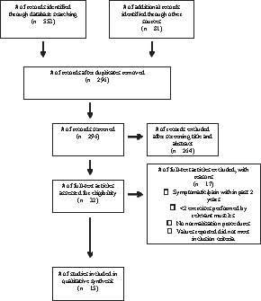
Prisma flowchart for research strategy.
Study Characteristics
Study Design
All 15 studies included were observational studies. For all studies, testing and EMG data collection occurred within the same day. Standardization of exercise techniques was used in 13 of the 15 studies included.5,6,10,11,12,13,14,16,17,18,20,21,22 Four studies provided a physical examination for the participants prior to the start of the study.5,6,18
Participants
Three studies included only male participants.11,14,22 None of the studies had any dropouts. Table 1 includes participant age characteristics for each study.
Risk of bias within studies
Three studies included a physical examination by a professional prior to the start of the exercises in order to assure normal scapulohumeral rhythm in the normal shoulders.5,6,18 Internal validity was compromised in the other studies, all of which did not control for the presence or absence of scapular dyskinesia. There were also biases among the studies for normalization and EMG standardization procedures, which could explain the differences in EMG values across studies during similar exercises. Two studies did not use a standardized technique (metronome) for exercise performance to assure continuity throughout the study.15,19 However, all studies allowed practice sessions for participants to become accustomed to the motions expected. One study,16 only reported EMG data for the eccentric phase of the exercises, leaving out the concentric and isometric phases. Although relevant for the purposes of this study, it may decrease the external validity by decreasing applicability to the general population, due to not comprising all phases of the exercise motion.25 Furthermore, five studies did not randomize the order in which exercises were performed, which is a risk for selection bias. This could induce fatigue, which could subsequently lower the %MVIC or promote compensation during the later exercises.26 None of the studies included blinded assessors, which is a risk for increasing biases. However, due to the observational nature of the studies included, the use of blinded assessors is not possible. Refer to Table 2 to view the quality assessment of internal and external validity among each study.
Results of individual muscles
Upper Trapezius. The UT muscle was analyzed in each of the studies. The most common exercises were variations of shoulder abduction. The UT was highly active during the rowing motion reported by Moseley et al.15 Other exercises in which the trapezius was highly active were abduction to 120 °, the shoulder/scapular shrug, and abduction in the scapular plane to 90 ° with the shoulder externally rotated.5,11,12,17,18,22
Exercises with ratios that favored the UT over the other scapular stabilizers include: Maximal forward flexion, shoulder shrug, and abduction with external rotation. See Tables 3, 4, and 5 for ratios for each exercise. Table 1 in Appendix 3 presents %MVIC values of the UT during each exercise, across all studies. (Available in Supplemental materials, linked on the IJSPT Website)
Table 3.
Table 3. Upper Trapezius/Middle Trapezius. Optimal Ratios.
| Cools, et al21 | Ekstrom, et al12 | Huang, et al6 | Kibler, et al13 | Marta, et al14 | Park, et al16 | |
|---|---|---|---|---|---|---|
| Abduction 60 °, Eccentric | _ | _ | _ | _ | _ | 0.46 |
| Abduction 180 °, Eccentric | _ | _ | _ | _ | _ | 0.38 |
| Flexion 180 °, Eccentric | _ | _ | _ | _ | _ | 0.12 |
| Prone ER at 90 ° abd, 90 ° elbow flexion | _ | 0.44 | _ | _ | 0.72 | _ |
| Side-lying ER with elbow at 90 ° | 0.37 | _ | 0.54* | _ | 0.38 | 0.44 |
Indicates ratios were reported in the study and were not calculated by authors
Studies were not included in table if they did not report on the above exercises
ER = external rotation
Values rounded to the nearest hundredth.
Table 5.
Upper Trapezius/Serratus Anterior.
| Optimal Ratios. | |||
|---|---|---|---|
| Ekstrom, et al12 | Uhl, et al19 | Wattanaprakornkul, et al20 | |
| Bench Press, Seated | _ | _ | 0.30§ |
| Diagonal Exercise | 0.66 | _ | _ |
| Bilateral Scapular Protraction | 0.13 | _ | _ |
| Supine Press | 0.11 | 0.06 | _ |
Indicates measurements were estimated from a graph
Studies were not included in table if they did not report on the above exercises
Values rounded to the nearest hundredth
| Least Optimal Ratios. | |||||
|---|---|---|---|---|---|
| Cools, et al21 | Ekstrom, et al12 | Pizzari, et al17 | Sciascia, et al18 | Wattanaprakornkul, et al20 | |
| Low Row 1 | 1.01 | _ | _ | _ | _ |
| Low Row 2 | 2.12 | _ | _ | _ | _ |
| Prone Abduction with ER | 3.66 | _ | _ | _ | _ |
| Prone Horiz. Abduction with ER (thumb up) | 3.61 | 7.33 | _ | 3.29 | _ |
| Row, Seated | 3.32 | _ | _ | _ | 1.11§ |
| Shoulder Shrug | _ | 4.41 | 4.93 | _ | _ |
| Unilateral Row, Prone | _ | 4.50 | _ | _ | _ |
Indicates measurements were estimated from a graph
Studies were not included in table if they did not report on the above exercises
ER = external rotation Horiz. = Horizontal
Low Row 1 = shoulders in 45 ° flexion; subject performs extension with elbows extended
Low Row 2 = shoulders in 45 ° flexion; subject performs extension with elbows flexed
Values rounded to the nearest hundredth
Middle Trapezius. The MT muscle was highly active during eccentric abduction and flexion.16 It was also highly active during prone overhead raise, prone unilateral row, and abduction in the scapular plane to 90 °.
Exercises with optimal ratios (ratios that favored MT activity with little UT activity) were eccentric exercises in the frontal and sagittal planes, especially flexion between 180 ° and 60 °. External rotation exercises with the elbow flexed to 90 ° also had optimal ratios for activating the MT in prone and side-lying positions. Table 3 displays data for all UT/MT ratios. See Table 2 in Appendix 3 for %MVIC values of the MT during each exercise. (Available in Supplemental materials, linked on the IJSPT Website)
Lower Trapezius. The LT muscle was highly active during prone flexion, prone overhead raise, and prone external rotation. Exercises in the scapular plane did not activate the LT as much as the other scapular stabilizers.
Exercises with optimal ratios for the LT were prone flexion, high scapular retraction, and prone external rotation with the shoulder abducted to 90 ° and elbow flexed. The least optimal ratios were during shoulder abduction and press-up exercises done in a standing or semi-reclined position. See Table 4 for all UT/LT ratios. See Table 3 in Appendix 3 for %MVIC values of the LT during each exercise. (Available in Supplemental materials, linked on the IJSPT Website)
Serratus Anterior. The SA muscle was most active during exercises that involved reaching across the body, such as, the dynamic hug and diagonal exercise. It was also the most active of the scapular muscles during side-lying forward flexion. Abduction in the scapular plane with external rotation activated the SA more if elevation exceeded 80 °.22
Exercises with optimal ratios for the SA were the diagonal exercises and scapular protraction. The bench press exercise and supine press also provided optimal ratios. Shoulder shrug, low row, and abduction with external rotation in prone provided the least optimal ratios for SA activation. See Table 5 for all UT/SA ratios. See Table 4 in Appendix 3 for %MVIC values for the SA during each exercise. (Available in Supplemental materials, linked on the IJSPT Website)
DISCUSSION
Summary of evidence
The results of this review illustrate the variations of common shoulder exercises, and their impact on muscle ratios in the scapular stabilizers. The authors performed a quality assessment in order to determine the risk of bias and interpret the quality of results. When interpreting significance of the results, the number of studies that examined a specific exercise and found similar results was considered. This was a consideration due the inclusion of 62 exercises, most of which were not examined in more than one study. Therefore, comparisons of EMG activity and ratios across studies were limited.
For the MT, this review revealed that the eccentric phase of flexion exercises from 180 ° and 60 ° promoted optimal ratios.16 However, when the average of all phases of shoulder flexion were analyzed in other studies, ratios exceeded 1.00.6,16 This indicates that the UT is more active than the MT. Therefore, if trying to activate the MT with the least amount of UT activity, only the eccentric phase should be performed. Isolating only one phase while performing an exercise is not typical practice of the general population. With relevance to clinical application, it is not functional to perform only one phase/type of contraction during dynamic exercises and activities as concentric and eccentric motions are often paired. During the prone unilateral row exercise, the MT was the only scapular stabilizer that was more activated than the UT. Another interesting finding represented by the data was the effect of the variations of the row exercise. This review shows that a rowing motion (shoulder extension) with the elbows extended promotes higher activation and a more favorable ratio in the MT muscle when compared to a traditional row (shoulder extension with elbow flexion). The UT/SA ratio during the low row exercise with elbows extended is approximately 1.00, which indicates that during this motion, the MT is the most active of the scapular muscles analyzed in this review.
Flexion in the prone position provided the best UT/LT ratio with a fairly high %MVIC of the LT, indicating isolation of the LT in comparison to the UT.20 This exercise also provided good UT/SA ratios, however, the contraction of the SA during this exercise was not as strong as the LT. Scapular retraction exercises in all positions reported in a study included in this review observed UT/LT ratios in favor of the LT muscle.10 Even though the scapular retraction exercises were only performed in a single study, the testing in various positions demonstrates consistency of results within the study. Scapular retraction is an exercise for strengthening the LT that can easily be adapted into an intervention or daily workout. In addition, the variation in positions, while still obtaining consistent results, makes it more generalizable.
Side-lying exercises (flexion and ER) provided optimal ratios for the MT and LT. In addition, side-lying external rotation yielded similar ratios across three studies for the MT and LT relative to the UT.6,14,21 This indicates that these scapular stabilizers can be strengthened together in the side-lying position with minimal activation of the UT.
Prone horizontal abduction at 100 ° forward flexion had ratios close to 1.00 for all of the trapezius muscles, possibly indicating this is a good exercise to activate all parts of the trapezius equally. Abduction in the scapular plane to 90 ° yielded ratios close to 1.00 for the LT and SA muscles relative to the UT, indicating they were relatively equally active. However, the same motion in the frontal plane activated the UT more than the LT and SA muscles. This demonstrates how minor variability amongst exercises could change the directional pull on the scapula.
This review also determined which exercises promoted optimal ratios for SA activation. All variations of scapular protraction exercises, including bench press, promoted optimal UT/SA ratios. Shoulder abduction in the scapular plane (with and without ER) above 90 ° produced greater activation of the SA when compared to the UT.22 However, the same exercises to 90 ° and lower produced greater UT activation.22 Prone and side-lying flexion and ER exercises also demonstrated greater SA muscle activity relative to the UT.6,12,18,20,21
In relation to the purpose of this review, exercises that promoted higher UT activity when compared to the other scapular stabilizers were also determined. If the target muscle is the SA, this review determined that prone horizontal abduction (with or without ER) and prone unilateral row exercises should be avoided. The UT was significantly more active than the LT during exercises in the scapular plane. The shoulder shrug exercises at 0 ° and 30 ° abduction produced UT muscle activation that was double to quadruple that of the comparison scapular muscles.12,17
A narrative review by Cricchio & Frazer reported similar findings in exercises that primarily activated the MT, LT, and/or SA. Consistent with the current findings, those authors also reported overhead arm raise at 125 ° activated the MT and LT, indicating less activation of the UT at elevation above 120 °. The narrative also determined prone exercises to be beneficial for activating the MT, as well as recommending side-lying and prone exercises for low UT/LT ratios. In terms of this review, low ratios reported in the narrative would be optimal.
In order to perform elevation activities, proper muscle activation is essential. Limiting UT muscle activation while the force couple of the MT, LT, and SA are activated is vital to prevent abnormal mechanics or symptoms. Appropriate exercise choices are vital in order to properly address muscle weakness that may be contributing to altered movement patterns. According to this review, best choices for the MT include prone external rotation and side-lying external rotation. During external rotation, the MT may be activated because of the need for retraction of the scapula as well as maintaining an optimal length of the external rotators as the movement is being performed.
The ideal exercises for the LT were prone flexion, high scapular retraction, and prone ER. These exercises are common utilized clinically and the movement is in proper alignment with the fiber direction of the LT.
The most effective SA exercises were the diagonal exercise, scapular protraction, bench press, and supine press. All of these exercises promote protraction and upward rotation of the scapula which are primary movements produced by the serratus anterior. Finally, clinicians should attempt to limit utilizing exercises that activate the UT excessively, such as the shoulder shrug, prone unilateral row and prone horizontal abduction.
Review Limitations
Although the %MVIC was used to calculate ratios, it could not be used to determine optimal exercises for individual muscles. This is due to the inconsistencies between normalization techniques and resistance used across studies that performed the same exercises. There were also differences in methodology across studies that make it difficult to compare similar exercises. The many variations of the exercises included in these studies also could account for discrepancies in muscles activity across the studies. Some studies recorded the concentric, isometric, and eccentric phases of the exercise separately.16 Averaging these values, rather than having the entire exercises recorded and averaged via EMG analysis, could account for variation from the true value. Furthermore, estimations made from the graphs in Wattanaprakornkul et al20 allowed for variation by interpretation.
Authors (AS, JQ, and JW) were unilingual and therefore unable to include studies in languages other than English. Many exercises were only reviewed in one article, giving us no aspect of inter-rater reliability or comparison across studies. Most studies only included %MVIC; therefore, ratios were calculated independently and not by the original researchers and standard deviations could not be calculated. Although load/resistance differences used between studies should not alter the biomechanics of the exercise, and therefore should not significantly alter the muscle activation ratios, compensation is more likely with increased loads, fatigue, or pathology.4 If compensation did occur, this may have impacted muscle activity and subsequent muscle ratios. Muscle ratios were calculated without consideration or separation of exercises according to muscle contraction type (eccentric, isometric, concentric). Therefore, caution should be noted in the selection of exercises based strictly on muscle contraction type. Due to no reporting of participants undergoing imaging prior to the studies, the authors do not know of any underlying pathologies that may have been present that could alter the biomechanics of the shoulder. Because tissue healing may take 1-3 years to gain 100% of normal tensile strength1 post injury exclusion criteria was set at two years, which may have allowed for decreased strength in previously injured participants included within these studies.
The recommendations from this review are based on studies and calculations made on healthy, non-pathological subjects. Therefore, the results of this review can only be used to inform guidelines for a rehabilitation program to be used with injured patients or clients. Further research is needed to determine the applicability of these results to a rehabilitation program for pathological shoulders. Future studies should also be performed with consistent parameters to improve continuity of results.
CONCLUSION
This review has identified optimal positions and exercises related to periscapular muscular recruitment and stability exercises. In general, standing exercises tend to activate the UT at a higher ratio than the MT, LT, and SA, especially during the 60-120 ° range. The UT was the least active, relative to the other scapular muscles examined, while performing exercises in prone, side-lying, and supine positions; and which one of these positions is recommended is dependent upon the exercise and whether the target muscle is the MT, LT, or SA. More studies need to be conducted to examine these exercises in greater detail and confirm their consistency in producing the optimal ratios determined in this review. Further investigation is required to determine the similarities and/or differences in the muscle ratios in subjects with healthy versus pathological shoulders.
APPENDIX 1: DETAILS OF SEARCH STRATEGY AND INCLUSION/EXCLUSION CRITERIA
PubMed Search Strategy
Trapezius [Text Word]
Serratus Anterior [Text Word]
1 OR 2
Exercise Therapy[MeSH Terms]
Exercise*[MeSH Terms]
Resistance Training[MeSH Terms]
Exercise*[Text Word]
4 OR 5 OR 6 OR 7
Electromyography[MeSH Terms]
Electromyography feedback[MeSH Terms]
Electromyography[Text Word]
EMG[Text Word]
9 OR 10 OR 11 OR 12
3 & 8 & 13
Filters: Humans, English
Discovery Layer (Walsh University) Search Strategy
Trapezius (AB abstract)
Resistance Exercise (TX All Text)
EMG (TX All Text)
1 & 2 & 3
Limit: English, Academic Journal
CIHNAL Search Strategy
Serratus Anterior OR Trapezius
Exercise
EMG
1 & 2 & 3
Limit: English, Academic Journal
SPORTDiscus Search Strategy
Trapezius (AB abstract)
Exercise (TX All Text)
Electromyography (TX All Text)
1 & 2 & 3
Limit: English & Academic Journal
Scopus Search Strategy
Trapezius OR Serratus Anterior
Resistance Training OR exercise
EMG OR electromyography
1 & 2 & 3
Limit: English, Human, Article, Review
Inclusion Criteria
English
Academic Journal
EMG used as primary tool
EMG analysis of the UT and at least one of the following muscles: MT, LT, or SA
Compare EMG activity of one or more of the above muscles during two or more active open-chain exercises
Normal, healthy, asymptomatic shoulder
Include %MVIC/MVC and/or ratio values for data standardization and continuity of measurement across studies
Method of normalization of EMG for improved quality and comparability of values
Detailed method of EMG analysis for all muscles tested or statement of guidelines followed for reproducibility, quality analysis, and continuity of appropriate usage and technology.
Exclusion Criteria
History of shoulder pathology within 2 years
History/current scapular pathology
Symptomatic/Pain within 2 years
Closed-Chain Exercises
No standardized approach for EMG normalization and analysis EMG not used as primary tool
Appendix 2
Upper Trapezius/Lower Trapezius.
| Exercise Descriptions | |
| Abduction | Shoulder abduction in the frontal plane with the shoulder in neutral position of rotation (palm down) |
| Abduction in scaption | Shoulder abduction in the scapular plane with the shoulder in a neutral position (palm down) |
| Abduction, maximal | Abduction in the frontal plane with the shoulder in neutral position (palm down) to the subject's maximum range |
| Bench Press, seated | Seated chest press using a weight machine; Starting position with shoulders abducted to 90 °, elbows flexed, scapular retraction, forearm pronation; Ending position with scapular protraction, horizontal shoulder adduction, elbow extension |
| Bilateral scapular protraction | Supine; Bilateral scapular protraction with the shoulder horizontally flexed to about 45 ° and the elbows flexed to about 45 ° |
| Diagonal exercise | Combination of shoulder flexion, horizontal flexion, and external rotation in the sitting position |
| Diagonal – eccentric | Shoulder neutral rotation and 30 ° horizontal flexion; starting position in 130 ° shoulder abduction; ending position with full shoulder internal rotation |
| Dynamic Hug | Standing with back toward the wall, knees slightly bent, and the feet shoulder-width apart; Being with elbow flexed 45 °, shoulder abducted 60 °, and shoulder internally rotated 45 °; Horizontally flex shoulder following an arc (hugging motion) until maximum protraction is attained |
| ER in scapular plane | Shoulder external rotation in the scapular plane (45 ° abduction, 30 ° horizontal abduction) with the elbow flexed to 90 ° |
| Extension, prone | Prone with shoulders resting in 90 ° forward flexion; Extension to neutral position with the shoulder in neutral rotation |
| Flexion – Eccentric | Shoulder in neutral rotation and 70 ° horizontal flexion; Start at 130 ° shoulder flexion; End at 40 ° shoulder flexion with full internal rotation |
| Forward flexion, maximal | Standing; Shoulder in neutral position; perform maximal forward flexion in the sagittal plane |
| Forward flexion, side-lying | Side-lying position; shoulder in neutral position; perform forward flexion in a horizontal plane to 135 ° |
| High row | Standing in front of a vertical pulley with shoulders in 135 ° forward flexion; performs shoulder extension to neutral position |
| High SR, sitting | Trunk supported, feet on the ground; 1m away from pulley apparatus; starting with scapular protraction, high scapular retraction was performed until the elbows were positioned at the lateral side of the trunk; maintain neutral spine |
| High SR, standing | Feet positioned shoulder width, legs straight; 1m away from pulley apparatus; starting with scapular protraction, high scapular retraction was performed until the elbows were positioned at the lateral side of the trunk; maintain neutral spine |
| High SR, static bipedal squat | Feet shoulder width apart with both knees positioned at 90 ° above the feet; 1m away from pulley apparatus; starting with scapular protraction, high scapular retraction was performed until the elbows were positioned at the lateral side of the trunk; maintain neutral spine |
| High SR, static lunge | Contralateral leg in front with the knee in a 90 ° angle. Distance between both feet determined by taking the distance between the ASIS and the medial malleolus of the dominant side; 1m away from pulley apparatus; starting with scapular protraction, high scapular retraction was performed until the elbows were positioned at the lateral side of the trunk; maintain neutral spine |
| High SR, static unipedal squat | Contralateral knee placed above the foot in a 45 ° angle. 1m away from pulley apparatus; starting with scapular protraction, high scapular retraction was performed until the elbows were positioned at the lateral side of the trunk; maintain neutral spine |
| High SR, dynamic bipedal squat | Starting position as static version. Concentric phase of arm movement during concentric squat |
Table 1.
Exercise Descriptions
| High SR, dynamic lunge | Starting position as static version. Concentric phase of arm movement during concentric squat |
| High SR, dynamic unipedal squat | Starting position as static version. Concentric phase of arm movement during concentric squat |
| Ipsilateral step-up with ball | Stand astride with the ipsilateral foot of the affected arm on a standard inch step and the other on the floor; a light plastic ball is held in both hands; shift weight forward and rises up onto the step as both arms are elevated overhead |
| Ispilateral shoulder flexion | Stands astride with the ipsilateral foot of the affected arm in front; shifts weight forward and elevates both arms overhead; motion should be rhythmic with the lower extremity preceding the upper extremity |
| Lawnmower | Diagonal pattern from the contralateral leg through trunk and ipsilateral arm; hip/trunk extension, trunk rotation, and scapular retraction |
| Low row 1 | Standing in front of pulley apparatus; shoulders in 45 ° forward flexion and neutral rotation; performs extension with elbows flexed |
| Low row 2 | Standing in front of pulley apparatus; shoulders in 45 ° forward flexion and neutral rotation; performs extension with elbows extended |
| Prone abduction with ER | Prone with shoulder in neutral position; performs shoulder abduction to 90 ° with external rotation in the horizontal plane |
| Prone horizontal abduction at 90 ° forward flexion | Prone with shoulders resting in 90 ° forward flexion; performs horizontal abduction to horizontal position |
| Prone horizontal abduction at 100 ° forward flexion | Prone horizontal abduction at 100 ° forward flexion with full external rotation |
| Prone horizontal abduction with ER | Prone with shoulders resting in 90 ° forward flexion; performs horizontal abduction to horizontal position with external rotation at the end of movement |
| Prone overhead raise | Arm raise above head with the upper extremity in line with the lower trapezius muscle fibers in prone position |
| Robbery | Begin in a standing position with the trunk flexed to approximately 40 ° to 50 ° with arms forward flexed and palms facing the thighs. While keeping elbows close to body, move into trunk and arm extension and elbow flexion; simultaneously pinch both scapulae toward back pockets |
| Row, seated | Seated in front of pulley apparatus with shoulders in 90 ° forward flexion: performs extension movement with elbows flexed in the horizontal plane |
| Scaption | Shoulder abduction in the scapular plane with the shoulder externally rotated (thumb up) |
| Side-lying ER with elbow at 90 ° | Side-lying with shoulder in neutral position and elbow flexed 90 °; performs external rotation of the shoulder with towel between trunk and elbow |
| Standing press-up | Standing; a can is pushed overhead toward the ceiling from a bent elbow resting position with the scapula protracted at the end |
| Supine shoulder press | Unilateral shoulder press with full scapular protraction with the shoulder flexed to 90 ° and the elbow fully extended in supine position |
| Unilateral row, prone | Prone with shoulder beginning in 90 ° forward flexion; extend shoulder to neutral and flex elbow to 90 ° |
| Wedge press-up | Supine, elevated on a 45 ° wedge; upper extremity pushes object from side up towards the ceiling with the scapula protracted at the end |
REFERENCES
- 1.Dutton M. Dutton's Orthopaedic Examination Evaluation and Intervention. 3rd ed New York: McGraw-Hill Medical; 2012. [Google Scholar]
- 2.Codman EA. The Shoulder: Rupture of the Supraspinatus Tendon and Other Lesions In or About the Subacromial Bursa. Boston: Thomas Todd Co.; 1934. [Google Scholar]
- 3.Cricchio M Frazer C. Scapulothoracic and scapulohumeral exercises: A narrative review of electromyographic studies. J Hand Ther. 2011; 24:322-333. [DOI] [PubMed] [Google Scholar]
- 4.Oatis CA. Kinesiology, The Mechanics and Pathomechanics of Human Movement, 2nd ed India: Lippincott Williams & Wilkins; 2009. [Google Scholar]
- 5.Cools AM Declercq GA Cambier DC Mahieu NN Witvrouw EE. Trapezius activity and intramuscular balance during isokinetic exercise in overhead athletes with impingement symptoms. Scand J Med Sci Sports. 2007; 17:25-33. [DOI] [PubMed] [Google Scholar]
- 6.Huang HY Lin JJ Guo YL Wang WTJ Chen YJ. EMG biofeedback effectiveness to alter muscle activity pattern and scapular kinematics in subjects with and without shoulder impingement. J Electromyogr Kinesiol. 2013; 23:267-274. [DOI] [PubMed] [Google Scholar]
- 7.Matsen FA III Arntz CT. Subacromial impingement. In: Rockwood CA Jr. Matsen FA III, eds. The Shoulder. Philadelphia, PA: WB Saunders, 1990. [Google Scholar]
- 8.Warner JJ Micheli LJ Arslanian LE Kennedy J Kennedy R. Scapulothoracic motion in normal shoulder and shoulders with glenohumeral instability and impingement syndrome. A study using Moire topographic analysis. Clin Orthop 1992; 285:191-199. [PubMed] [Google Scholar]
- 9.Sharkey NA Marder RA Hanson PB. The role of the rotator cuff in elevation of the arm. Trans Orthop Res Soc 1993; 18:137. [DOI] [PubMed] [Google Scholar]
- 10.De Mey K Danneels L Cagnie B Lotte VB Johan F Cools AM. Kinetic chain influences on upper and lower trapezius muscle activation during eight variations of a scapular retraction exercise in overhead athletes. J Sci Med Sport. 2013; 16:65-70. [DOI] [PubMed] [Google Scholar]
- 11.Decker MJ Hintermeister RA Faber KJ Hawkins RJ. Serratus anterior muscle activity during selected rehabilitation exercises. Am J Sports Med. 1999; 27:784-791. [DOI] [PubMed] [Google Scholar]
- 12.Ekstrom RA Donatelli RA Soderberg GL. Surface electromyographic analysis of exercises for the trapezius and serratus anterior muscles. J Orthop Sports Phys Ther. 2003; 33:247-258. [DOI] [PubMed] [Google Scholar]
- 13.Kibler WB Sciascia AD Uhl TL Tambay N Cunningham T. Electromyographic analysis of specific exercises for scapular control in early phases of shoulder rehabilitation. Am J Sports Med. 2008; 36:1789-98. [DOI] [PubMed] [Google Scholar]
- 14.Marta S Pezarat-Coreia P Fernandes O Carita A Cabri J Moraes AC. A. Electromyographic analysis of posterior deltoid, posterior rotator cuff and trapezius musculature in different shoulder exercises. Int Sport Med J. 2013; 14:1-15. [Google Scholar]
- 15.Moseley JB Jobe FW Pink M Perry J Tibone J. EMG analysis of the scapular muscles during a shoulder rehabilitation program. Am J Sports Med. 1992; 20:128-134. [DOI] [PubMed] [Google Scholar]
- 16.Park S Nho H Chang MJ Kim JK. Electromyography activites for shoulder muscles over various movements on different torque changes. Euro J of Sport Sci. 2012; 12:408-417. [Google Scholar]
- 17.Pizzari T Wickham J Balster S Ganderton C Watson L. Modifying a shrug exercise can facilitate the upward rotator muscles of the scapula. Clin Biomech. 2014; 29:201-205. [DOI] [PubMed] [Google Scholar]
- 18.Sciascia A Kuschinsky N Nitz AJ Mair SD Uhl TL. Electromyographical comparison of four common shoulder exercises in unstable and stable shoulders. Rehabil Res Pract. 2012; 2012:783824. [DOI] [PMC free article] [PubMed] [Google Scholar]
- 19.Uhl TL Muir TA Lawson L. Electromyographical assessment of passive, active assistive, and active shoulder rehabilitation exercises. PM R. 2010; 2:132-41. [DOI] [PubMed] [Google Scholar]
- 20.Wattanaprakornkul D Halaki M Cathers I Ginn KA. Direction-specific recruitment of rotator cuff muscles during bench press and row. J Electromyogr Kinesiol. 2011; 21:1041-1049. [DOI] [PubMed] [Google Scholar]
- 21.Cools AM Dewitte V Lanszweert F, et al. Rehabilitation of scapular muscle balance: which exercises to prescribe? Am J Sports Med. 2007; 35:1744-1751. [DOI] [PubMed] [Google Scholar]
- 22.de Oliveira V Batista L Pirauá A Pitangui A Araujo R. Electromyographic activity and scapular dyskinesia in atheletes with and without shoulder impingement syndrome. Brazil J of Kinanthropometry Hum Perf. 2013; 15:193-203. [Google Scholar]
- 23.Siegfried N Muller M Deeks J, et al. HIV and male circumcision – a systematic review with assessment of the quality of studies. Lancet Infect Dis. 2005; 5:165-173. [DOI] [PubMed] [Google Scholar]
- 24.Ganderton C Pizzari T. A systematic literature review of the resistance exercises that promote maximal muscle activity of the rotator cuff in normal shoulders. Shoulder and Elbow. 2013; 5:120-135. [Google Scholar]
- 25.O’Sullivan S. Physical Rehabilitation. 6th ed F A Davis Company; 2013. [Google Scholar]
- 26.Dimitrova NA Dimitrov GV. Interpretation of EMG changes with fatigue: facts, pitfalls, and fallacies. J Electromyogr Kinesiol. 2003; 13:13-36. [DOI] [PubMed] [Google Scholar]