Abstract
Erlotinib, an epidermal growth factor receptor tyrosine kinase inhibitor, is an oral targeted anticancer drug that is used to treat non-small cell lung cancer (NSCLC). Previous studies have confirmed that erlotinib is safe and is well-tolerated by patients. The most common adverse reactions observed following erlotinib treatment include a rash and mild diarrhea. In the current study, the first case of acute myocardial infarction following one month of treatment with erlotinib in a 63-year-old male NSCLC patient is presented. The present study highlights the importance of clinicians remaining cautious following erlotinib administration. In elderly NSCLC patients and those with a history of coronary heart disease, cardiac function must be carefully monitored following erlotinib treatment so that serious adverse reactions, such as myocardial infarction, may be identified early and treated quickly.
Keywords: lung cancer, targeted therapy, erlotinib, adverse reactions, myocardial infarction
Introduction
At present, lung cancer is the leading cause of cancer-associated mortality worldwide (1). Therefore, treatment strategies for lung cancer are of significant interest. The standard approach to lung cancer treatment is multidisciplinary and includes surgery, chemotherapy, radiotherapy and targeted therapy. Targeted therapy is a novel and promising therapeutic modality. The Chinese guidelines on the diagnosis and treatment of primary lung cancer (2011 version) (2) state that systemic therapy improves quality of life and prolongs the survival of stage IV non-small cell lung cancer (NSCLC) patients. Targeted treatment of NSCLC patients with epidermal growth factor receptor (EGFR) mutations is more likely to be effective than in patients without EGFR mutations; therefore, gefitinib or erlotinib are recommended as the first-line treatment for stage IV NSCLC patients with sensitive EGFR mutations (2).
In 2006, oral erlotinib (150 mg, daily) was approved by the China Food and Drug Administration for the treatment of NSCLC in China (3). Erlotinib, an EGFR tyrosine kinase inhibitor, inhibits the phosphorylation of EGFR, which is expressed on the surface of normal and tumor cells. In non-clinical experimental models, the inhibition of EGFR phosphorylation has been demonstrated to cause cell growth arrest and cell death (3). The most common adverse reactions following erlotinib administration are rash and mild diarrhea. Additional adverse reactions include severe rash, severe paronychia, interstitial pneumonia, myocardial ischemia and acute hepatitis (3).
The present study reports the first case of acute myocardial infarction occurring after one month of treatment with erlotinib in an NSCLC patient, and highlights the importance of careful monitoring following this treatment.
Case report
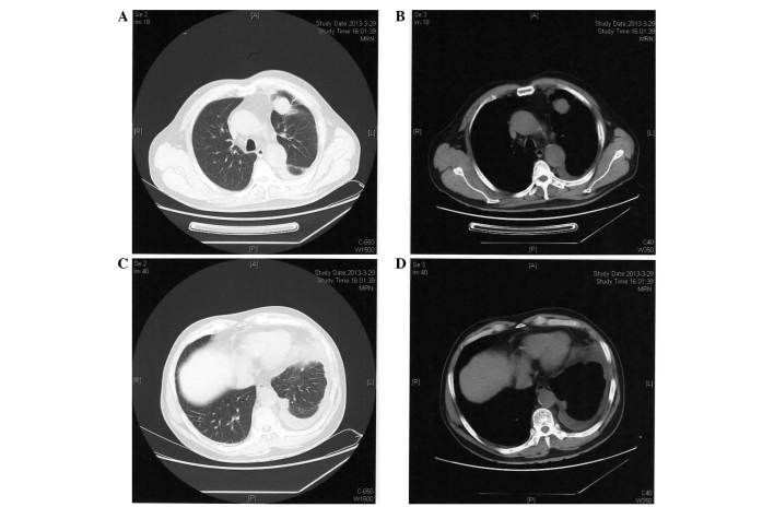
On March 25, 2013, a 63-year-old male patient was admitted to the Provincial Hospital Affiliated to Shandong University (Jinan, China) with chest tightness that had lasted for 14 days. The patient had smoked for 40 years (≤20 cigarettes per day) and reported a 30-year history of hypertension. A computed tomography (CT; Discovery CT750 HD; GE Healthcare Life Sciences, Shanghai, China) scan identified a mass in the upper lobe of the left lung and left pleural effusions (Fig. 1). Cytology of pleural effusions was clearly positive at malignant cells, with tumor marker carcinoembryonic antigen (CEA) levels of >1,000 ng/ml (normal range, 0–10 ng/ml). A percutaneous lung biopsy was performed in April 2013, which confirmed the diagnosis of histologically invasive adenocarcinoma. Subsequently, the patient received 7 cycles of chemotherapy with pemetrexed (0.8 g on day 1 every 21 days) and nedaplatin (40 mg on days 2–4 every 21 days) and experienced significant bone marrow suppression during chemotherapy. During the chemotherapy treatment, the patient complained of chest discomfort. However, repeated electrocardiogram (ECG; 9310P; Nihon Kohden, Tokyo, Japan) examinations revealed no obvious abnormalities. An echocardiographic examination performed on October 8, 2013 indicated that the cardiac structures were normal (Fig. 2A) with a left ventricular ejection fraction (LVEF) of 63%.
Figure 1.
Computed tomography scan performed on March 29, 2013, prior to chemotherapy treatment with pemetrexed and nedaplatin. (A) Lung and (B) mediastinal windows showing a mass in the upper lobe of the left lung and (C) lung and (D) mediastinal windows showing pleural effusions in the left side.
Figure 2.
Echocardiography and ECG examinations performed prior to erlotinib treatment. (A) Echocardiography performed on October 18, 2013, prior to the administration of targeted treatment. (B) ECG performed on April 14, 2014, prior to the administration of targeted treatment. ECG, electrocardiogram.
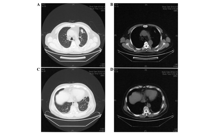
In April 2014, the patient was admitted to the Provincial Hospital Affiliated to Shandong University for review. ECG examination demonstrated sinus rhythm and no evidence of arrhythmia or myocardial ischemia (Fig. 2B). A CT scan revealed that the mass had decreased in size compared with that prior to the 7 cycles of chemotherapy (Fig. 3). Emission CT revealed increased imaging agent (iodinated contrast) uptake in the 6th, 7th and 8th left front ribs compared with the contralateral ribs. Although no significant progression of the lesions was evident following 7 cycles of pemetrexed and nedaplatin, evidence of bone metastasis was identified using bone imaging examination, and the patient had experienced adverse reactions to chemotherapy; the blood cell count of the patient was significantly reduced, demonstrating bone marrow suppression. Due to the limited efficacy and undesirable side effects of chemotherapy, and the presence of sensitive EGFR mutations in the tumor, demonstrated by genetic testing of pathological specimens, erlotinib maintenance treatment (150 mg orally, once daily) was selected.
Figure 3.
Computed tomography scan performed on April 15, 2014 following 7 cycles of chemotherapy with pemetrexed and nedaplatin. (A) Lung and (B) mediastinal windows showing a mass in the upper lobe of the left lung and (C) lung and (D) mediastinal windows showing pleural effusions in the left side, which are decreased in size compared to those observed in Figure 1.
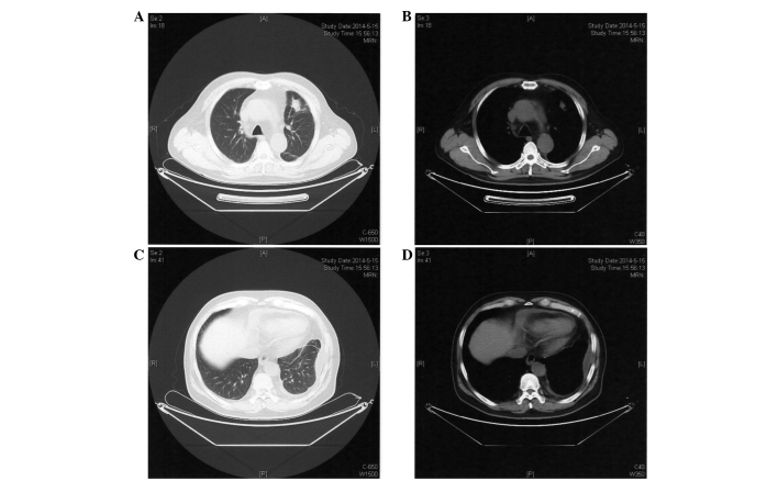
In May 2014, after 1 month of treatment with erlotinib, the patient was admitted for follow-up, complaining of mild nausea and occasional pain in the left chest area, without radiating pain, vomiting, fever or expectoration. Physical examination revealed a body temperature of 35.6°C (normal range, 36.0–37.0°C), pulse rate of 92 bpm (normal range, 60–100 bpm), respiratory rate of 23 times/min (normal range, 16–20 times/min), blood pressure of 115/74 mmHg (normal range, 90–130/60–90 mmHg), and a rash located on the face and lower jaw. A CT scan performed on May 15, 2014 (Fig. 4) revealed no significant differences in the mass located in the upper lobe of the left lung compared with the previous CT scan. ECG showed that ST-segments of V1-V5 were arched upward and inverted T waves were present (Fig. 5A). High-sensitivity troponin T assay revealed troponin T levels of 23.77 pg/ml (normal range, 3–14 pg/ml) and N-terminal pro-brain natriuretic peptide levels of 2,771 pg/ml (normal range, 0–125 pg/ml). Echocardiography indicated extensive infarction of the anterior myocardial tissue (Fig. 5B) and a LVEF of 54%. The patient was considered to have experienced acute myocardial infarction and thus erlotinib treatment was discontinued immediately. Subsequently, the patient's blood pressure was controlled and anticoagulant and diuretic drugs were administered (asprin enteric-coated tablets, 0.1 g every day; clopidogrel hydrogen sulphate tablets, 75 mg every day; furosemide tablets, 20 mg twice a day).
Figure 4.
Computed tomography scan performed on May 15, 2014 following treatment with erlotinib for 1 month. (A) Lung and (B) mediastinal windows showing a mass in the upper lobe of the left lung and (C) lung and (D) mediastinal windows showing pleural effusions in the left side, which are not considerably different to those observed in Figure 3.
Figure 5.
Echocardiography and ECG examinations performed following erlotinib treatment. (A) ECG performed on May 15, 2014, following targeted treatment. (B) Echocardiography performed on May 19, 2014, following 1 month of targeted treatment. ECG, electrocardiogram.
During follow-up, a computed tomographic angiography of the coronary arteries identified multiple plaques in the right and left coronary arteries with corresponding luminal stenosis. Currently, the patient is well and his heart function is controlled; however, pulmonary metastasis was identified in 2015.
Written informed consent was obtained from the patient's family for the publication of this study.
Discussion
Recently, certain drugs administered for the treatment of tumors, including target drugs such as imatinib, sunitinib and nilotinib, have been reported to cause cardiac toxicity (4). A cardiotoxicity study revealed that sunitinib treatment resulted in mitochondrial damage and myocardial apoptosis in mice and cultured rat cardiomyocytes (5). Erlotinib has been demonstrated as a safe and well-tolerated treatment for NSCLC (6–9), with no cases of myocardial infarction reported to date.
The patient in the present case report had a history of hypertension, and the patient's triglyceride, total cholesterol and CEA levels at various time points are shown in Table I. Prior to treatment with erlotinib, the cardiac ultrasound and repeated ECG examinations were normal. Following treatment with erlotinib for 1 month, commencing in April 2014, the lesion in upper lobe of the left lung exhibited no significant changes and the CEA level of the patient was lower than before the treatment (Table I). During targeted therapy, the patient developed a facial rash, and subsequently experienced acute myocardial infarction following 1 month of erlotinib treatment. During this treatment period, no other chemotherapy drugs that may cause myocardial ischemia or myocardial infarction were administered. Based on these findings, erlotinib was considered to be the cause of the adverse reactions observed.
Table I.
Triglyceride, total cholesterol and CEA serum levels of the patient.
| Date | Triglycerides (mmol/l) | Total cholesterol (mmol/l) | CEA (ng/ml) |
|---|---|---|---|
| 26/03/2013 | 2.06 | 5.31 | 86.19 |
| 14/01/2014 | 2.50 | 4.75 | 61.51 |
| 18/02/2014 | 3.71 | 4.96 | – |
| 27/03/2014 | 2.96 | 4.94 | 32.86 |
| 15/04/2014 | 2.36 | 4.63 | – |
| 16/05/2014 | 2.25 | 3.61 | 14.77 |
Normal reference values: Triglycerides, 0.4–1.8 mmol/l; total cholesterol, 3.6–6.2 mmol/l; and CEA, 0–10 ng/ml. CEA, carcinoembryonic antigen.
We postulate that erlotinib may lead to myocardial infarction; however, the mechanism remains unclear. Erlotinib exerts anticancer effects via the inhibition of EGFR phosphorylation. EGFR is found on the surface of cancer cells, but is also present on various kinds of normal cells. Doherty et al (10) investigated the toxicity of crizotinib, sunitinib, erlotinib and nilotinib in vitro and revealed that treatment with 10 µM erlotinib caused human cardiomyocyte cell death. It was also demonstrated in a phase I study that the steady-state plasma concentration of erlotinib (150 mg, administered daily) is 1.37–1.64 µg/ml (11). Notably, Wu et al (11) reported that the plasma concentration of erlotinib in patients with interstitial lung disease following treatment for 6 days (150 mg, daily) was 3.62 µg/ml. Consequently, it is possible that the in vivo concentration of erlotinib is so high that it may trigger myocardial cell death, resulting in myocardial infarction in addition to tumor cell apoptosis. Additionally, Doherty et al (10) demonstrated that erlotinib marginally reduced lipid deposition of human cardiac cells. Notably, coronary plaque deposition was observed in the patient of the present study; thus, we hypothesize that erlotinib may cause plaque instability, subsequently leading to acute myocardial infarction.
Furthermore, erlotinib may partially inhibit a certain type of potassium channel (10), causing prolongation of QT intervals and leading to arrhythmia, thus inducing myocardial ischemia or myocardial infarction.
Although this hypothesis requires further study for validation, cardiac toxicity caused by the administration of erlotinib requires serious attention. When administering targeted drugs, such as erlotinib, to patients with advanced NSCLC, particularly elderly patients or those with a history of coronary heart disease, clinicians must be cautious and a comprehensive assessment of the general condition of patients should be performed. ECG, echocardiography and myocardial enzymes testing must be performed regularly so that serious adverse reactions may be identified early and treated quickly.
References
- 1.Gaga M, Powell CA, Schraufnagel DE, Schönfeld N, Rabe K, Hill NS, Sculier JP. ATS/ERS Task Force on the Role of the Pulmonologist in the Management of Lung Cancer: An official American Thoracic Society/European Respiratory Society statement: The role of the pulmonologist in the diagnosis and management of lung cancer. Am J Respir Crit Care Med. 2013;188:503–507. doi: 10.1164/rccm.201307-1269ST. [DOI] [PMC free article] [PubMed] [Google Scholar]
- 2.Zhi X, Wu Y, Ma S, Wang T, Wang C, Wang J, Shi Y, Lu Y, Liu L, Liu D, et al. Chinese guidelines on the diagnosis and treatment of primary lung cancer (2011 version) Zhongguo Fei Ai Za Zhi. 2012;15:677–688. doi: 10.3779/j.issn.1009-3419.2012.12.01. (In Chinese) [DOI] [PMC free article] [PubMed] [Google Scholar]
- 3.Li HR, Sun F. Erlotinib serious adverse reactions and preventive measures. Chin J Pharmacoepidemiol. 2010;19:232–233. (In Chinese) [Google Scholar]
- 4.Orphanos GS, Ioannidis GN, Ardavanis AG. Cardiotoxicity induced by tyrosine kinase inhibitors. Acta Oncol. 2009;48:964–970. doi: 10.1080/02841860903229124. [DOI] [PubMed] [Google Scholar]
- 5.Chu TF, Rupnick MA, Kerkela R, Dallabrida SM, Zurakowski D, Nguyen L, Woulfe K, Pravda E, Cassiola F, Desai J, et al. Cardiotoxicity associated with tyrosine kinase inhibitor sunitinib. Lancet. 2007;370:2011–2019. doi: 10.1016/S0140-6736(07)61865-0. [DOI] [PMC free article] [PubMed] [Google Scholar]
- 6.Reck M, Mok T, Wolf J, Heigener D, Wu YL. Reviewing the safety of erlotinib in non-small cell lung cancer. Expert Opin Drug Saf. 2011;10:147–157. doi: 10.1517/14740338.2011.540799. [DOI] [PubMed] [Google Scholar]
- 7.Merimsky O, Cheng CK, Au JS, von Pawel J, Reck M. Efficacy and safety of first-line erlotinib in elderly patients with advanced non-small cell lung cancer. Oncol Rep. 2012;28:721–727. doi: 10.3892/or.2012.1824. [DOI] [PubMed] [Google Scholar]
- 8.Reck M, van Zandwijk N, Gridelli C, Baliko Z, Rischin D, Allan S, Krzakowski M, Heigener D. Erlotinib in advanced non-small cell lung cancer: Efficacy and safety findings of the global phase IV Tarceva Lung Cancer Survival Treatment study. J Thorac Oncol. 2010;5:1616–1622. doi: 10.1097/JTO.0b013e3181f1c7b0. [DOI] [PubMed] [Google Scholar]
- 9.Wu YL, Kim JH, Park K, Zaatar A, Klingelschmitt G, Ng C. Efficacy and safety of maintenance erlotinib in Asian patients with advanced non-small-cell lung cancer: A subanalysis of the phase III, randomized SATURN study. Lung cancer. 2012;77:339–345. doi: 10.1016/j.lungcan.2012.03.012. [DOI] [PubMed] [Google Scholar]
- 10.Doherty KR, Wappel RL, Talbert DR, Trusk PB, Moran DM, Kramer JW, Brown AM, Shell SA, Bacus S. Multi-parameter in vitro toxicity testing of crizotinib, sunitinib, erlotinib and nilotinib in human cardiomyocytes. Toxicol Appl Pharmacol. 2013;272:245–255. doi: 10.1016/j.taap.2013.04.027. [DOI] [PubMed] [Google Scholar]
- 11.Wu X, Gao G, Ren S, Zhou C. Four cases of interstitial lung disease induced by erlotinib and a review of the literatures. Zhongguo Fei Ai Za Zhi. 2012;15:494–498. doi: 10.3779/j.issn.1009-3419.2012.08.08. (In Chinese) [DOI] [PMC free article] [PubMed] [Google Scholar]


