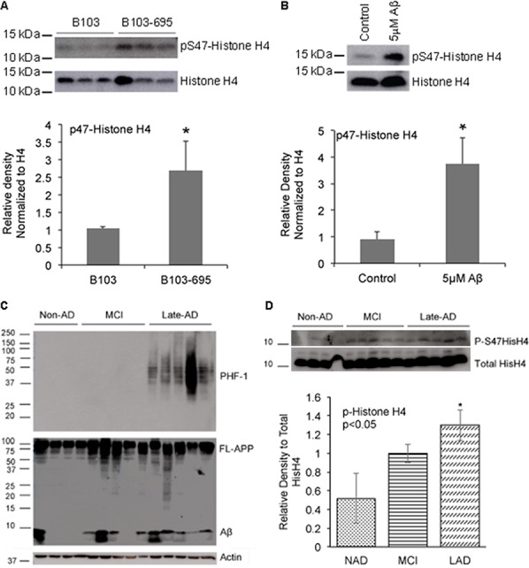
Figure 3. APP expressing cells, Aβ-treated neurons and AD samples show increased phosphorylation of Histone H4 at Serine 47.

A. B103-695 cells show increased phosphorylation of Ser47 Histone H4: Nuclear fractions isolated from B103 and B103-695 cells were analyzed using P-Histone H4 Ser47 (top panel) antibodies. Blot was reprobed with total Histone H4 antibody for normalization (bottom panel). The bar graph shows ratio of P-Histone H4 to total Histone H4. B. Neurons treated with Aβ show increased phosphorylation of Histone H4 at Ser 47: Primary cortical neurons were treated with 5μM Aβ for 24hr and samples were analyzed by western blot using Ser 47 P-Histone H4 antibody (top panel). Blots were reprobed with total Histone H4 antibody for normalization (bottom panel). The bar graph shows ratio of P-Histone H4 to total histone H4. C. Human MCI and AD samples show increased levels of P-Histone H4 compared to non-AD: Equal amounts of proteins from human brain samples (shown in Table 2) analyzed using PHF-1 (top panel) and 6E10 (middle panel) antibodies show increased levels of P-tau (top panel) and Aβ (middle panel) in LAD samples compared to MCI and non-AD. Actin antibody was used for normalization (bottom panel). D. P-Histone H4 Ser47 is significantly increased in LAD brain samples: Blots from the human samples were reprobed with P-Histone H4 (top panel) and total Histone H4 (bottom panel) antibodies, which showed a tendency towards increase in Ser47 P-Histone H4 in MCI and significant increase in LAD; bar graph shows ratio of P-Histone H4 to total Histone H4. *p value < 0.05.
