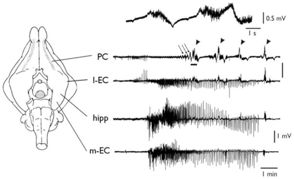
Figure 1.
Independent epileptiform patterns induced by arterial perfusion of 4AP in the olfactory and limbic systems. Extracellular recordings from piriform cortex (PC), lateral entorhinal cortex (l-EC), CA1 region of the hippocampus (hipp), and medial entorhinal cortex (m-EC), after arterial perfusion of 50 μM 4AP in the in vitro isolated guinea pig brain. Electrode position is illustrated in the scheme on the left. Runs of fast activity and seizure-like events are marked by the arrows and arrowheads, respectively. The upper trace illustrates an expansion of the PC recording underlined by the thick tract showing details of FRs.
Epilepsia © ILAE

