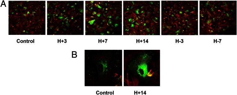
Figure 4.
A and B, Immunofluorescence ICC confirms hormone-induced changes in Cx43 protein expression. Immunofluorescence ICC in ESC monolayers showed Cx43 up- and down-regulation after the E/P/c-induced decidualization (H+3, H+7, H+14) and withdrawal (H-3, H-7, panel A). Green fluorescence represents Cx43 and red fluorescence is β-actin. Panel B shows a high-magnification view (×480) demonstrating intracellular distribution of Cx43 (green) under control conditions and after 14 days of hormone stimulation.

