Abstract
A commonly accepted paradigm of molecular biology is that transcription factors control gene expression by binding sites at the 5' end of a gene. However, there is growing evidence that transcription factor targets can occur within genes or between convergent genes. In this work, we have investigated one such target for the cyclic AMP receptor protein (CRP) of enterotoxigenic Escherichia coli. We show that CRP binds between two convergent genes. When bound, CRP regulates transcription of a small open reading frame, which we term aatS, embedded within one of the adjacent genes. Our work demonstrates that non-canonical sites of transcription factor binding can have hidden functionality.
Introduction
A long standing view is that promoters, and transcription factor binding sites, should locate to the 5' end of an annotated gene [1]. Unexpectedly, recent genome-wide studies of RNA polymerase distribution, and transcription factor binding, have shown that the situation is more complex [2]. Notably, transcription factor binding sites are frequently found within genes or between convergent genes. For example, RutR (a regulator of pyrimidine catabolism) binds predominantly at sites within genes [3]. Similarly, LeuO (a regulator of leucine biosynthesis) binds to numerous targets between convergent genes [4]. A major challenge is to understand if, and how, such transcription factor binding sites contribute to gene regulation.
In this work we have focused on the the cAMP receptor protein (CRP). Widely conserved in bacteria, CRP is global regulator of transcription that responds to cAMP levels [1]. When bound to DNA, CRP regulates transcription by one of two distinct mechanisms. For example, at class I promoters, CRP binds upstream of the promoter -35 element and interacts with the C-terminal domain of the RNA polymerase α-subunit [5–7]. Conversely, at class II promoters, CRP binds close to the promoter -35 element and interacts with the N-terminal domain of the RNA polymerase α-subunit [8, 9]. In both cases, CRP stimulates transcription [5–9]. Recently, we have shown that CRP binds numerous sites that are not close to the 5' end of an annotated gene [10, 11]. In this work we have focused on one such atypical target; a CRP site between two convergent genes. Our data show that CRP activates expression of a small open reading frame (ORF) completely embedded within one of these convergent genes. Activation occurs by a standard class II CRP-dependent mechanism. Thus, although the genomic context of the CRP binding site is unusual, gene regulation proceeds via a well-defined mechanism.
Methods and Materials
Strains, plasmids and oligonucleotides
Strains and plasmids used are listed in Table 1. Oligonucleotides used are listed in Table 2.
Table 1. Strains and plasmids used in this study.
| Strain or plasmid | Description | Source |
|---|---|---|
| Strains | ||
| Escherichia coli M182 | Δlac galK galU strA | [39] |
| Escherichia coli M182Δcrp | [39] | |
| Plasmids | ||
| pRW50 | Broad-host-range lac fusion vector for cloning promoters on EcoRI–HindIII fragments: contains the RK2 origin of replication and encodes TcR. | [40] |
| pSR | pBR322-derived plasmid containing an EcoRI–HindIII fragment upstream of the λoop transcription terminator | [17] |
| pRW225 | pRW50-derived plasmid in which the ribosome binding site upstream of lacZ has been deleted. | [23] |
| pDCRP | crp gene preceding its native promoter (located on EcoRI-SalI flanked fragment). pBR322 derived. Encodes AmpR. ColE1 origin. | [41] |
Table 2. Oligonucleotides used in this study.
| Oligonucleotide name | Sequence (5'-3') | Source |
|---|---|---|
| Oligonucleotides used to generate PaatS derivatives | ||
| aatS1 For | GGCTGCGAATTCATAAAGTGATAAAAATCACATAAAATTTTTATTAAAAGGATATAACCTTCATATCACTTGTAATTAAATTTGTGTCA | This study |
| aatS1 Rev | GCCCGAAGCTTCATGGATATACTTCTTAAGTATTATAAACAAGGTGGAACAGTTGTTATGGTAACCCATGACACAAATTTAATTACAA | This study |
| aatS2 Rev | GCCCGAAGCTTCATGGATATTGAAGAAAAGTATTATAAACAAGGTGGAACAGTTGTTATGGTAACCCATGACACAAATTTAATTACAA | This study |
| Oligonucleotides used for primer extension experiments | ||
| D49724 | GGTTGGACGCCCGGCATAGTTTTTCAGCAGGTCGTTG | [13] |
β-galactosidase assays
β-galactosidase assays were done according to the method of Miller [12]. Cells were grown in M9 minimal media supplemented with 1% fructose to stationary phase as specified in figure legends. Since PaatS was 2-fold more active in stationary phase cultures compared to log phase cultures the cells from the former phase of growth were used in all experiments. Values shown are the mean of three independent experiments. Error bars represent the standard deviation of three independent experiments. To calculate P we used a paired, two-tailed, student’s t test.
Primer extension
Primer extension was done as described by Lloyd et al. [13]. Briefly, RNA was extracted from M182 cells (or the Δcrp derivative) harbouring pRW50 containing the aatS1 promoter fragment. Extractions were done using a Qiagen RNeasy mini kit according to the manufacturer’s instructions and residual DNA was removed using a Turbo DNA-free kit (Ambion). RNA integrity was then checked by visualisation following agarose gel electrophoresis. The ratio of absorbance at 260 nm and 280 nm was used to assess the purity of the RNA. Primer extension products were analysed on a 6% denaturing polyacrylamide gel alongside size standards generated by manual sequencing of M13 phage DNA. Full gel images are provided in the supporting information (S1 Fig).
Proteins
CRP and σ70 proteins were purified as previously described [14, 15]. RNA polymerase core enzyme was purchased from Cambio, and was incubated at 37°C with a ten-fold molar excess of σ70 before use to generate RNA polymerase holoenzyme.
DNAse I footprinting
DNA templates were generated by excision of AatII-HindIII fragments from maxipreps of pSR plasmid containing the 134 bp aatS1 promoter fragment. The resulting 212 bp AatII-HindIII fragment was labelled at the HindIII end using γ-dATP and T4 polynucleotide kinase. Footprinting reactions were done as previously described [15] in buffer containing 120 mM KCl, 100 μM EDTA, 20 mM Tris pH 7, 10 mM MgCl2 and ~10 nM of PaatS DNA. Reactions also containing 12.5 μg ml-1 Herring Sperm DNA as a competitive inhibitor. Footprints were analysed on denaturing 6% polyacrylamide gels alongside calibrating Maxam-Gilbert G/A ladders [16]. Full gel images are provided in the supporting information (S1 Fig).
Multi-round in vitro transcription assays
In vitro transcription reactions were carried out as described by Savery et al., [14] using the method of Kolb et al. [17]. Supercoiled DNA was extracted from M182 cultures harbouring pSR containing the aatS1 fragment. Reaction buffer contained 20 mM Tris pH 7.9, 200 mM GTP/ATP/CTP, 10 mM UTP, 5 μCi (α32P) UTP, 500 mM DTT, 5 mM MgCl2, 100 μg ml-1 BSA and 0.2 mM cAMP. Template DNA (at a final concentration of 16 μg ml-1) was incubated with CRP at 37°C for 5 minutes in reaction buffer, prior to the addition of RNA polymerase holoenzyme to start the reaction. Full gel images are provided in the supporting information (S1 Fig).
Bioinformatics
NCBI ORF finder was used to search for putative genes [18]. NCBI BLAST was used to search for aatS homologues. A list of DUF1602-containing proteins was obtained using the InterPro tool [19], the list is correct as of 14/01/16.
Results
Identification of promoter-like DNA sequence elements between two convergent genes
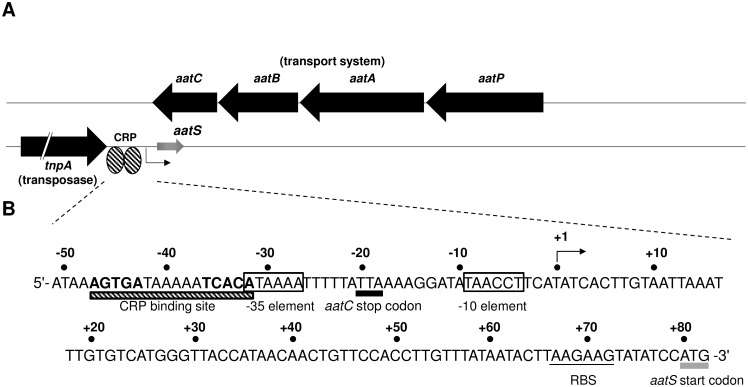
The starting point for this work was our previous analysis of global CRP binding in enterotoxigenic Escherichia coli (ETEC) [11]. This work predicted a CRP target between the convergent genes aatC (encoding a subunit of a transport system) and tnpA (encoding a transposase). The genomic context of the CRP site is shown in Fig 1A and the surrounding DNA sequence is shown in Fig 1B. We searched the locus for promoters that may be under the control of CRP. A sequence (5'-TAACCT-3') that resembled a promoter -10 hexamer was found close to the CRP site within the aatC coding region (Fig 1B). A 189 bp ORF, which we named aatS, and a ribosome binding site (RBS) were also identified (Fig 1B). Note that aatS fully overlaps aatC but is in the opposite orientation. We considered that the overlap might result from aatC being annotated incorrectly. However, on close examination, this seems unlikely; there is no alternative stop codon within the aatC open reading frame. Furthermore, the full aatC sequence is conserved in numerous bacteria.
Fig 1. The aatPABC operon of ETEC H10407.
Schematic of the aatPABC operon and adjacent tnpA gene. The two DNA strands are shown as black lines. Known genes are shown as black arrows and the predicted aatS gene as a grey arrow. Gene names are shown in italic and gene function in parenthesis. The position of a putative CRP binding site is indicated by striped ovals. A. Sequence if the tnpA-aatC intergenic region. The CRP site is highlighted as a striped rectangle with the two half sites highlighted bold. The start codon of the aatS open reading frame is highlighted with a grey rectangle. The transcription start site, as determined by mRNA primer extension is denoted “+1” and indicated by a bent arrow. Distances upstream (-) and downstream (+) of this start site are numbered. The -35 and -10 hexamers are boxed, and the ribosome binding site (RBS) is underlined.
PaatS is a CRP-dependent promoter in vivo
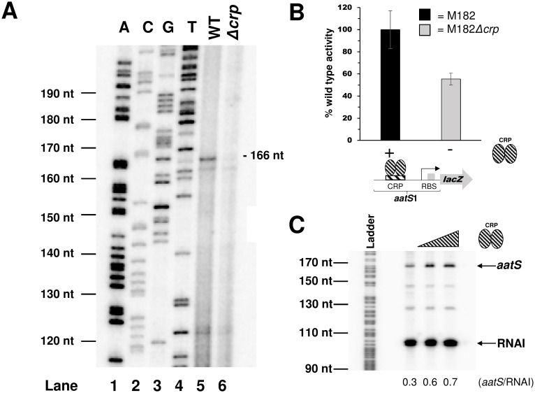
Our next aim was to determine if the putative promoter upstream of aatS (PaatS) was functional. Thus, a 134 bp DNA fragment containing the sequence shown in Fig 1B, flanked by EcoRI and HindIII restriction sites, was generated. The DNA fragment, named aatS1, was cloned upstream of lacZ in plasmid pRW50 to create a PaatS::lacZ fusion. Next, E. coli M182 and the Δcrp derivative were transformed with the resulting plasmid. Transformants were cultured in liquid media and RNA was isolated as a template for primer extension. A 166 nucleotide primer extension product was observed using RNA from M182. Conversely, little extension product was observed using RNA from the Δcrp derivative (compare lanes 5 and 6 in Fig 2A). Thus, the aatS transcription start site (marked “+1” in Fig 1B) is located 4 bp downstream of the putative aatS promoter -10 hexamer. Furthermore, PaatS is poorly active in the absence of CRP. Consistent with our primer extension analysis, PaatS controlled expression of lacZ was also significantly reduced in cells lacking CRP (P = 0.018; Fig 2B).
Fig 2. Characterisation of the PaatS promoter.
A. Primer extension analysis of the aatS transcript. Lanes 1–4 on the gel are arbitrary size standards, used for calibration, generated by sequencing of M13mp18 phage DNA. Lane 5 shows the primer extension product generated using RNA from wildtype M182 cells carrying the aatS1::lacZ fusion. Lane 6 shows the primer extension product generated using RNA from M182Δcrp cells carrying the aatS1::lacZ fusion. The transcription start site is indicated in Fig 1B. B. β-galactosidase activity determined using lysates of M182 wildtype or M182Δcrp cells carrying PaatS cloned upstream of lacZ in plasmid pRW50. Values shown are percentages of activity observed in strain M182 (92 Miller units). We obtained 7 and 3 Miller units of activity from lysates of M182 or M182Δcrp carrying promoterless pRW50. Error bars represent the standard deviation of three independent experiments. C. Multi-round in vitro transcription assay using PaatS. The aatS1 DNA fragment was cloned into pSR upstream of a λoop terminator. Purified, supercoiled pSR plasmid was incubated with purified CRP at 37°C, and the reaction started by the addition of 400 nM σ70- RNA polymerase holoenzyme. CRP concentrations are; 0 nM, 200 nM, or 400 nM. The 108 nt RNAI transcript from the pSR replication origin, and the 169 nt transcript from PaatS, are indicated. The gel is calibrated with an arbitrary G+A DNA sequencing reaction as a size standard.
PaatS is a CRP dependent promoter in vitro
We next confirmed activation of PaatS by CRP using in vitro transcription assays. The aatS1 DNA fragment was cloned upstream of the λoop transcription terminator in plasmid pSR. Thus, the transcript generated from PaatS is expected to be 169 nucleotides (nt) in length. Note that, pSR also encodes RNAI; a 108 nt factor independent transcript derived from the plasmid replication origin. Thus, we observed two bands on denaturing PAGE gels (Fig 2C). As expected, synthesis of RNAI did not require CRP. However, production of the larger 169 nt aatS transcript was stimulated by CRP.
CRP binds a site overlapping the PaatS -35 element
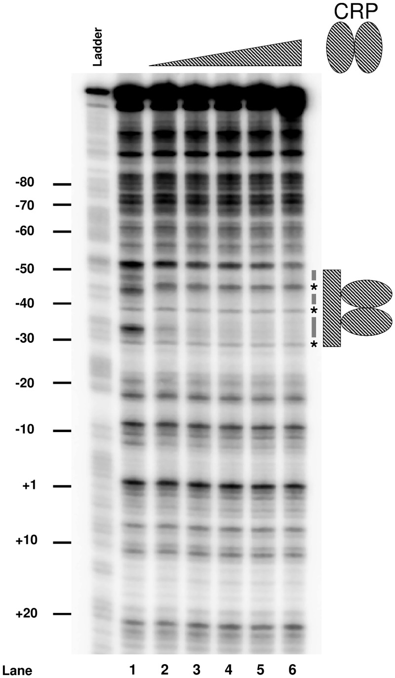
To confirm CRP binding to the predicted target upstream of PaatS we used DNAse I footprinting. The result of the experiment is shown in Fig 3. Lane 1 shows the banding pattern resulting from DNAse I cleavage of aatS1 in the absence of CRP. Alterations to this pattern are evident in lanes 2–6 as increasing concentrations of CRP are added. Thus, CRP induced the appearance of three hypersensitive bands (starred) and protected the flanking DNA regions (underlined). The footprint aligns precisely with the predicted DNA target for CRP. The CRP site is 39.5 bases upstream of the PaatS transcription start site (Fig 1B). However, we note that the aatS mRNA start is 4 bp, rather than the usual 7 bp, downstream of the promoter -10 element. Hence, CRP is well positioned to interact with RNA polymerase via a standard class II mechanism.
Fig 3. Binding of CRP to the PaatS region.
DNAse I footprint analysis of the PaatS region. The lane labelled ‘G+A’ is a Maxim/Gilbert G+A sequencing reaction. Lane 1 shows the cleavage pattern obtained from aatS1 DNA digested with DNAse I in the absence of CRP. Lanes 2–6 show DNAse I cleavage patterns generated in the presence of increasing concentrations of CRP (0.35 μM, 0.7 μM, 1.05 μM, 1.4 μM and 2.1 μM). The predicted CRP site is indicated by a hashed grey bar. DNA protection is indicated by black dashes, hypersensitive bends are highlighted by stars.
The aatS coding region is preceded by a functional ribosome binding site
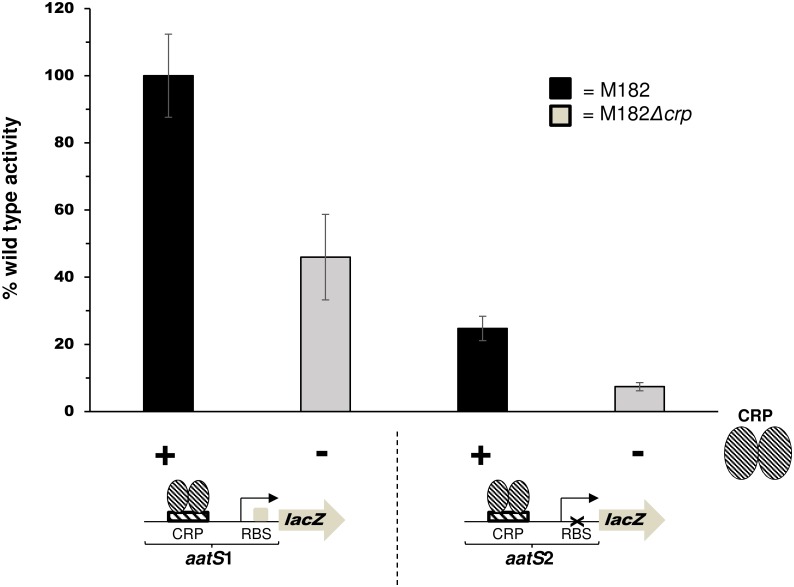
The predicted aatS ORF is located 82 bp downstream of the aatS transcription start site, within the coding region of aatC. We noticed that the 5' end of the aatS mRNA contains a sequence, 5'-UUAAGAAGU-3', that resembles a RBS (5'-UAAGGAGGU-3') [20, 21]. In addition, the predicted aatS RBS is located 6 bp upstream of the aatS start codon, a position suitable for translation initiation [22]. To determine if the RBS was functional, we created a translational aatS1::lacZ fusion and explored the effects of mutating the RBS on lacZ expression. Thus, we generated a derivative of the aatS1 fragment, called aatS2, where the sequence of the RBS was altered to 5'-UUUUCUUCA-3' (Fig 4). Both aatS1 and aatS2 were translationally fused to lacZ by cloning into pRW225 [23]. Resulting plasmids were used to transform M182 and the Δcrp derivative. LacZ activities were then determined in lysates of transformants grown in liquid culture (Fig 4). LacZ expression was significantly reduced when the aatS RBS was mutated (P = 0.011). Residual LacZ expression, driven by the aatS2::lacZ fusion in M182Δcrp cells, was not significantly different from background LacZ activity (P = 0.096). Background LacZ activity was obtained using M182Δcrp transformed with pRW225 having no promoter insert.
Fig 4. The aatS mRNA contains a functional ribosome binding site.
The graph shows activity of different aatS:lacZ translational fusions. The wildtype ribosome binding site (5'-AAGAAG-3') in the aatS1 fragment was mutated to (5'-TTCTTC-3') in aatS2. LacZ activites was determined using the lysates of stationary phase M182 or M182Δcrp. In M182 cells crp was supplied in trans by plasmid pCRP that encodes crp under the control of its own promoter. Values shown are percentages of activity observed in strain M182 (5 Miller units). We obtained 0.25 and 0.26 Miller units of activity from lysates of M182 or M182Δcrp, carrying promoterless pRW225, respectively. Error bars represent the standard deviation of three independent experiments.
AatS contains a conserved domain of unknown function
We cannot exclude the possibility that PaatS drives transcription of a small or antisense RNA. However, the presence of a functional RBS, appropriately positioned upstream of the aatS open reading frame, is consistent with the transcript being an mRNA. If this is the case, translation would result in the production of a 62 amino acid AatS peptide. To better understand AatS we used BLAST to identify 38 microbial proteins with a significantly similar sequence (E-value <10) in the NCBI blastp database. The search also reveals that AatS shares 67% identity with a conserved domain of unknown function (DUF1602, E-value: 4.07e-7). Genes encoding DUF1602 are found in diverse species in all kingdoms of life (Table 3). Strikingly, as in the case of aatS, genes encoding DUF1602 are often genetically associated with, and occasionally overlap, genes encoding transport systems. We conclude that aatS may encode a small protein that could be an ancillary subunit of the Aat transport system in ETEC.
Table 3. Phylogentic distribution of DUF1602-containing proteins.
| Kingdom/Species | Number of DUF1602-containing proteins |
|---|---|
| Archaea (all Euryarchaeota) | 5 |
| Bacteria:Actinobacteria:Chlamydiae:Cyanobacteria:Firmicutes:Proteobacteria: | 146202218100 |
| EukaryotesFungi:Metazoa:Chlorophyta:Streptophyta: | 59141143 |
| Unclassified sequences | 4 |
Discussion
In this study we have characterised the function of a predicted CRP binding site located between two convergent genes. We show that CRP activates transcription of aatS; a small ORF embedded within the much larger aatC gene. The aatPABC operon encodes a type I secretion system (the Aat system) found in many pathogenic bacteria [24, 25]. The presence of a functional RBS, correctly positioned upstream of aatS, suggests that the gene encodes a small protein rather than a regulatory RNA. Interestingly, many small proteins in bacteria localise to the membrane and function as accessory factors in transport systems [26]. Consistent with such a function, aatS is genetically associated with genes encoding transport systems in many bacteria. Furthermore, a potential transmembrane helix is predicted between residues 4 and 21 of AatS [27].
Documented instances of overlapping, protein-encoding, genes in bacteria are rare [28]. We are aware of only two examples; rpmH is encoded within rnpA in Thermus thermophilus and setAB resides inside the pic gene of E. coli 042 and Shigella flexneri [29, 30]. However, we speculate that further overlapping transcription units may become evident as unusual transcription factor targets are examined in detail. For instance, in a study of 154 Mycobacterium tuberculosis transcription factors, 75% of binding targets were located inside genes [31]. Similarly, a study of 116 transcription factors in E. coli, identified many intragenic binding events [32]. Presumably, some of these targets will control production of unannotated transcripts. Thus, in the case we have examined, the position of the CRP site is only surprising on first inspection. Detailed investigation of the aat locus reveals that CRP acts via a well-established mechanism and it is the position of aatS, embedded within aatC, which confounds the situation. In summary, whilst some bacterial transcription factors bind primarily in expected locations [33–35] many have unusually situated targets [3,10,11,36,37]. Surprising binding sites are often ignored [36] or dismissed as artefacts [38]. This work demonstrates that careful genetic and biochemical analysis can identify regulatory function for such targets.
Supporting Information
Data Availability
All relevant data are within the paper and its Supporting Information files.
Funding Statement
This work was supported by the Wellcome Trust Award Number WT085092MA to DCG and BBSRC Award Number BB/N005961/1 to DCG. The funders had no role in study design, data collection and analysis, decision to publish, or preparation of the manuscript.
References
- 1.Browning DF, Busby SJW. The regulation of bacterial transcription initiation. Nat Rev Microbiol. 2004; 2: 57–64. [DOI] [PubMed] [Google Scholar]
- 2.Wade JT, Grainger DC. Pervasive transcription: illuminating the dark matter of bacterial transcriptomes. Nat Rev Microbiol. 2014; 12: 647–653. 10.1038/nrmicro3316 [DOI] [PubMed] [Google Scholar]
- 3.Shimada T, Ishihama A, Busby SJW, Grainger DC. The Escherichia coli RutR transcription factor binds at targets within genes as well as intergenic regions. Nucleic Acids Res. 2008; 36: 3950–3955. 10.1093/nar/gkn339 [DOI] [PMC free article] [PubMed] [Google Scholar]
- 4.Shimada T, Bridier A, Briandet R, Ishihama A. New roles of LeuO in transcription regulation of E. coli genome: antagonistic interplay with the universal silencer H-NS. Mol Microbiol. 2011; 82: 378–397. 10.1111/j.1365-2958.2011.07818.x [DOI] [PubMed] [Google Scholar]
- 5.Blatter EE, Ross W, Tang H, Gourse RL, Ebright RH. Domain organization of RNA polymerase α subunit: C-terminal 85 amino acids constitute a domain capable of dimerization and DNA binding. Cell. 1994; 78: 889–896. [DOI] [PubMed] [Google Scholar]
- 6.Gaston K, Bell A, Kolb A, Buc H, Busby S. Stringent spacing requirements for transcription activation by CRP. Cell. 1990; 62: 733–743. [DOI] [PubMed] [Google Scholar]
- 7.Savery NJ, Lloyd GS, Busby SJW, Thomas MS, Ebright RH, Gourse RL. Determinants of the C-terminal domain of the Escherichia coli RNA polymerase α subunit important for transcription at Class I cyclic AMP receptor protein-dependent interactions. J Bacteriol. 2002; 184: 2273–2280. [DOI] [PMC free article] [PubMed] [Google Scholar]
- 8.Niu W, Kim Y, Tau G, Heyduk T, Ebright RH. Transcription activation at class II CAP-dependent promoters: two interactions between CAP and RNA polymerase. Cell. 1996; 87: 1123–1134. [DOI] [PMC free article] [PubMed] [Google Scholar]
- 9.Rhodius VA, West DM, Webster CL, Busby SJW, Savery NJ. Transcription activation at class II CRP-dependent promoters: the role of different activating regions. Nucleic Acids Res. 1997; 25: 326–332. [DOI] [PMC free article] [PubMed] [Google Scholar]
- 10.Grainger DC, Hurd D, Harrison M, Holdstock J, Busby SJW. Studies of the distribution of Escherichia coli cAMP-receptor protein and RNA polymerase along the E. coli chromosome. Proc Natl Acad Sci. 2005; 102: 17693–17698. [DOI] [PMC free article] [PubMed] [Google Scholar]
- 11.Haycocks JRJ, Sharma P, Stringer AM, Wade JT, Grainger DC. The molecular basis for control of ETEC enterotoxin expression is response to environment and host. PLoS Pathog. 2015; 11(1): e1004605 10.1371/journal.ppat.1004605 [DOI] [PMC free article] [PubMed] [Google Scholar]
- 12.Miller JH. Experiments in molecular genetics. Cold Spring Harbor, NY, Cold Spring Harbor Laboratory Press; 1972. [Google Scholar]
- 13.Lloyd GS, Hollands K, Godfrey RE, Busby SJW. Transcription initiation in the Escherichia coli K-12 malI-malX intergenic region and the role of the cyclic AMP receptor. FEMS Microbiol Lett. 2008; 288: 250–257. 10.1111/j.1574-6968.2008.01365.x [DOI] [PubMed] [Google Scholar]
- 14.Savery NJ, Lloyd GS, Kainz M, Gaal T, Ross W, Ebright RH, et al. Transcription activation at class II CRP-dependent promoters: identification of determinants in the C-terminal domain of the RNA polymerase α subunit. EMBO J. 1998; 17: 3439–3447. [DOI] [PMC free article] [PubMed] [Google Scholar]
- 15.Grainger DC, Goldberg MD, Lee DJ, Busby SJW. Selective repression by Fis and H-NS at the Escherichia coli dps promoter. Mol Microbiol. 2008; 68: 1366–1377. 10.1111/j.1365-2958.2008.06253.x [DOI] [PubMed] [Google Scholar]
- 16.Maxam AM, Gilbert W. A new method for sequencing DNA. Proc Natl Acad Sci U S A. 1977; 74: 560–564. [DOI] [PMC free article] [PubMed] [Google Scholar]
- 17.Kolb A, Kotlarz D, Kusano S, Ishihama A. Selectivity of the Escherichia coli RNA polymerase Eσ38 for overlapping promoters and ability to support CRP activation. Nucleic Acids Res. 1995; 23: 819–826. [DOI] [PMC free article] [PubMed] [Google Scholar]
- 18.Wheeler DL, Church DM, Federhen S, Lash AE, Madden TL, Pontius JU, et al. Database resources of the National Center for Biotechnology. Nucleic Acids Res. 2003; 31: 28–33. [DOI] [PMC free article] [PubMed] [Google Scholar]
- 19.Mitchell A, Chang H, Daugherty L, Fraser M, Hunter S, Lopez R, et al. The InterPro protein families database: the classification resource after 15 years. Nucleic Acids Res. 2015; 43: D213–D221. 10.1093/nar/gku1243 [DOI] [PMC free article] [PubMed] [Google Scholar]
- 20.Shine J, Dalgarno L. The 3’-Terminal sequence of Escherichia coli 16S ribosomal RNA: Complementarity to nonsense triplets and ribosome binding sites. Proc Natl Acad Sci. 1974; 71: 1342–1346. [DOI] [PMC free article] [PubMed] [Google Scholar]
- 21.Shultzaberger RK, Bucheimer RE, Rudd KE, Schneider TD. Anatomy of Escherichia coli ribosome binding sites. J Mol Biol. 2001; 313: 215–228. [DOI] [PubMed] [Google Scholar]
- 22.Chen H, Bjerknes M, Kumar R, Jay E. Determination of the optimal aligned spacing between the Shine-Dalgarno sequence and the translation initiation codon of Escherichia coli mRNAs. Nucleic Acids Res. 1994; 22: 4953–4957. [DOI] [PMC free article] [PubMed] [Google Scholar]
- 23.Islam MD, Shaw RK, Frankel G, Pallen MJ, Busby SJW. Translation of a minigene in the 5’ leader sequence of the enterohaemorrhagic Escherichia coli LEE1 transcription unit affects expression of the neighbouring downstream gene. Biochem J. 2012; 441: 247–253. 10.1042/BJ20110912 [DOI] [PMC free article] [PubMed] [Google Scholar]
- 24.Crossman LL, Chaudhuri RR, Beatson SA, Wells TJ, Desvaux M, Cunningham AF, et al. A Commensal Gone Bad: Complete Genome Sequence of the Prototypical Enterotoxigenic Escherichia coli Strain H10407. J Bacteriol. 2010; 192: 5822–5831. 10.1128/JB.00710-10 [DOI] [PMC free article] [PubMed] [Google Scholar]
- 25.Nishi J, Sheikh J, Mizuguchi K, Luisi B, Burland V, Boutin A, et al. The export of coat protein from enteroaggregative Escherichia coli by a specific ATP-binding cassette transporter system. J Biol Chem. 2003; 278: 45680–45689. [DOI] [PubMed] [Google Scholar]
- 26.Storz G, Wolf YI, Ramamurthi KS. Small proteins can no longer be ignored. Annu Rev Biochem. 2014; 83: 753–77. 10.1146/annurev-biochem-070611-102400 [DOI] [PMC free article] [PubMed] [Google Scholar]
- 27.Tusnády GE, Simon I. The HMMTOP transmembrane topology prediction server. Bioinformatics. 2001. 17; 849–850. [DOI] [PubMed] [Google Scholar]
- 28.Ellis JC, Brown JW. Genes within genes in bacteria. Trends Biochem Sci. 2003; 28: 521–523. [DOI] [PubMed] [Google Scholar]
- 29.Feltens R, Gößringer M, Willkomm DK, Urlaub H, Hartmann RK. An unusual mechanism of bacterial gene expression revealed for the RNase P protein of Thermus strains. Proc Natl Acad Sci. 2003; 100: 5724–5729. [DOI] [PMC free article] [PubMed] [Google Scholar]
- 30.Behrens M, Sheikh J, Nataro JP. Regulation of the overlapping pic/set locus of Shigella flexneri and Enteroaggregative Escherichia coli. Infect Immun. 2002; 70: 2915–2925. [DOI] [PMC free article] [PubMed] [Google Scholar]
- 31.Minch KJ, Rustad TR, Peterson EJR, Winkler J, Reis DJ, Ma S, et al. The DNA-binding network of Mycobacterium tuberculosis. Nat Commun. 2014; 6: 5829. [DOI] [PMC free article] [PubMed] [Google Scholar]
- 32.Ishihama A, Shimada T, Yamazaki Y. Transcription profile of Escherichia coli: genomic SELEX search for regulatory targets of transcription factors. Nucleic Acids Res. 2016; 44: 2058–74. 10.1093/nar/gkw051 [DOI] [PMC free article] [PubMed] [Google Scholar]
- 33.Grainger DC, Aiba H, Hurd D, Browning DF, Busby SJW. Transcription factor distribution in Escherichia coli: studies with FNR protein. Nucleic Acids Res. 2007; 35: 269–278. [DOI] [PMC free article] [PubMed] [Google Scholar]
- 34.Grainger DC, Overton TW, Reppas N, Wade JT, Tamai E, Hobman JL, Constantinidou C, Struhl K, Church G, Busby SJW. Genomic studies with Escherichia coli MelR protein: applications of chromatin immunoprecipitation and microarrays J. Bacteriol. 2004; 186: 6938–6943. [DOI] [PMC free article] [PubMed] [Google Scholar]
- 35.Yamamoto K, Ishihama A, Busby SJW, Grainger DC. The Escherichia coli K-12 MntR miniregulon includes dps, which encodes the major stationary-phase DNA-binding protein. J. Bacteriol. 193; 1477–1480. 10.1128/JB.01230-10 [DOI] [PMC free article] [PubMed] [Google Scholar]
- 36.Partridge JD, Bodenmiller DM, Humphrys MS, Spiro S. NsrR targets in the Escherichia coli genome: new insights into DNA sequence requirements for binding and a role for NsrR in the regulation of motility. Mol Microbiol. 2009; 73: 680–694. 10.1111/j.1365-2958.2009.06799.x [DOI] [PubMed] [Google Scholar]
- 37.Efromovich S, Grainger D, Bodenmiller D, Spiro S. Genome-wide identification of binding sites for the nitric oxide-sensitive transcriptional regulator NsrR. Methods in Enzymology. 2008; 437; 211–233. 10.1016/S0076-6879(07)37012-2 [DOI] [PubMed] [Google Scholar]
- 38.Waldminghaus T, Skarstad K. ChIP on Chip: surprising results are often artifacts. BMC genomics. 2010; 11: 414 10.1186/1471-2164-11-414 [DOI] [PMC free article] [PubMed] [Google Scholar]
- 39.Busby S, Kotlarz D, Buc H. Deletion mutagenesis of the Escherichia coli galactose operon promoter region. J Mol Biol. 1983; 167: 259–274. [DOI] [PubMed] [Google Scholar]
- 40.Lodge J, Fear J, Busby S, Gunasekaran P, Kamini NR. Broad host range plasmids carrying the Escherichia coli lactose and galactose operons. FEMS Microbiol Lett. 1992; 95: 271–276. [DOI] [PubMed] [Google Scholar]
- 41.Bell A, Gaston K, Williams R, Chapman K, Kolb A, Buc H, et al. Mutations that alter the ability of the Escherichia coli cyclic AMP receptor protein to activate transcription. Nucleic Acids Res. 1990; 18: 7243–7250. [DOI] [PMC free article] [PubMed] [Google Scholar]
Associated Data
This section collects any data citations, data availability statements, or supplementary materials included in this article.
Supplementary Materials
Data Availability Statement
All relevant data are within the paper and its Supporting Information files.


