Abstract
The type III histone deacetylase silent information regulator 1 (SIRT1) is an enzyme that is critical for the modulation of immune and inflammatory responses. However, the data on its role in rheumatoid arthritis (RA) are limited and controversial. To better understand how SIRT1 regulates adaptive immune responses in RA, we evaluated collagen-induced arthritis (CIA) in myeloid cell-specific SIRT1 knockout (mSIRT1 KO) and wild-type (WT) mice. Arthritis severity was gauged on the basis of clinical, radiographic and pathologic scores. Compared with their WT counterparts, the mSIRT1 KO mice exhibited less severe arthritis, which was less destructive to the joints. The expression levels of inflammatory cytokines, matrix metalloproteinases and ROR-γT were also reduced in the mSIRT1 KO mice compared with the WT mice and were paralleled by reductions in the numbers of Th1 and Th17 cells and CD80- or CD86-positive dendritic cells (DCs). In addition, impaired DC maturation and decreases in the Th1/Th17 immune response were observed in the mSIRT1 KO mice. T-cell proliferation was also investigated in co-cultures with antigen-pulsed DCs. In the co-cultures, the DCs from the mSIRT1 KO mice showed decreases in T-cell proliferation and the Th1/Th17 immune response. In this study, myeloid cell-specific deletion of SIRT1 appeared to suppress CIA by modulating DC maturation. Thus, a careful investigation of DC-specific SIRT1 downregulation is needed to gauge the therapeutic utility of agents targeting SIRT1 in RA.
Introduction
Rheumatoid arthritis (RA) is a chronic inflammatory disease marked by progressive disability, systemic complications and a high socioeconomic toll.1 The seven mammaliansirtuin members, SIRT1 to SIRT7, are class III histone deacetylases that regulate senescence, stress resistance, metabolism and inflammation.2 Silent information regulator 1 (SIRT1), in particular, is known to deacetylate the p65 subunit of nuclear factor-κB (NF-κB), thus interrupting this pathway and exerting an anti-inflammatory effect.3, 4 The NF-κB pathway is a central signaling node for the stimulation of inflammatory cytokines and production of matrix metalloproteinase (MMP) in RA.5, 6 This affiliation prompted us to investigate the impact of SIRT1 on a passive K/BxN serum transfer model of arthritis using myeloid cell-specific SIRT1 knockout (mSIRT1 KO) mice.7 These mice exhibit enhanced macrophage activation and profound inflammatory arthritis through the hyperacetylation and subsequent hyperactivation of the NF-κB pathway.
A variety of cell types, including macrophages, mast cells, dendritic cells (DCs), T cells, B cells and fibroblast-like synoviocytes (FLSs), are intricately involved in RA.8 The Janus-like behavior of SIRT1 in tumorigenesis, in which its suppressor or promoter activity is dictated by the cancer cell type,9 may also apply to autoimmune diseases (including RA). We and others have demonstrated that SIRT1 acts as a negative regulator of macrophage activation via suppressing the NF-κB pathway.4, 7 Of note, Zhang et al.10 and Gao et al.have shown that SIRT1 is a critical suppressor of T-cell immunity and is required for T-cell immune tolerance; therefore, SIRT1 deletion in macrophages or T cells may indeed trigger flare-ups of RA. Conversely, Niederer et al.12 have reported that SIRT1 prolongs chronic inflammation by increasing the levels of pro-inflammatory cytokines and inhibiting apoptosis in rheumatoid fibroblast-like synoviocytes. Furthermore, Yang et al.13 have recently shown that DC-specific SIRT1 deletion protects mice from experimental autoimmune encephalomyelitis by suppressing Th17 differentiation. In terms of DCs and fibroblast-like synoviocytes, the loss of SIRT1 functions may ultimately suppress RA activity.
In a passive K/BxN serum transfer model of arthritis, the disease is mediated by pathogenic autoantibodies against glucose-6-phosphate isomerase, a ubiquitously expressed glycolytic enzyme that is deposited on joint surfaces.14 Although this model has many clinical and histologic similarities to human RA, it does not reflect adaptive immunity (i.e., DCs and T cells). As an alternate model, collagen-induced arthritis (CIA) requires intact DC and T-cell dynamics, complete with sequential antigen presentation and T-cell activation.15 Hence, we studied CIA in mSIRT1 KO mice as a model of RA that incorporates adaptive immunity to assess the impact of SIRT1 deletion on the successive polarization of DCs and T cells. Surprisingly, and in contrast to symptoms of passive K/BxN serum transfer arthritis, comparatively less inflammation and joint destruction were evident in the mSIRT1 KO mice with CIA. These findings were attributed to impaired DC maturation and the related decrease in the Th1/Th17 immune response, ultimately suggesting that the effects of SIRT1 in RA may differ as a result of the relative contributions of the innate and adaptive immune effector cells.
Materials and methods
Mice and CIA induction
SIRT1loxP/loxPLysM-Cre+ (mSIRT1 KO) and SIRT1loxP/loxPLysM-Cre− (WT) mice were generated, and the selective deletion of SIRT1 in myeloid cells was confirmed as previously described.7 For CIA induction, male mSIRT1 KO and littermate WT mice (14–18 weeks old) were immunized with 150 μg of chicken type II collagen (CII; Chondrex, Redmond, WA, USA) emulsified in 4 mg ml−l of complete Freund's adjuvant (CFA; Chondrex) in equal volumes.15 The point of initial immunization was designated Day 0. Immunization was boosted by an equal volume of emulsion of CII and incomplete Freund's adjuvant (IFA; Chondrex) on Day 21 and by an intraperitoneal injection of lipopolysaccharide (LPS, 5 μg 100 ml−1) on Day 28. The clinical arthritis scores (0–4 scale) were registered for each limb, with a total possible score of 16.16 A caliper was placed across the ankle joint at the widest point to measure ankle thickness. All animal experiments adhered to a protocol approved by the Chonbuk National University Institutional Animal Care and Use Committee (CBU 2014-00053).
Radiographic and pathologic analyses
Plain radiographs of the hindpaws were obtained using a mammographic imager with a direct detection, flat panel-array design (Mammomat NovationDR; Siemens Healthcare, Erlangen, Germany) at constant exposure (30 kVp and 90 mA). All images were semiquantitatively scored according to a previously described system.5 The joint tissues were fixed in 10% formalin, decalcified for 3–4 weeks in 10% ethylenediaminetetraacetic acid (EDTA), dehydrated and embedded in paraffin. The sections (6 μm) were stained with hematoxylin and eosin or Safranin-O for light microscopy. The joint areas were scored (0–5 scale) for the intensity of synovial inflammation and the severity of the cartilage damage.17 The macrophages and neutrophils were immunohistochemically detected with rat monoclonal antibodies to F4/80 (Abcam, Cambridge, MA, USA) and LY6G (LifeSpan Biosciences, Seattle, WA, USA), respectively. The numbers of macrophages and neutrophils in four randomly selected high-power fields (× 400) were counted in each ankle joint.
Patients and DC isolation
Blood samples were collected from five healthy individuals and from five patients with RA who had taken no medications, such as glucocorticoids, disease-modifying antirheumatic drugs or biologics. The peripheral blood mononuclear cells were isolated through standard Ficoll density gradient centrifugation (GE Healthcare Life Science, Marlborough, MA, USA). The CD11c+ DCs were isolated from the peripheral blood mononuclear cells by magnetic-activated cell sorting using a CD11c+ DC isolation kit (Miltenyi Biotec, Gmbh, Bergisch Gladbach, Germany).
Real-time reverse transcriptase–PCR and enzyme-linked immunosorbent assay
Ankle RNA was isolated using an RNeasy Lipid Tissue Mini Kit (Qiagen, Hilden, Germany). The RNA from the lymph nodes was isolated using TRIzol reagent (Life Technologies, Carlsbad, CA, USA), precipitated with isopropanol and dissolved in purified diethylpyrocarbonate (DEPC)-treated water. The total RNA (2 μg) was then treated with RNase-free DNase, and the first-strand cDNAs were generated using the random hexamer primers from a First-Strand cDNA Synthesis Kit (Applied Biosystems, Foster City, CA, USA). The primer designs for each specific gene relied on Primer Express Software (Table 1; Applied Biosystems). The real-time reverse transcriptase–PCR mixtures consisted of 10 ng of the reverse transcribed total RNA, 167 nmol l−1 of the forward and reverse primers, and a PCR master mixture in a final volume of 10 μl. The reactions were performed in 384-well plates using an ABI Prism 7900HT Sequence Detection System (Applied Biosystems). The levels of interferon-γ (IFN-γ), interleukin (IL)-17, IL-6, IL-12p70, IL-10, IL-1β and anti-CII antibodies in the sera and cell culture supernatants were assayed with the appropriate enzyme-linked immunosorbent assay kits (IFN-γ, IL-17 and IL-1β from eBioscience, San Diego, CA, USA; anti-CII antibodies from Biolegend, San Diego, CA, USA; all others from Life Technologies).
Table 1. Sequences and accession numbers for the primers (forward, FOR; reverse, REV) used in real-time RT–PCR.
| Gene | Primer sequences | Accession no. |
|---|---|---|
| SIRT1 | FOR:GACACAGAGACGGCTGGAA REV: CAGACCCTCAAGCCATGTTT | NM_ 019812 |
| TNF-α | FOR: GCTGTCCCTGCGCTTCA REV:CTCGTCCCCAATGACATCCT | NM_ 013693 |
| IL-1β | FOR:CCATGGCACATTCTGTTCAAA REV:GCCCATGAGAGGCAAGGA | NM_ 008361 |
| IL-2 | FOR:AACTCCCCAGGATGCTCAC REV:CGCAGAGGTCCAAGTTCATC | NM_008366 |
| IL-4 | FOR:TCGGCATTTTGAACGAGGTC REV:GAAAAGCCCGAAAGAGTCTC | NM_021283 |
| IL-6 | FOR: CCACGGCCTTCCCTACTTC REV: TTGGGAGTGGTATCCTCTGTGA | NM_ 031168 |
| IL-13 | FOR: CGCAAGGCCCCCACTAC REV:AAAGTGGGCTACTTCGATTTTGG | NM_008355 |
| IL-17 | FOR: TGAGCTTCCCAGATCACAGA REV: TCCAGAAGGCCCTCAGACTA | NM_ 010552 |
| IL-23 | FOR: GCTCCCCTTTGAAGATGTCA REV: GACCCACAAGGACTCAAGGA | NM_ 031252 |
| IL-10 | FOR: TGTCAAATTCATTCATGGCCT REV: ATCGATTTCTCCCCTGTGAA | NM _010548 |
| IFN-γ | FOR: TGAGCTCATTGAATGCTTGG REV: ACAGCAAGGCGAAAAAGGAT | NM_ 008337 |
| MCP-1 | FOR: TGTCACCCTTGGAGCTCATG REV: TTTTTCGACTTTTATCCTCTGTTG | NM_ 011333 |
| MMP-3 | FOR: CCCCTGATGTCCTCGTGGTA REV: GCACATTGGTGATGTCTCAGGTT | NM_ 010809 |
| MMP-13 | FOR: GGTCCTTGGAGTGATCCAGA REV: TGATGAAACCTGGACAAGCA | NM_ 008607 |
| T-bet | FOR: TTCCCATTCCTGTCCTTCAC REV: CCACATCCACAAACATCCTG | NM_ 019507 |
| ROR-γT | FOR: TGAGGCCATTCAGTATGTGG REV: CTTCCATTGCTCCTGCTTTC | NM_011281 |
| GATA3 | FOR: GGAAACTCCGTCAGGGCTA REV: AGAGATCCGTGCAGCAGAG | NM_008091 |
| Foxp3 | FOR: CCTGCCTTGGTACATTCGTG REV: TGTTGTGGGTGAGTGCTTTG | NM_054039 |
| GAPDH | FOR: CGTCCCGTAGACAAAATGGT REV: TTGATGGCAACAATCTCCAC | NM_008084 |
Abbreviations: GAPDH, glyceraldehyde-3-phosphate dehydrogenase; MCP-1, monocyte chemoattractant protein-1.
Flow cytometry
For cytokine detection, single-cell lymph node suspensions were cultured (4 h) in triplicate at 1 × 10⁶ cells/well in a 96-well plate and then stimulated with 50 ng ml−1 phorbol 12-myristate 13-acetate and 1 μM ionomycin in the presence of brefeldin A (3 μg ml−1). Thereafter, the cells were stained with the PerCP-Cy5.5-conjugated anti-CD4 antibody (eBioscience) and washed twice in FACS buffer (97% phosphate-buffered saline and 3% fetal bovine serum). The cells were then fixed in 4% paraformaldehyde solution, washed twice in permeabilization buffer and stained with fluorochrome-conjugated antibodies directed against IL-17 and IFN-γ (eBioscience). For cell surface marker detection, the cells were incubated on ice (30 min), stained with fluorochrome-conjugated antibodies directed against CD11c+CD11b+CD86+ or CD11c+CD11b+CD80+ (eBioscience), and again incubated on ice (30 min), followed by a wash in FACS buffer. The stained cells were evaluated by flow cytometry (Accuri; BD Biosciences, San Jose, CA, USA).
T-cell proliferation assay
Single-cell suspensions were prepared from the lymph nodes of the mSIRT1 KO or WT mice. The lymphocytes were plated in 96-well plates, allowed to adhere in the presence or absence of CII (10 or 50 μg ml−1) and stained with carboxyfluorescein diacetate succinimidyl ester (CFSE; eBioscience; 1 μM). The CFSE-labeled cells (1 × 10⁶ cells perwell) were cultured in uncoated or anti-CD3 antibody-coated 96-well plates, with or without an anti-CD28 antibody (eBioscience). The plates were incubated (72 h, 37 °C) in a 5% CO2 incubator, and cellular proliferation was evaluated by flow cytometry.
Differentiation, culture and assessment of the bone marrow-derived dendritic cells
The bone marrow-derived dendritic cells (BMDCs) were isolated and cultured as previously described.18 Briefly, the tibial and femoral bone marrow cells collected from six-week-old female C57BL/6 mice were incubated (37 °C, 5% CO2) in 12-well cell culture plates in RPMI 1640 containing 10% inactivated fetal bovine serum, 1% penicillin/streptomycin, granulocyte-macrophage colony-stimulating factor (20 ng ml−1) and IL-4 (10 ng ml−1). To assess their antigen-uptake capacity, the BMDCs (2 × 10⁵ cells) were incubated (37 or 4 °C, 45 min) with 1 mg ml−1 of fluorescein isothiocyanate (FITC)-conjugated dextran. The harvested cells were then washed in phosphate-buffered saline and stained with a PE-conjugated anti-CD11c antibody. To detect the surface molecules, the LPS (50 ng ml−1)-treated BMDCs were harvested, washed in cold phosphate-buffered saline, treated with a PE-conjugated mouse monoclonal antibody directed against CD80, CD86, MHC class I or MHC class II and a FITC-conjugated mouse anti-CD11c antibody (30 min, 4 °C), fixed in 4% paraformaldehyde and analyzed by flow cytometry.
Co-culture of T cells and CII-pulsed DCs
The WT C57BL/6 mice were immunized using an emulsion of chicken CII (150 μg) and CFA (4 mg ml−1) in equal volumes. Eight days later, the mice were intraperitoneally injected with equal volumes of the CII and incomplete Freund's adjuvant emulsion. After 3 days, the CD4+ T cells were isolated using a Dynabeads FlowComp Mouse CD4 Kit (Life Technologies) and labeled using CFSE (1 μM). The BMDCs from the WT and mSIRT1KO mice were seeded in 96-well plates (1 × 10⁵ per well) and incubated with LPS (50 ng ml−1) alone or with LPS plus CII (20 μg ml−1) overnight. Following the incubation, the BMDCs were thoroughly washed and co-cultured with the CFSE-labeled CD4+ T cells (3 days; 1:10 ratio). The co-cultured cells were then collected, washed in FACS buffer and stained with the PerCP-Cy5.5-conjugated anti-CD4-antibody. The percentages of CD4+ T cells were determined by flow cytometry.
Western blot analysis
The cells were lysed in RIPA buffer (50 mM Tris, pH 7.2, 150 mM NaCl, 1% NP-40 and 0.1% sodium dodecyl sulfate) supplemented with a protease inhibitor cocktail. The cells were then sonicated and centrifuged (12 000 g, 10 min, 4 °C) to remove the insoluble debris. The protein concentration was determined using the BCA assay (Thermo Scientific, Pittsburgh, PA, USA). The total proteins (30 μg) were resolved on a sodium dodecyl sulfate polyacrylamide gel, which was then electroblotted onto a nitrocellulose membrane. After blocking with a 5% skim milk solution, the membranes were incubated with antibodies directed against SIRT1 (Cell Signaling Technology, Danvers, MA, USA), phospho-c-jun (Cell Signaling Technology), β-actin (Sigma-Aldrich, St Louis, MO, USA), p-65 (Santa Cruz Biotechnology, Dallas, TX, USA) or lamin B (Santa Cruz Biotechnology), and the proteins were detected with the ECL Detection System (GE Healthcare Life Science).
Statistical analysis
The data were expressed as the means±s.e. Statistical comparisons were performed using two-tailed Student's t-tests. P-values less than 0.05 were considered statistically significant.
Results
Myeloid deletion of SIRT1 suppresses synovial inflammation and bone destruction
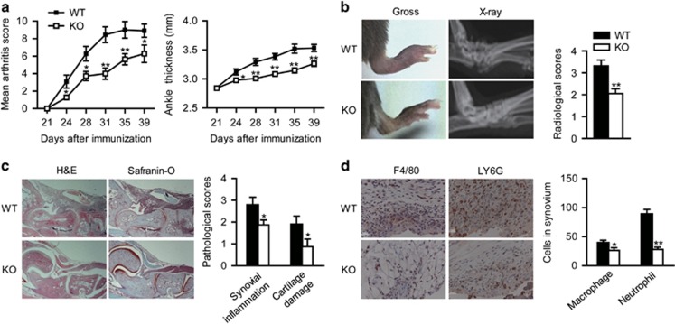
As a model of chronic adaptive immunity, the CIA mice were studied to determine the impact of myeloid cell-specific SIRT1 deletion. Interestingly, the disease severity, including the arthritis score and ankle thickness, was significantly reduced in the mSIRT1 KO mice (Figure 1a). Likewise, the bone destruction was less severe, and the radiographic scores were lower in the mSIRT1 KO mice as compared with the severe bone erosion and joint damage observed in the WT mice (Figure 1b). In the pathologic analysis, the synovial inflammation and cartilage damage were less pronounced in the mSIRT1 KO mice (Figure 1c). The influx of macrophages and neutrophils in the arthritic joints of the mSIRT1 KO mice was significantly reduced as well (Figure 1d). These results suggest that the myeloid cell-specific SIRT1 deficiency attenuates joint inflammation and bone destruction in CIA.
Figure 1.
Assessment of the clinical deterioration, radiographic bone destruction and pathologic abnormalities in collagen-induced arthritis (CIA). CIA was induced in male mSIRT1 KO (KO) and wild-type (WT) littermates at 14–18 weeks old. (a) The mean clinical scores and ankle thickness were determined on the indicated days after primary immunization. (b) The gross hindpaw appearance and plain ankle radiographs of representative mice at Day 39 are shown (left panel). Semiquantification of bone destruction with the radiographic scoring system (right panel). (c) Ankle joints in representative hematoxylin and eosin - and safranin-O-stained sections (× 100) (left panel) are shown, along with the mean pathologic scores for synovial inflammation and cartilage damage (right panel). The results of three independent experiments were pooled (total of 10 mice per group). (d) Arthritic joints in representative sections stained for F4/80 or LY6G (left panel). The mean macrophage and neutrophil counts (right panel) from four randomly selected high-power fields (× 400) in each ankle joint (n=5 mice for each group) are shown. The values are expressed as the means±s.e., *P<0.05, **P<0.01 vs WT.
Myeloid deletion of SIRT1 suppresses proinflammatory gene expression in the CIA ankle and lymph nodes
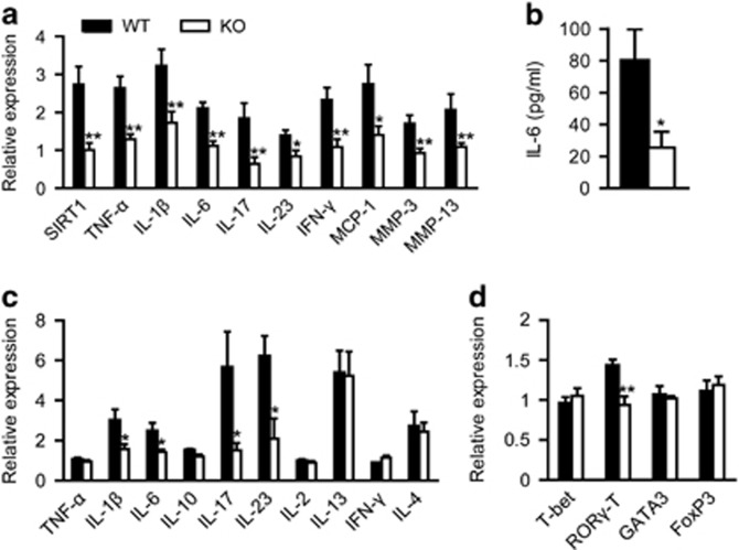
To gauge the inflammatory response and T-cell function, the mRNA expression levels were measured in the ankles and lymph nodes of mice with CIA. The expression levels of TNF-α, IL-1β, IL-6, monocyte chemoattractant protein-1, MMP-3 and MMP-13 were significantly decreased in the ankles of mSIRT1 KO mice, as were the levels of IFN-γ, IL-17 and IL-23 (key cytokines in Th1 or Th17 differentiation) (Figure 2a). In addition, the IL-6 expression in the serum was reduced in the mSIRT1 KO mice (Figure 2b). The cytokines and transcription factors that influence T-cell differentiation were also assayed in the lymph nodes of mice with CIA. Again, the expression levels of IL-1β, IL-6, IL-17 and IL-23 were decreased in the lymph nodes of the mSIRT1 KO mice (Figure 2c). These findings were consistent with a decreased level of ROR-γT, a transcription factor that is important for the differentiation of Th17 cells (Figure 2d). The levels of the transcription factors that govern the differentiation of other T-cell subsets were unaffected. Hence, the subdued form of arthritis induced in the mSIRT1 KO mice is associated with reduced levels of proinflammatory cytokines and a diminished Th17 cell response.
Figure 2.
Levels of cytokines and mediators related to inflammatory responses and T-cell function in the CIA mouse model. The levels of cytokines and inflammatory mediators in the ankles, sera and lymph nodes of the mice were measured at Day 39 by real-time reverse transcriptase–PCR and enzyme-linked immunosorbent assay. (a) mRNA levels of SIRT1, TNF-α, IL-1β, IL-6, IL-17, IL-23, IFN-γ, MCP-1, MMP-3 and MMP-13 in the ankle joints of the mSIRT1 KO (KO) and wild-type (WT) mice. (b) Comparison of the IL-6 concentrations in the sera from the mSIRT1 KO and WT mice. (c, d) mRNA levels of the cytokines and transcription factors related to T-cell differentiation in the mouse lymph nodes. The values are expressed as the means±s.e. (10 mice per group), *P<0.05, **P<0.01 vs WT.
Myeloid deletion of SIRT1 impairs Th1 and Th17 cell differentiation and DC maturation in CIA
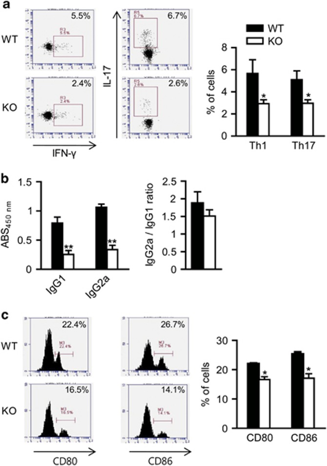
We observed DC maturation and T-cell differentiation in the early phase of CIA, as reported by other groups.19, 20 We harvested the lymph nodes from the arthritic mSIRT1 KO and WT mice on Day 28 to analyze the T-cell and DC populations by flow cytometry. The serum levels of the anti-CII IgG1 and IgG2a antibodies were also measured. The percentages of CD4+ Th1 and CD4+ Th17 cells were significantly reduced in the mSIRT1 KO mice compared with their WT counterparts (Figure 3a). The autoantibody levels and the IgG2a-to-IgG1 ratio were also reduced in the mSIRT1 KO mice (Figure 3b). Because the myeloid-specific deletion of SIRT1 in the mSIRT1 KO mice is selective for DCs, macrophages and neutrophils, but not T cells, we analyzed the mature DCs, which are critical for driving Th1/Th17 response. During the active phase of CIA, the percentages of mature DCs expressing CD80+ or CD86+ were significantly reduced in the lymph nodes of the mSIRT1 KO mice (Figure 3c). Thus, the downregulated Th1 and Th17 responses and reduced number of mature DCs are closely linked in the mSIRT1 KO mice.
Figure 3.
Phenotypes of the T cells and DCs and levels of anti-type II collagen (CII) antibodies in the CIA mouse model. The regional lymph nodes were harvested on Day 28, and the sera were collected on Day 39 from the mSIRT1 KO (KO) and wild-type (WT) mice with CIA. The T cell and DC phenotypes were determined by flow cytometry, and the levels of anti-CII antibodies were measured by enzyme-linked immunosorbent assay. (a) Dot plots (left panel) and proportions (right panel) of Th1 and Th17 cells. (b) The serum levels of IgG2a and IgG1 and IgG2a-to-IgG1 ratio were determined. (c) Histogram (left panel) and proportions (right panel) of CD80- and CD86-positive DCs. The values are expressed as the means±s.e. (4–8 mice per group), *P<0.05, **P<0.01 vs WT.
CII-specific T-cell proliferation and cytokine production are reduced in the lymph nodes of the mSIRT1 KO mice
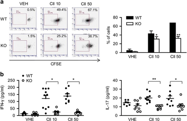
The cells were harvested from the mouse lymph nodes on Day 28 of CIA and re-stimulated with CII to characterize antigen-specific T-cell proliferation and cytokine production. The proliferative response to CII stimulation was significantly weaker in the lymph node cells from the mSIRT1 KO mice (Figure 4a). Moreover, the IFN-γ and IL-17 production by the antigen-stimulated cells from the lymph nodes of the mSIRT1 KO mice compared with the WT mice was significantly decreased (Figure 4b). These results suggest that myeloid cell-derived SIRT1 is critical for modulating T-cell proliferation and Th1/Th17 cytokine production during CIA.
Figure 4.
Effects of SIRT1 deletion on type II collagen (CII)-specific T-cell proliferation and cytokine production. The cells from the regional lymph nodes of the mSIRT1 KO (KO) and wild-type (WT) mice were isolated on Day 28 and incubated with vehicle (VEH) or CII (10 or 50 μg ml−l) for 72 h. (a) The proliferative response was assessed by the fluorescence of 5(6)-carboxyfluorescein diacetate succinimidyl ester (CFSE). The dot plots (left panel) represent one of four experiments, and the graph (right panel) shows proportion of CFSE-positive cells (4 mice per group). (b) The IFN-γ and IL-17 levels in the culture supernatants were determined by enzyme-linked immunosorbent assay (7–10 mice per group). The values are expressed as the means±s.e., *P<0.05, **P<0.01 vs WT.
LPS-induced maturation of DCs is diminished in the mSIRT1 KO mice
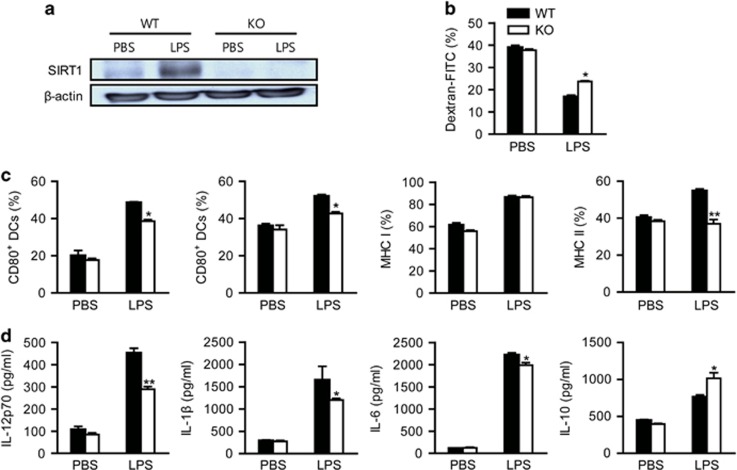
In a previous study, we have demonstrated that SIRT1 is selectively deleted in myeloid cells, such as macrophages and osteoclasts, but not in DCs.7 Before investigating the impact of SIRT1 on DC maturation, we confirmed the complete deletion of SIRT1 in the DCs of the mSIRT1 KO mice (Figure 5a). Generally, immature DCs have higher antigen endocytic capacity than mature DCs, thus prompting us to examine the influence of SIRT1 on the endocytic capacity of DCs via dextran-FITC uptake. The LPS-stimulated SIRT1 KO DCs had a higher endocytic capacity than that of the WT DCs (Figure 5b).
Figure 5.
Effect of SIRT1 deletion on LPS-induced DC maturation. The bone marrow-derived DCs from the mSIRT1 KO (KO) and wild-type (WT) mice were treated with phosphate-buffered saline or LPS (50 ng ml−l) for 24 h. (a) Western blot analysis of SIRT1 expression in the DCs before and after LPS stimulation. (b) The endocytic capacity of the DCs was determined by FITC-conjugated dextran uptake. The bar graph shows the amount of FITC-dextran (%) in the CD11c+ cells. (c) The expression of the surface molecules was determined by flow cytometry and staining with anti-CD80, anti-CD86, anti-MHC class I or anti-MHC class II antibodies in the CD11c+-gated cells. (d) Enzyme-linked immunosorbent assay was performed to determine the levels of IL-12p70, IL-1β, IL-6 and IL-10 in the LPS-treated DCs from the KO and WT mice. The values are expressed as the means±s.e. of three experiments. *P<0.05, **P<0.01 vs WT.
We also measured the levels of surface molecules displayed by the LPS-stimulated DCs from the mSIRT1 KO or WT mice. As shown in Figure 5c, the stimulation of the WT DCs with LPS resulted in the upregulation of surface molecular markers of DC maturity, such as CD80, CD86 and the MHC class II molecule. However, these markers were expressed at significantly lower levels in the LPS-treated SIRT1 KO DCs. The LPS-induced expression of the MHC class I molecule was not inhibited in the SIRT1 KO DCs.
Finally, the production of pro- and anti-inflammatory cytokines by the LPS-stimulated DCs was assessed. The levels of IL-12p70, IL-1β and IL-6 were significantly reduced in the LPS-stimulated SIRT1 KO DCs compared with the WT DCs (Figure 5d). However, the secretion of IL-10, which inhibits Th1 immune functions, was significantly increased in the LPS-treated SIRT1 KO DCs compared with the WT DCs. Consequently, it appears that SIRT1 is essential for DC maturation and function.
The proliferation and proinflammatory differentiation of T cells are reduced when the cells are co-cultured with SIRT1 KO DCs
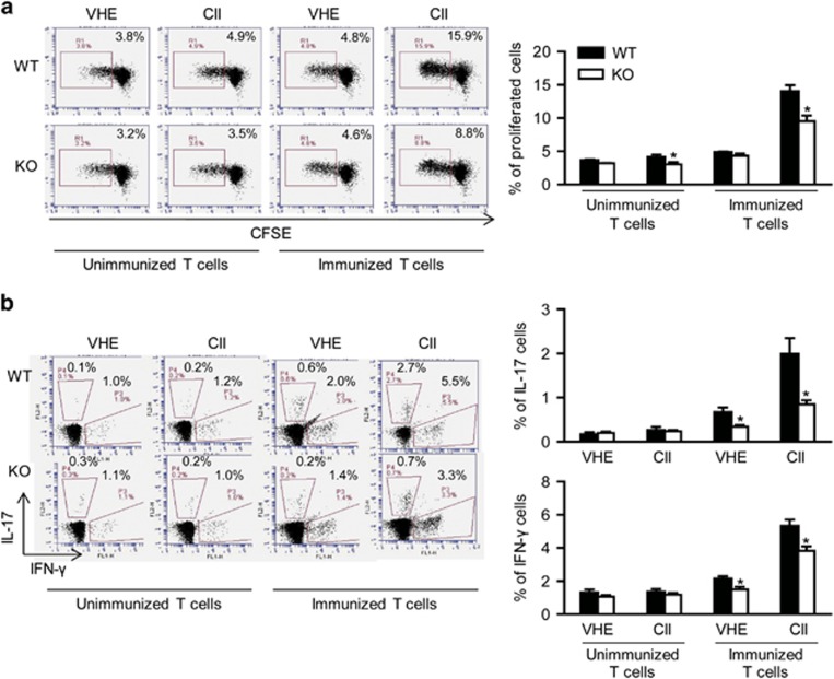
To clearly discern whether the observed deficit in T-cell proliferation was due to the deletion of SIRT1 in DCs, we generated CII-pulsed and mature DCs from the mSIRT1 KO and WT mice for co-culture with the CD4+ T cells isolated from the CII-immunized mice. The proliferation rate of the CD4+ T cells (Figure 6a), as well as the percentages of Th1 and Th17 cells (Figure 6b), was significantly reduced when the cells were co-cultured with the DCs from the mSIRT1 KO mice, confirming that SIRT1 deficiency attenuates DC maturation, which in turn limits T-cell proliferation and Th1/Th17 differentiation.
Figure 6.
Impacts of SIRT1-deficient DCs on T-cell proliferation and Th1/Th17 differentiation in co-culture. The CII-pulsed and LPS-stimulated DCs were generated from the immature bone marrow-derived DCs isolated from the mSIRT1 KO (KO) and wild-type (WT) mice. Activated and matured DCs were co-cultured with the T cells from the non-immunized or immunized CIA mice. (a) The T cells' proliferative response was assessed by the fluorescence of 5(6)-carboxyfluorescein diacetate succinimidyl ester (CFSE) after stimulation with vehicle (VEH) or CII (20 μg ml−1) for 72 h. The dot plots (left panel) represent one of three experiments, and show the proportion (right panel) of CFSE-positive cells. (b) The T cells' phenotype was determined by flow cytometry. Dot plots (left panel) and proportion (right panel) of Th1 or Th17 cells. The values are expressed as the means±s.e. of three experiments. *P<0.05, vs WT.
Increased NF-κB and decreased AP-1 activity in the SIRT1 KO DCs
Because transcription factors from both the NF-κB and activator protein-1 (AP-1) pathways are involved in DC maturation, we assessed the p65 and phospho-c-jun levels in the DCs. The nuclear translocation of p65 was enhanced in the SIRT1-deleted DCs compared with the WT DCs (Supplementary Figure S1). In addition, c-jun phosphorylation was reduced in the SIRT1-deleted DCs. These data suggest that reduced AP-1 activation contributes to the impaired maturation of the SIRT1-deleted DCs.
The expression levels of SIRT1 are increased in the DCs from RA patients
To further explore the link between SIRT1 expression and DC activation in the setting of RA, we compared the SIRT1 levels in the DCs from both treatment-naive patients with RA and healthy individuals. The levels of SIRT1 mRNA were higher in the CD11c+ DCs from patients with RA (P<0.01; Supplementary Figure S2).
Discussion
In this study, the impact of myeloid deletion of SIRT1 on the development of RA was assessed in a CIA mouse model. Clinical arthritis and bone destruction were clearly ameliorated in the mSIRT1 KO mice, as demonstrated by the reduced numbers of Th1 and Th17 cells and downregulation of both inflammatory cytokines and ROR-γT. Consistently, CII-specific T-cell proliferation and cytokine production were inhibited in the cells extracted from the lymph nodes of the mSIRT1 KO mice. The maturation and cytokine production (IL-1, IL-6 and IL-12p70) in the SIRT1 KO DCs were also diminished, and T-cell proliferation and Th1/Th17 differentiation were restricted in co-cultures with the SIRT1 KO DCs. These findings indicate that myeloid deletion of SIRT1 inhibits immune-mediated inflammation in RA, presumably by impairing DC maturation and producing subsequent deficits in Th1 and Th17 cells.
Our previous investigations have underscored the findings that the NF-κB pathway actively contributes to the pathogenesis of RA and is considered a potential therapeutic target.5, 16 SIRT1 is known to suppress NF-κB-dependent transcription through the deacetylation of the p65 subunits that complex with the NF-κB target-gene promoters.3, 4 Consistently with these findings, we have demonstrated that myeloid cell-specific deletion of SIRT1 intensifies passive K/BxN serum transfer arthritis due to macrophage hyperactivation via the NF-κB pathway.7 However, another study has shown that high expression of SIRT1 directly enhances proinflammatory cytokine production and prolongs RA-fibroblast-like synoviocyte lifespan;12 recently, SIRT1-mediated suppression of anti-inflammatory IL-27 expression in DCs has been documented, which leads to increased Th17 differentiation and the development of inflammatory disease.13 Conversely, Wendling et al.21 have reported a correlation between SIRT1 activity and the expression levels of TNF and IL-6, despite the similar levels of SIRT1 activity in the peripheral blood mononuclear cells from patients with RA and healthy controls.21 Consequently, the role of SIRT1 in the context of RA is open to debate. Here, we further assessed the roles of SIRT1 in a CIA model of RA by focusing on adaptive immunity. Surprisingly, myeloid-deficiency of SIRT1 resulted in significantly lower scores for arthritis and joint destruction in the CIA mice, accompanied by reduced expression of proinflammatory cytokines and MMPs.
Given these contradictory results, we explored the gene expression profiles of the mSIRT1 KO mice. We discovered that specific cytokines (IL-1β, IL-6, IL-17 and IL-23) were diminished in the joint and lymphoid tissues and that ROR-γT, a transcription factor that is critical for Th17 cellular differentiation,22 was deficient in the lymph nodes from the mSIRT1 KO mice. Additionally, the percentages of Th1 and Th17 cells in the regional lymph nodes and the autoantibody levels and IgG2a-to-IgG1 ratio in the sera of mSIRT1 KO were lower than those in the WT mice. The CII-reactive T-cell responses in vitro were also similar, with deficient T-cell proliferation and reduced levels of Th1/Th17 cytokines. Overall, these outcomes suggest that SIRT1 is pivotal for the antigen-specific proinflammatory T-cell responses.
The behavior of DCs is an important focus of this study. Unlike the passive K/BxN serum transfer model of arthritis, intact DCs are essential for driving the T-cell responses in the CIA model.23, 24 DCs are antigen-presenting cells that are highly equipped to activate naive T cells and instigate effective T-cell immunity. They are also important for the induction and maintenance of peripheral T-cell tolerance.25 This dual function of DCs is determined in part by their maturational stage.26 In this investigation, higher levels of SIRT1 were registered in the DCs from patients with RA, whereas fewer mature (CD80- or CD86-positive) DCs populated the lymph nodes of the mSIRT1 KO mice with CIA. Additional detailed in vitro experiments showed a similar tendency: the SIRT1 KO DCs displayed immature phenotypes that were marked by reduced expression of the MHC class II molecule, co-stimulatory molecules and pro-inflammatory cytokines and an increased antigen endocytic capacity. Our results agreed with those of a previous report showing that DC-specific SIRT1 deletion confers resistance to experimental autoimmune encephalomyelitis.13 Together, these results imply that the inhibition of SIRT1 expression in DCs blocks their phenotypic maturation, thereby protecting the mice from developing RA.
With respect to the role of SIRT1 in T cells, our findings differ from those of a previous study showing that SIRT1 deletion in T cells results in increased T-cell activation and a breakdown of CD4+ T-cell tolerance.10 We used LysM-Cre mice to specifically produce Cre-mediated deletion of the loxP-flanked SIRT1 gene in myeloid cells (such as myeloid DCs, macrophages and neutrophils) but not in non-myeloid cells (including T cells). Because the immature SIRT1 KO DCs appear to be impaired in their ability to affect T-cell proliferation and Th1/Th17 differentiation, we isolated T cells from mice with CIA and co-cultivated them with preactivated DCs. In this instance, the SIRT1 KO DCs were still less efficient than the WT DCs in inducing T-cell proliferation. This direct evidence emphasizes the essential role of SIRT1 in sequential DC maturation and the antigen-specific T-cell response.
SIRT1 is a known negative regulator of NF-κB in macrophages, T cells and lung cancer cell lines.4, 27, 28 Although the transcriptional activity of AP-1 is attenuated by SIRT1 in macrophages and T cells,10, 29 SIRT1 enhances AP-1 activity in mouse cutaneous epithelial cells.30 Our findings indicate that NF-κB activity is increased and AP-1 activity is decreased in the SIRT1-deleted DCs. Thus, SIRT1 may suppress DC maturation by inhibiting the AP-1 pathway. The loss of SIRT1 functions in DCs, which disables the acetylation of interferon regulatory factor-1 and generates anti-inflammatory IL-27 and interferon β, offers an alternate mechanism for explaining the observed inhibition of CIA by the SIRT-1 deleted DCs.13
In summary, the myeloid deletion of SIRT1 impaired DC maturation and reduced Th1 and Th17 differentiation, which in turn reduced the synovial inflammation and bone destruction in a CIA model of RA. Recently, tolerogenic DCs have emerged as a promising means of reinstating immune tolerance, and two related trials have already been completed in RA.31 As such, therapeutics based on DC-selective downregulation of SIRT1 may be pivotal in treating RA.
Acknowledgments
This work was supported by grants (no. 2008-0062279, NRF-2014R1A2A1A11051360, NRF-2015R1C1A1A01054628 and NRF-2015R1A5A2008833) from the National Research Foundation of Korea (NRF) funded by the Korean government (MSIP).
The authors declare no conflict of interest.
Footnotes
Supplementary Information accompanies the paper on Experimental & Molecular Medicine website (http://www.nature.com/emm)
Supplementary Material
References
- Firestein GS. Evolving concepts of rheumatoid arthritis. Nature 2003; 423: 356–361. [DOI] [PubMed] [Google Scholar]
- Michan S, Sinclair D. Sirtuins in mammals: insights into their biological function. Biochem J 2007; 404: 1–13. [DOI] [PMC free article] [PubMed] [Google Scholar]
- Lee JH, Song MY, Song EK, Kim EK, Moon WS, Han MK et al. Overexpression of SIRT1 protects pancreatic β-cells against cytokine toxicity by suppressing the nuclear factor-κB signaling pathway. Diabetes 2009; 58: 344–351. [DOI] [PMC free article] [PubMed] [Google Scholar]
- Schug TT, Xu Q, Gao H, Peres-da-Silva A, Draper DW, Fessler MB et al. Myeloid deletion of sirt1 induces inflammatory signaling in response to environmental stress. Mol Cell Biol 2010; 30: 4712–4721. [DOI] [PMC free article] [PubMed] [Google Scholar]
- Hah YS, Lee YR, Jun JS, Lim HS, Kim HO, Jeong YG et al. A20 suppresses inflammatory responses and bone destruction in human fibroblast-like synoviocytes and in mice with collagen-induced arthritis. Arthritis Rheumatol 2010; 62: 2313–2321. [DOI] [PubMed] [Google Scholar]
- Tak PP, Firestein GS. NF-κB: a key role in inflammatory diseases. J Clin Invest 2001; 107: 7–11. [DOI] [PMC free article] [PubMed] [Google Scholar]
- Hah YS, Cheon YH, Lim HS, Cho HY, Park BH, Ka SO et al. Myeloid deletion of SIRT1 aggravates serum transfer arthritis in mice via nuclear factor-κB activation. PLoS ONE 2014; 9: e87733. [DOI] [PMC free article] [PubMed] [Google Scholar]
- McInnes IB, Schett G. The pathogenesis of rheumatoid arthritis. N Engl J Med 2011; 365: 2205–2219. [DOI] [PubMed] [Google Scholar]
- Song NY, Surh YJ. Janus-faced role of SIRT1 in tumorigenesis. Ann NY Acad Sci 2012; 1271: 10–19. [DOI] [PMC free article] [PubMed] [Google Scholar]
- Zhang J, Lee SM, Shannon S, Gao B, Chen W, Chen A et al. The type III histone deacetylase Sirt1 is essential for maintenance of T cell tolerance in mice. J Clin Invest 2009; 119: 3048–3058. [DOI] [PMC free article] [PubMed] [Google Scholar]
- Gao B, Kong Q, Kemp K, Zhao YS, Fang D. Analysis of sirtuin 1 expression reveals a molecular explanation of IL-2-mediated reversal of T-cell tolerance. Proc Natl Acad Sci USA 2012; 109: 899–904. [DOI] [PMC free article] [PubMed] [Google Scholar]
- Niederer F, Ospelt C, Brentano F, Hottiger MO, Gay RE, Gay S et al. SIRT1 overexpression in the rheumatoid arthritis synovium contributes to proinflammatory cytokine production and apoptosis resistance. Ann Rheum Dis 2011; 70: 1866–1873. [DOI] [PubMed] [Google Scholar]
- Yang H, Lee SM, Gao B, Zhang J, Fang D. The histone deacetylase Sirtuin 1 deacetylates IRF1 and programs dendritic cells to control Th17 differentiation during autoimmune inflammation. J Biol Chem 2013; 288: 37256–37266. [DOI] [PMC free article] [PubMed] [Google Scholar]
- Monach P, Hattori K, Huang H, Hyatt E, Morse J, Nguyen L et al. The K/BxN mouse model of inflammatory arthritis: theory and practice. Methods Mol Med 2007; 136: 269–282. [DOI] [PubMed] [Google Scholar]
- Inglis JJ, Simelyte E, McCann FE, Criado G, Williams RO. Protocol for the induction of arthritis in C57BL/6 mice. Nat Protoc 2008; 3: 612–618. [DOI] [PubMed] [Google Scholar]
- Lee HS, Woo SJ, Koh HW, Ka SO, Zhou L, Jang KY et al. Regulation of apoptosis and inflammatory responses by insulin-like growth factor binding protein 3 in fibroblast-like synoviocytes and experimental animal models of rheumatoid arthritis. Arthritis Rheumatol 2014; 66: 863–873. [DOI] [PubMed] [Google Scholar]
- Joosten LA, Helsen MM, Saxne T. van de Loo FA, Heinegard D, van den Berg WB. IL-1αβ blockade prevents cartilage and bone destruction in murine type II collagen-induced arthritis, whereas TNF-α blockade only ameliorates joint inflammation. J Immunol 1999; 163: 5049–5055. [PubMed] [Google Scholar]
- Jung ID, Lee MG, Chang JH, Lee JS, Jeong YI, Lee CM et al. Blockade of indoleamine 2,3-dioxygenase protects mice against lipopolysaccharide-induced endotoxin shock. J Immunol 2009; 182: 3146–3154. [DOI] [PubMed] [Google Scholar]
- Nakahama T, Kimura A, Nguyen NT, Chinen I, Hanieh H, Nohara K et al. Aryl hydrocarbon receptor deficiency in T cells suppresses the development of collagen-induced arthritis. Proc Natl Acad Sci USA 2011; 108: 14222–14227. [DOI] [PMC free article] [PubMed] [Google Scholar]
- Brand DD, Kang AH, Rosloniec EF. Immunopathogenesis of collagen arthritis. Springer Semin Immunopathol 2003; 25: 3–18. [DOI] [PubMed] [Google Scholar]
- Wendling D, Vidon C, Abbas W, Guillot X, Toussirot E, Herbein G. Sirt1 activity in peripheral blood mononuclear cells from patients with rheumatoid arthritis. Joint Bone Spine 2014; 81: 462–463. [DOI] [PubMed] [Google Scholar]
- Miossec P, Korn T, Kuchroo VK. Interleukin-17 and type 17 helper T cells. N Engl J Med 2009; 361: 888–898. [DOI] [PubMed] [Google Scholar]
- Khan S, Greenberg JD, Bhardwaj N. Dendritic cells as targets for therapy in rheumatoid arthritis. Nat Rev Rheumatol 2009; 5: 566–571. [DOI] [PMC free article] [PubMed] [Google Scholar]
- Moret FM, Hack CE, van der Wurff-Jacobs KM, Radstake TR, Lafeber FP, van Roon JA. Thymic stromal lymphopoietin (TSLP): a novel proinflammatory mediator in rheumatoid arthritis that potently activates CD1c+ myeloid dendritic cells to attract and stimulate T cells. Arthritis Rheumatol 2014; 66: 1176–1184. [DOI] [PubMed] [Google Scholar]
- Steinman RM, Nussenzweig MC. Avoiding horror autotoxicus: the importance of dendritic cells in peripheral T cell tolerance. Proc Natl Acad Sci USA 2002; 99: 351–358. [DOI] [PMC free article] [PubMed] [Google Scholar]
- Moser M. Dendritic cells in immunity and tolerance - do they display opposite functions? Immunity 2003; 19: 5–8. [DOI] [PubMed] [Google Scholar]
- Yeung F, Hoberg JE, Ramsey CS, Keller MD, Jones DR, Frye RA et al. Modulation of NF-κB-dependent transcription and cell survival by the SIRT1 deacetylase. EMBO J 2004; 23: 2369–2380. [DOI] [PMC free article] [PubMed] [Google Scholar]
- Kong S, Kim S-J, Sandal B, Lee S-M, Gao B, Zhang DD et al. The type III histone deacetylase Sirt1 protein suppresses p300-mediated histone H3 lysine 56 acetylation at Bclaf1 promoter to inhibit T cell activation. J Biol Chem 2011; 286: 16967–16975. [DOI] [PMC free article] [PubMed] [Google Scholar]
- Zhang R, Chen HZ, Liu JJ, Jia YY, Zhang ZQ, Yang RF et al. SIRT1 suppresses activator protein-1 transcriptional activity and cyclooxygenase-2 expression in macrophages. J Biol Chem 2010; 285: 7097–7110. [DOI] [PMC free article] [PubMed] [Google Scholar]
- Dey S, Bakthavatchalu V, Tseng MT, Wu P, Florence RL, Grulke EA et al. Interactions between SIRT1 and AP-1 reveal a mechanistic insight into the growth promoting properties of alumina (Al2O3) nanoparticles in mouse skin epithelial cells. Carcinogenesis 2008; 29: 1920–1929. [DOI] [PMC free article] [PubMed] [Google Scholar]
- Hilkens CM, Isaacs JD. Tolerogenic dendritic cell therapy for rheumatoid arthritis: where are we now? Clin Exp Immunol 2013; 172: 148–157. [DOI] [PMC free article] [PubMed] [Google Scholar]
Associated Data
This section collects any data citations, data availability statements, or supplementary materials included in this article.


