To the Editor: Pulmonary Langerhans cell histiocytosis (PLCH) in adults is a rare disease occurring almost exclusively in smokers. The characteristic high-resolution computed tomography (HRCT) manifestation of PLCH is a combination of cysts (or cavities) and nodules mainly in the upper lung zone.[1] However, not all HRCT patterns of PLCH are typical. Few treatments are effective in current practice regarding PLCH. Targeted therapy with an inhibitor of mutated BRAF (vemurafenib) has been proved effective in Langerhans cell histiocytosis (LCH) harboring BRAF valine at position 600 (V600E) mutation.[2] MAP2K1 mutations are mutually exclusive with BRAF mutations and might have implications for the use of BRAF targeted therapy.[3] Here, we reported a case of PLCH proven by lung biopsy.
A 61-year-old man was referred to our institution for evaluation of intermittent cough with white phlegm. He had been a former smoker (cumulative dose = 20 pack/years) and quit approximately one year before. A chest HRCT scan [Figure 1a] revealed, besides emphysema, diffuse nodules up to 1.5 cm in diameter spread in both lung fields without associated lymphadenopathy or pleural effusions. Positron emission tomography-computed tomography (CT) showed diffuse nodules in lung, many of which revealed increased 18F-deoxyglucose uptake (the average standardized uptake value [SUV]: 2.3; maximum SUV: 3.0). Pulmonary function test revealed moderate mixed restriction and obstruction pattern. Routine laboratory tests, ultrasound cardiogram, and bronchoscopy were unremarkable. CT-guided percutaneous transthoracic fine-needle aspiration (FNA) of pulmonary lesions was performed twice, showing atypical epithelial cells. The patient was discharged upon his own request.
Figure 1.

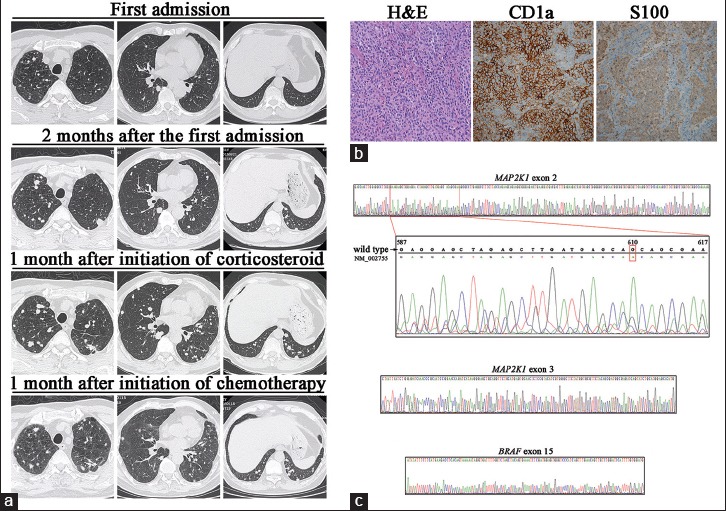
(a) Chest high-resolution computed tomography findings of the patient. (b) Histopathological findings of the lung biopsy specimen. H and E stain showed cellular nodule with aggregates of Langerhans cells (original magnification ×200); immunohistochemistry for CD1a and S100 demonstrated positive staining in Langerhans cells (original magnification ×200). (c) Sequencing of MAP2K1 and BRAF. One silent mutation (code “CAG” to “CAA” at position 610) in exon 2 of MAP2K1 was detected while no mutations in exon 3 of MAP2K1 or in exon 15 of BRAF were detected.
Two months later, reexamined chest HRCT [Figure 1a] showed a marked increase in the number and size of nodules, with diameter up to 2.0 cm. The patient underwent video-assisted thoracoscopic surgical (VATS) biopsy of the left upper and lower lobes. During the procedure, many palpable nodules were noted in lung parenchyma with diameters ranging from 0.3 to 1.8 cm. A key morphological feature of LCH cells on microscopic examination was noted, which was their highly convoluted nuclear membranes, and the cells strongly expressed Cluster of Differentiation (CD)1a and S100 [Figure 1b]. PLCH diagnosis was made. Treatment started with prednisone (PDN) (0.5 mg·kg−1·d−1 for 1 month). Unfortunately, chest HRCT obtained one month later showed an increase in the number and size of nodular lesions [Figure 1a]. Based on the results of LCH-III,[4] a randomized international clinical trial embarked by the Histiocyte Society, an adjusted chemotherapeutic regimen of methotrexate (MTX) + vindesine (VDS) + PDN was pursued, i.e., MTX 500 mg/m2 once every two weeks intravenous (i.v.), VDS 3 mg/m2 once a week i.v., PDN 30 mg/m2 d1-30 orally (afterward weekly reduction). He experienced WHO Grade 1/2 toxicities including hepatotoxicity, nausea, and vomiting. One month later, chest HRCT showed a partial clearing of nodules [Figure 1a], and his cough resolved. To explore whether the patient would benefit from the BRAF inhibitor therapy, BRAF and MAP2K1 mutations were further tested. Deoxyribonucleic acid (DNA) was extracted from formalin-fixed and paraffin-embedded (FFPE) samples of the surgical biopsy after histological detection of histiocyte-rich areas, using QIAmp DNA FFPE tissue kit (QIAGEN, Germany) according to instructions. Exon 2 and exon 3 of MAP2K1 and exon 15 of BRAF were amplified by polymerase chain reaction and sequenced with primers as listed in Table 1. As indicated in Figure 1c, one silent mutation (code “CAG” to “CAA” at position 610) in exon 2 of MAP2K1 was detected. However, there were no mutations in exon 3 of MAP2K1 and exon 15 of BRAF. The experiment was approved by the Ethics Committee of Drum Tower Hospital, and the patient gave written informed consent.
Table 1.
Polymerase chain reaction and sequencing primers for MAP2K1 (exon 2 and exon 3) and BRAF (exon 15)
| Template | Sequence (5’-3’) | Product size (bp) |
|---|---|---|
| MAP2K1 exon 2 | 530 | |
| Forward | CTCTCTAGCCTCCCACTTTGATT | |
| Reverse | GTTTGAACCCAGGAGATGGA | |
| MAP2K1 exon 3 | 356 | |
| Forward | CCTTCCTCCCTCTTTCTTTCATA | |
| Reverse | CCCAAGCTCTACAGTTAGACTTCC | |
| BRAF exon 15 | 587 | |
| Forward | TACTATCTGCAGCATCTTCATTCC | |
| Reverse | TACTATAGTTGAGACCTTCAATGAC |
Several findings of this case were quite unique and rare. First, the chest HRCT manifestation of this patient was diffuse nodules alone throughout the lung, without formation of cysts or cavities. Second, the onset of disease was nearly one year after smoking cessation. Diffuse lung nodules progressed both in number and size despite smoking cessation. Third, corticosteroid monotherapy failed to control disease progression while intensified combination chemotherapy (MTX + VDS + PDN) showed efficacy. Finally, and particularly, BRAF and MAP2K1 of the lung lesions were sequenced. One MAP2K1 mutation was observed while BRAF V600E mutation was not detected. To the best of our knowledge, the report regarding MAP2K1 mutations in single-system PLCH was seldom and a new mutation site was identified (code “CAG” to “CAA” at position 610 in exon 2 of MAP2K1). These findings differentiate this case from the usual case of PLCH seen in clinical practice.
PLCH usually has little systemic damage. In this case, we did not find extrapulmonary disease manifestations. In elder patients with pure nodular forms in chest HRCT, it is especially important to exclude malignancy, which is why this patient received twice CT-guided percutaneous transthoracic FNA followed by VATS lung biopsy. It is generally thought that lung bases are spared in PLCH. However, in this patient, nodules extended into the lower lung zones and costophrenic angles were infiltrated, which might associate with the severity of disease.
Persisting uncertainty about the pathogenesis of PLCH has limited current treatment alternatives. Smoking cessation has been regarded as a treatment; however, worsening conditions despite cessation was observed in this case, indicating that PLCH might be induced by antigens in cigarette smoke but not terminated by smoke cessation. The role of smoking cessation on disease progress seems indifferent. In this case, corticosteroid monotherapy failed to control the disease. This patient further received chemotherapy (MTX + VDS + PDN) according to LCH-III.[4] HRCT imaging showed improvement one month later. Even though this case is one-system LCH, involving the lung might associate with rapid progression and worse prognosis, thus a more intensive chemotherapeutic regimen might be necessary as in multisystem LCH. The most common activating somatic mutation in BRAF gene is a substitution of glutamic acid for V600E, which has been identified in a part of LCH cases. This mutation results in constitutive activation of the mitogen-activated protein kinase (MAPK) pathway. MAP2K1 encodes the dual-specificity kinase of the MAPK pathway. MAP2K1 mutations have been demonstrated to confer resistance to BRAF inhibitor therapy in LCH and other neoplasms.[5] A mutation in exon 2 of MAP2K1 was positive in this patient. Even though it is a silent mutation, it indicates there might be another mechanism of MAPK pathway activation in BRAF V600E-negative PLCH. There is clearly a necessity for better therapies for PLCH.
Financial support and sponsorship
This work was supported by the grants from The Natural Science Foundation of Jiangsu Province (No. BK20130089), and the National Natural Science Foundation of China (No. 81501972).
Conflicts of interest
There are no conflicts of interest.
Acknowledgment
We would like to thank Qing Ye, M.D., and Fanqing Meng, M.D., (Department of Pathology, Nanjing Drum Tower Hospital, The Affiliated Hospital of Nanjing University Medical School, Nanjing, China) for their technical support in the evaluation of the pathological samples.
Footnotes
Edited by: Ning-Ning Wang
REFERENCES
- 1.Ling CH, Ji C, Raymond DP, Bourne PA, Xu TC. Uncommon features of pulmonary Langerhans'cell histiocytosis: Analysis of 11 cases and a review of the literature. Chin Med J. 2010;123:498–501. doi:10.3760/cma.j.issn.0366-6999.2010.04.020. [PubMed] [Google Scholar]
- 2.Haroche J, Cohen-Aubart F, Emile JF, Arnaud L, Maksud P, Charlotte F, et al. Dramatic efficacy of vemurafenib in both multisystemic and refractory Erdheim-Chester disease and Langerhans cell histiocytosis harboring the BRAF V600E mutation. Blood. 2013;121:1495–500. doi: 10.1182/blood-2012-07-446286. doi:10.1182/blood-2012-07-446286. [DOI] [PubMed] [Google Scholar]
- 3.Brown NA, Furtado LV, Betz BL, Kiel MJ, Weigelin HC, Lim MS, et al. High prevalence of somatic MAP2K1 mutations in BRAF V600E-negative Langerhans cell histiocytosis. Blood. 2014;124:1655–8. doi: 10.1182/blood-2014-05-577361. doi:10.1182/blood-2014-05-577361. [DOI] [PubMed] [Google Scholar]
- 4.Gadner H, Minkov M, Grois N, Pötschger U, Thiem E, Aricò M, et al. Therapy prolongation improves outcome in multisystem Langerhans cell histiocytosis. Blood. 2013;121:5006–14. doi: 10.1182/blood-2012-09-455774. doi:10.1182/blood-2012-09-455774. [DOI] [PubMed] [Google Scholar]
- 5.Emery CM, Vijayendran KG, Zipser MC, Sawyer AM, Niu L, Kim JJ, et al. MEK1 mutations confer resistance to MEK and B-RAF inhibition. Proc Natl Acad Sci U S A. 2009;106:20411–6. doi: 10.1073/pnas.0905833106. doi:10.1073/pnas.0905833106. [DOI] [PMC free article] [PubMed] [Google Scholar]


