Abstract
CHARGE syndrome is an autosomal dominant syndrome in which ocular coloboma (C), heart defects (H), choanal atresia (A), growth retardation (R), genital hypoplasia (G), ear abnormalities (E), and tracheoesophageal fistula, dysphagia, cleft palate, micrognathia, facial paralysis, hypopituitarism, and brain abnormalities may be seen in patients. The patients with CHARGE syndrome face surgical procedures many times from birth. Especially, the problems we meet in the airway may be special. In this case report, we aimed to share our experience of endotracheal intubation performed with Glidescope video laryngoscopy for a patient at the age of 20 months, weight 7.5 kg and height 70 cm, with CHARGE syndrome who was undergoing cochlear implantation.
Keywords: CHARGE syndrome, difficult airway, Glidescope video laryngoscopy
Introduction
CHARGE syndrome (CS) is an autosomal dominant inherited syndrome that was first described by two independent physicians, Hall and Hittner, in 1979 and presents with many systemic anomalies (1, 2). Six main components of CS were systemized by Pagon et al. (3): ocular coloboma (C), heart defects (H), choanal atresia (A), growth retardation (R), genital hypoplasia (G), ear abnormalities (E) and/or deafness findings. In addition to these components, other findings, including tracheo-oesophageal fistula, dysphagia, cleft palate, micrognathia, facial paralysis, hypopituitarism and brain anomalies, can be seen (1–3). Blake et al. (4) re-classified CS into major and minor criteria in 1998. Choanal atresia, iris coloboma, external ear anomalies and cranial nerve dysfunctions were described as major criteria, and congenital heart defects, growth retardation, tonus changes, deformities in hands and orofacial anomalies were described as minor criteria.
Children with CS often undergo surgical procedures in newborn age. In this case, serious airway problems may occur at the beginning of and following anaesthesia (5). Since craniofacial anomalies, micrognathia, higher location of the larynx, cleft palate, adenoids and enlarged tonsils in CHARGE syndrome can lead to difficulty in intubation, it is vital to evaluate the airway carefully before anaesthesia and to make the necessary preparations for challenging intubation (6, 7).
In this case report, we aimed to emphasize the advantage of the Glidescope video laryngoscope (GVL) in patients with CS.
Case Presentation
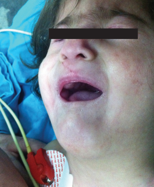
The parents were informed that the clinical condition of the patient would be shared in a scientific journal, and their written informed consent was obtained. The patient was a 20-month-old, 7.5 kg and 70 cm-long girl who had been diagnosed with CS and planned to have a cochlear implantation device inserted under general anaesthesia due to hearing loss. Her physical examination revealed a syndromic facial appearance, growth retardation, cleft palate, bilateral ear anomaly and decreased hearing, exophthalmos, hypertelorism, congenital hip dislocation, hepatosplenomegaly, increased muscle tonus in the whole body, and 2/6 degree pansystolic murmur in the mesocardiac and apical focus (Figure 1). In the echocardiographic examination, secundum-type ASD, MVP and first-degree mitral insufficiency were observed. In the laboratory workup, the value of haemoglobin was 12.3 g dL−1, the value of haematocrit was 36.6%, glucose was 115 mg dL−1 and other findings were found to be within normal reference ranges. PA chest radiography revealed that the mediastinum and heart shadow were expanded. Because our case had a small mouth, micrognathia, cleft palate and hypertrophic tonsils, the possibility of a difficult intubation was considered, and a laryngeal mask, video laryngoscope, fibreoptic laryngoscope and urgent tracheostomy requirements were kept ready. Infective endocarditis prophylaxis was applied preoperatively with an ampicillin sulbactam and gentamicin combination. Vascular access was established with a 24 gauge Branule on the back of the left hand of the patient under operation, and 500 cc 1/3 izodeks fluid was initiated at a rate of 20 mL kg−1 hr−1. The patient, who underwent routine monitoring (electrocardiography, non-invasive blood pressure, peripheral oxygen saturation), was administered anaesthesia induction with 2 μg kg−1 fentanyl, 2 mg kg−1 propofol and 0.5 mg kg−1 atracurium following preoxygenation with 100% O2 for 2–3 minutes. In the direct laryngoscopy (DL), cleft palate, hypertrophic tonsil and high location of the larynx were demonstrated, and the Cormack-Lehane (CL) score was found to be III. The intubation procedure failed in the first attempt. A GVL (Verathon Medical, Bothell, WA, USA) was used for the second attempt of intubation. In the video laryngoscopy performed with a number 2blade, the CL score was found to be I, and intubation was performed in a single intervention with an endotracheal tube without a cuff (No: 4, 5). The maintenance of anaesthesia was provided with controlled ventilation with 2.5% sevoflurane in 50% O2 air. The haemodynamic parameters of the patient were stable during the operation, and she was extubated without any problem following the surgery, lasting for 3.5 hours. Since the patient did not display any complications in the postoperative follow-up examinations, she was discharged from the hospital on the 3rd day.
Figure 1.

Typical appearance of a patient with CHARGE syndrome
Discussion
Although children with CS, including multisystemic problems, have a high possibility of undergoing operations in newborn age, a few number of cases are available in the literature. A carefully performed evaluation and follow-up are required in every stage of anaesthesia for patients having CHARGE syndrome, due to their upper and lower respiratory tract abnormalities and cardiac problems (5).
The airway management of patients with CS is important for the anaesthesia.
The craniofacial anomalies, micrognathia, high location of the larynx, cleft palate, adenoids and enlarged tonsils seen in CHARGE syndrome can cause difficulties in the intubation (7).
The studies conducted suggested that 10%–30% of patients with CS could require a tracheostomy and that difficulty in intubation increases in parallel with age (8).
In a series including 9 cases conducted by Blake et al. (5), while postoperative airway problems of patients with CS after the first anaesthesia intervention was 39%, airway problems decreased with repeated anaesthesia interventions. The rate of airway problems after cardiovascular interventions was 65%, while it was 39% after gastrointestinal tract interventions and 36% after interventions performed for evaluating the airway. The most common problem is desaturation, but non-extubated patients and airway problems requiring intensive care can also be encountered (5).
The Glidescope video laryngoscope is a system developed for difficult airways; it has a camera on its tip and a 60° curvature, it is made of hard plastic and it consists of a blade, light source and monitor on which the view is transferred. The studies conducted demonstrated the superiority of DL in difficult intubation conditions (9). Shimuzi et al. (10) reported that by using the GVL, they easily intubated CS patients having a DL and CL score of IV, who could not be intubated by DL and a Pentax airway scope. In our case, the non-intubated patient with a DL and CL score of III was successfully intubated in the first intervention through the GVL, with a CL score of I.
Lee et al. (11) used a GVL blade (GLVw) fitting the patient’s weight and a blade one size smaller (GLVs) for a patient whose DL and CL score was ≥III in their study, and they found that the GLVs improved intubation conditions better than the DL and GLVw and that the GLVw improved them better than the DL. The possibility of difficult intubation is high in patients having craniofacial anomalies, as well as CS (12). For these patients, the GVL can be effectively used with the appropriate blade.
Hara et al. (13) reported that they provided the airway of a non-intubated patient with DL and CL score of IV by using a Proseal LMA. The LMA can be an alternative for cases that can not be intubated. However, in patients with CS, the upper airway anomalies and smaller mouth, oropharynx and larynx can make the placement of an LMA difficult, and it can lead to aspiration of intraoral secretions and stomach contents.
In patients with CHARGE syndrome, oral respiration is in the forefront, instead of nasal respiration, because of the developmental delay in the nasopharyngeal tract (14). Due to the reasons associated with cranial nerve IX and X anomalies, including dysphagia, gastro-oesophageal reflux and tracheo-oesophageal fistula, in patients with CS, the risk for aspiration of oral secretions, nutritional products and stomach contents is higher. Therefore, it is essential to evaluate and to follow up carefully with regard to aspiration pneumonia in the preoperative and postoperative periods (5, 6, 15).
Conclusion
Airway management of patients with CS is important for anaesthesiologists. All preparations should be completed for the possibility of a difficult intubation, and preoxygenation should be carried out adequately. Video laryngoscopes, which have much higher intubation success compared to DLs, should be kept ready as an alternative in case of a difficult intubation. The patient should be followed up closely for possible airway problems after extubation. As seen in our case, we suggest that the GVL can be used as a non-invasive and effective alternative in the airway management of patients with CS.
Footnotes
Informed Consent: Written informed consent was obtained from patients’ parents who participated in this case.
Peer-review: Externally peer-reviewed.
Author Contributions: Concept - V.S.; Design - V.S., A.M., S.G.; Supervision - M.C., S.G.; Funding - V.S., M.Ş.; Materials - V.S., R.G.; Data Collection and/or Processing - V.S.; Analysis and/or Interpretation - V.S., A.M., S.G., R.G.; Literature Review - V.S., M.Ş.; Writer - V.S.; Critical Review - R.G., A.M.
Conflict of Interest: No conflict of interest was declared by the authors.
Financial Disclosure: The authors declared that this study has received no financial support.
References
- 1.Hall BD. Choanal atresia and associated multipl anomalies. J Pediatr. 1979;95:395–8. doi: 10.1016/s0022-3476(79)80513-2. http://dx.doi.org/10.1016/S0022-3476(79)80513-2. [DOI] [PubMed] [Google Scholar]
- 2.Hittner HM, Hirsch NJ, Kreh GM, Rudolph AJ. Colobomatous microphthalmia, heart disease, hearing loss, and mental retardation-a syndrome. J Pediatr Ophthalmol Strabismus. 1979;16:122–8. doi: 10.3928/0191-3913-19790301-10. [DOI] [PubMed] [Google Scholar]
- 3.Pagon RA, Graham JM, Jr, Zonana J, Yong SL. Coloboma, conjenital heart disease, and choanal atresia with multiple anomalies: CHARGE assosicition. J Pediatr. 1981;99:223–7. doi: 10.1016/s0022-3476(81)80454-4. http://dx.doi.org/10.1016/S0022-3476(81)80454-4. [DOI] [PubMed] [Google Scholar]
- 4.Blake KD, Davenport SL, Hall BD, Hefner MA, Pagon RA, Williams MS, et al. CHARGE association: an updateand review for the primary pediatrician. Clin Pediatr (Phila) 1998;37:159–73. doi: 10.1177/000992289803700302. http://dx.doi.org/10.1177/000992289803700302. [DOI] [PubMed] [Google Scholar]
- 5.Blake K, MacCuspie J, Hartshorne TS, Roy M, Davenport SL, Corsten G. Postoperative airway events of individuals with CHARGE syndrome. Int J Pediatr Oto Rhinolaryngol. 2009;73:219–26. doi: 10.1016/j.ijporl.2008.10.005. http://dx.doi.org/10.1016/j.ijporl.2008.10.005. [DOI] [PubMed] [Google Scholar]
- 6.Tellier AL, Cormier-Daire V, Abadie V, Amiel J, Sigaudy S, Bonnet D, et al. CHARGE syndrome: report of 47 cases and review. Am J Med Genet. 1998;76:402–9. doi: 10.1002/(sici)1096-8628(19980413)76:5<402::aid-ajmg7>3.0.co;2-o. http://dx.doi.org/10.1002/(SICI)1096-8628(19980413)76:5<402::AID-AJMG7>3.0.CO;2-O. [DOI] [PubMed] [Google Scholar]
- 7.Roger G, Morisseau-Durand MP, Van Den Abbeele T, Nicollas R, Triglia JM, Narcy P, et al. The CHARGE Association, The Role of Tracheotomy. Arch Otolaryngol Head Neck Surg. 1999;125:33–8. doi: 10.1001/archotol.125.1.33. http://dx.doi.org/10.1001/archotol.125.1.33. [DOI] [PubMed] [Google Scholar]
- 8.Stack CG, Wyse RK. Incidence and management of air- way problems in the CHARGE Association. Anaesthesia. 1991;46:582–5. doi: 10.1111/j.1365-2044.1991.tb09664.x. http://dx.doi.org/10.1111/j.1365-2044.1991.tb09664.x. [DOI] [PubMed] [Google Scholar]
- 9.Griesdale DE, Liu D, McKinney J, Choi PT. Glidescope video-laryngoscopy versus direct laryngoscopy for endotracheal intubation: a systematic review and meta-analysis. Can J Anaesth. 2012;59:41–52. doi: 10.1007/s12630-011-9620-5. http://dx.doi.org/10.1007/s12630-011-9620-5. [DOI] [PMC free article] [PubMed] [Google Scholar]
- 10.Shimizu S, Koyama T, Mizota T, Fukuda K. Successful tracheal intubation with the GlideScope® in a patient with CHARGE syndrome. J Anesth. 2013;27:965–6. doi: 10.1007/s00540-013-1631-7. http://dx.doi.org/10.1007/s00540-013-1631-7. [DOI] [PubMed] [Google Scholar]
- 11.Lee JH, Park YH, Byon HJ, Han WK, Kim HS, Kim CS, et al. A comparative trial of the GlideScope(R) video laryngoscope to direct laryngoscope in children with difficult direct laryngoscopy and an evaluation of the effect of blade size. Anesth Analg. 2013;117:176–81. doi: 10.1213/ANE.0b013e318292f0bf. http://dx.doi.org/10.1213/ANE.0b013e318292f0bf. [DOI] [PubMed] [Google Scholar]
- 12.Sinkueakunkit A, Chowchuen B, Kantanabat C, Sriraj W, Wongswadiwat M, Bunsangjaroen P, et al. Outcome of anesthetic management for children with craniofacial deformities. Pediatr Int. 2013;55:360–5. doi: 10.1111/ped.12080. http://dx.doi.org/10.1111/ped.12080. [DOI] [PubMed] [Google Scholar]
- 13.Hara Y, Hirota K, Fukuda K. Successful airway management with use of a laryngeal mask airway in a patient with CHARGE syndrome. J Anesth. 2009;23:630–2. doi: 10.1007/s00540-009-0791-y. http://dx.doi.org/10.1007/s00540-009-0791-y. [DOI] [PubMed] [Google Scholar]
- 14.Sert H, Gözdemir M, Çimen NK, Muslu B. Airway management of CHARGE syndome (CASE REPORT) Turk J Anaesth Reanim. 2011;39:149–52. http://dx.doi.org/10.5222/JTAICS.2011.149. [Google Scholar]
- 15.Işık B, Arslan M, Doğan A, Akçabay M. Anaesthetic Approach in Charge Syndrome (Case Report) Turkiye Klinikleri J Anest Reanim. 2004;2:153–6. [Google Scholar]


