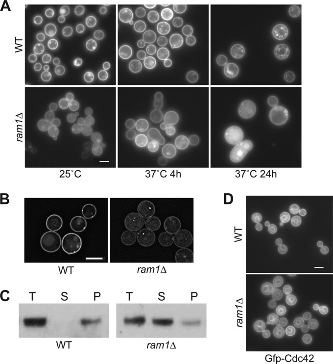
FIG 2 .

Ras1 is a specific target of Ram1. (A and B) Ras1 localization to the PM is reduced in the ram1Δ mutant strain. To compare the patterns and intensities of mCherry-Ras1 fusion protein localization, wild-type (CBN121) and ram1Δ mutant (SKE17) strains expressing mCh-RAS1 under the control of the histone H3 promoter were incubated in rich medium, diluted 10-fold, and subsequently incubated for 4 or 24 h at 25°C and 37°C. (A) Live cells grown at 25°C or 37°C for 4 h and at 37°C for 24 h were imaged by fluorescence microscopy (Zeiss Axio Imager A1) using the appropriate filter. Bar, 5 µm. (B) Live cells incubated at 25°C for 4 h were also imaged using DeltaVision deconvolution microscopy with the appropriate filter. Images were deconvolved using softWoRx software. Bar, 5 µm. Images were taken using identical exposures. (C) Ras1 protein accumulates in the cytoplasm in the ram1Δ mutant strain. To compare the relative amounts of mCherry-Ras1 protein on cellular membranes and in the cytoplasm, total lysates from strains CBN121 and SKE17 were separated into soluble (cytoplasmic) and insoluble (membrane-associated) fractions by ultracentrifugation. Total lysate (T), soluble (S), and insoluble pellet (P) samples were assessed by Western blotting with an antibody specific to the mCherry fusion protein. (D) Cdc42 localization is not dependent on Ram1. Wild-type (CBN302) and ram1Δ mutant (SKE19) strains expressing GFP-CDC42 under the control of the histone H3 promoter were incubated overnight in YPD, diluted 10-fold, and grown for 4 h at 25°C. Live cells were imaged (Zeiss Axio Imager A1) using the appropriate filter. Bar, 5 µm.
