Abstract
Background:
Cerebrospinal fluid (CSF) rhinorrhea, when left untreated, can lead to meningitis and other serious complications. Treatment traditionally has entailed an open craniotomy, although the paradigm has now evolved to encompass endoscopic procedures. Trauma, both accidental and iatrogenic, causes the majority of leaks, and trauma involving skull base and facial fractures is most likely to cause CSF rhinorrhea. Diagnosis is aided by biochemical assay and imaging studies.
Methods:
We reviewed the literature and summarized current practice regarding the diagnosis and management of CSF rhinorrhea.
Results:
Management of CSF leaks is dictated by the nature of the fistula, its location, and flow volume. Control of elevated intracranial pressure may require medical therapy or shunt procedures. Surgical reconstruction utilizes a graduated approach involving vascularized, nonvascularized, and adjunctive techniques to achieve closure of the CSF leak. Endoscopic techniques have an important role in select cases.
Conclusion:
An active surgical approach to closing CSF leaks may provide better long-term outcomes in some patients compared to more conservative management.
Keywords: Cerebrospinal fluid, cerebrospinal fluid leak, cerebrospinal fluid rhinorrhea, endoscopy, skull base
INTRODUCTION
Cerebrospinal fluid (CSF) rhinorrhea is a condition that results from an acquired communication between the central nervous system and external environment. CSF rhinorrhea may present spontaneously or following trauma and if untreated may lead to ascending meningitis and other complications. Management often requires the collaborative care of multiple clinicians. Although CSF rhinorrhea has been traditionally treated with open surgical procedures, the introduction of the rigid endoscope for minimally invasive endonasal procedures has revolutionized treatment for many patients. We review current practice regarding the diagnosis and management of CSF rhinorrhea.
CSF PHYSIOLOGY
CSF is the end product of active and passive filtration of plasma at the choroid plexus with minor contribution from ependymal cells and parenchymal capillaries. The majority of CSF production occurs in the choroid plexus of the lateral ventricles and the third and fourth ventricles. After circulation through the subdural space, CSF is reabsorbed into the venous system by arachnoid granulations in the dural surface of the superior sagittal sinus. Arachnoid villi use 1-way valves that rely on hydrostatic pressure to resorb CSF flowing in a pulsatile manner into the venous system. Resorption occurs when intracranial pressure (ICP) is 1.5-7.0 cmH2O greater than central venous pressure.1
The hourly production of CSF is approximately 20 mL, with a daily production rate of 400-600 mL that can increase in response to chronic loss of CSF volume. Normal CSF volume averages 150 mL in adults, with turnover 3-4 times daily. While normal ICP is 5-15 cmH2O, diurnal and positional variations in ICP can also affect CSF dynamics. Factors that can alter ICP include changes in the rate of CSF production or reabsorption.1 An ICP consistently >20 cmH2O is considered elevated and requires prompt evaluation.2
ETIOLOGY OF CSF RHINORRHEA
Trauma, both iatrogenic and noniatrogenic, contributes to 80%-90% of CSF fistulas.3 The most common cause of iatrogenic rhinorrhea is functional endoscopic sinus surgery.4 The lateral lamella of the cribriform plate, which is thinner than the more lateral portions of the ethmoid roof, has been described as being most susceptible to damage.2 The posterior ethmoid skull base is another common location for injury because it slopes inferiorly toward the sphenoid rostrum. CSF rhinorrhea resulting from noniatrogenic trauma has been reported in 2% of all head traumas, 12%-30% of skull base fractures, and 25% of facial fractures.5
Leaks may be identified during the early posttraumatic period, or leak presentation may be delayed. An active surgical approach to closing CSF leaks from defects at the skull base may provide better long-term outcomes compared to more conservative management, particularly in patients who have a prior history of ascending bacterial meningitis.6 Delay in leak presentation may be attributed to resolution of associated brain edema, devascularization of tissue, formation of the fistula tract, and resolution of blood products, all of which can result in increased ICP.
Spontaneous CSF leaks often result from skull base dehiscence and bony erosion from sinonasal and intracranial lesions. While the pathophysiology of spontaneous CSF rhinorrhea may vary, the underlying etiology has implications for prognosis and treatment, particularly in patients with occult increased ICP. Patients with a history of major trauma may have acquired a skull base defect at the time of injury that has slowly expanded from normal intracranial pulsation, rendering the location of dehiscence vulnerable to leakage. Patients with a likely normal CSF physiology—and therefore higher surgical success rates—can have spontaneous CSF leaks resulting from congenital paranasal sinus pneumatization with skull base dehiscence.
A theory of incomplete embryologic fusion of the sphenoid bone, albeit controversial, would account for meningoencephalocele and CSF leak development in both the central7 and the lateral craniopharyngeal canals.8 Expansive pneumatization in the sphenoid sinus can predispose patients to dehiscence of the floor of the middle cranial fossa under the influence of intracranial pulsations.9 An elevated ICP can contribute to and exacerbate any of these conditions. Management can include acetazolamide treatment or CSF shunt procedures.10 Patients with spontaneous CSF fistulas often require more attentive counseling and postoperative care given their lower rate of successful CSF leak closure.11
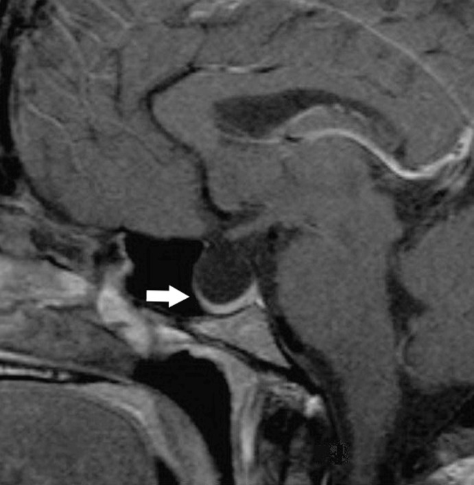
In patients with nontraumatic increased ICP, a CSF leak can often be attributed to an intracranial lesion with mass effect, hydrocephalus, or benign intracranial hypertension (Figure 1).12 Diagnostic findings include the absence of localizing neurologic signs, increased CSF pressure, normal CSF count and chemistries, papilledema on fundus examination, and a lack of other identifying etiology of increased ICP.13 An ICP pressure finding >20-25 cmH2O often requires treatment.14 Clinical symptoms are sensitive to postural changes and the Valsalva maneuver.
Figure 1.
Sagittal magnetic resonance imaging shows an empty sella in a patient with benign intracranial hypertension. The pituitary gland is compressed inferiorly (arrow).
DIAGNOSIS OF CSF LEAK
Patients presenting with unilateral clear nasal discharge associated with nonspecific headache symptoms raise suspicions for rhinorrhea of cerebrospinal origin. Patients can also present with mental status changes, seizure, and meningitis, thereby requiring a high level of suspicion for accurate diagnosis.15,16 Rhinorrhea secondary to a CSF fistula can be provoked by placing the patient's face in a downward position and observing for leakage for several minutes. Patients with a CSF leak and benign intracranial hypertension may also display bilateral papilledema.
Beta-2 transferrin immunofixation is currently the gold standard for diagnosing CSF rhinorrhea.1 Beta-2 transferrin is found in CSF, perilymph, and the vitreous fluid of the eye. Perilymph is produced in small amounts, and contamination of other fluid with vitreous eye fluid is highly unlikely, allowing beta-2 transferrin to serve as a highly specific assay for the presence of CSF. This assay has been noted to have a sensitivity of 100% and a specificity of 71% for detection of CSF leaks.17
Localization of the leak may be accomplished by direct visualization with diagnostic nasal endoscopy, although this technique is typically insufficient, particularly in patients without a history of sinonasal surgery. Radiologic studies can aid diagnosis with plain films, coronal computed tomography (CT) images, or magnetic resonance imaging (MRI). While plain-film radiography has limited applicability, it can be used to identify fractures and pneumocephalus in critically unstable patients. A thin-cut noncontrasted CT of the paranasal sinuses is the most common initial study. Findings of skull base dehiscence, air-fluid levels in adjacent sinuses, and pneumocephalus may be suggestive enough when correlated with clinical presentation to proceed with treatment planning. False-negative results can occur in patients with small bony defects, while false-positive results can occur from volume averaging. Some bony defects seen on CT may not correlate with the actual site of the leak. CT is superior for its visualization of the craniofacial skeleton and for calcified tissues at the base of the skull.1
MRI is particularly useful in distinguishing tissue and tumor density by signal intensity readings.1 Further, MRI can be used to characterize soft tissue opacification within the paranasal sinuses. MRI is less definitive near the bony anatomy of the skull base and paranasal sinuses where resolution suffers. In patients with multiple skull base defects, MRI is best used to assess for the presence of meningoencephalocele in addition to CSF rhinorrhea, a finding that is often present in this group of patients.18
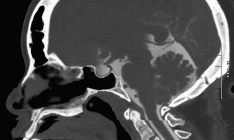
Cisternography with intrathecal radioactive isotope is a well-established method of confirming and localizing CSF fistulas when clinical suspicion of CSF rhinorrhea is present, but the leak has not been localized on CT or MRI. This situation often occurs with patients who have low-volume or intermittent CSF leaks. The technique for CT cisternography has evolved from metrizamide to water-soluble iodine contrast material (Figure 2).19 Magnetic resonance cisternography uses T2-weighted images with fat suppression and image reversal to highlight CSF.20 A positive finding of CSF leak is supported by visualization of contrast material flowing through a skull base defect site. Supportive findings include the pooling of contrast within a related paranasal sinus. However, cisternography studies have variable sensitivities that are often correlated with the rate of the CSF leak. Other limitations of cisternography include invasiveness, radiation exposure, and poor anatomic localization.21 The sensitivity and specificity for this test in determining the presence of CSF rhinorrhea have been documented to be 92% and 80%, respectively.22
Figure 2.
Computed tomography cisternogram shows enhancement of the subarachnoid space in a patient with elevated intracranial pressure.
Intrathecally injected fluorescein performed with or without blue-light endoscopy is an alternative method for localization of CSF leaks. Traditionally, a lumbar puncture is required, followed by nasal endoscopy for inspection of green fluid to confirm the presence of a CSF leak. Potential side effects include cardiac arrhythmias, seizures, headaches, and cranial nerve defects. Therefore, intrathecal use of fluorescein is considered an off-label use in the United States, requiring detailed patient counseling and informed consent prior to utilization.23 Sensitivity and specificity of this diagnostic procedure have been noted to be as high as 73.8% and 100%, respectively.24
APPROACHES TO REPAIR
Once a CSF fistula has been identified, management is dictated by the etiology of the leak, its location, and its flow volume. High-flow CSF leaks rarely close spontaneously and often require surgical intervention. For low-volume leaks, conservative measures may be employed.
Spontaneous leaks with low or intermittent volume may be managed conservatively with bed rest, head elevation, avoidance of straining activities, and temporary CSF diversion with a lumbar drain.25 Because the majority of CSF leaks caused by closed head injuries resolve spontaneously, trauma patients may be conservatively managed unless they experience neurologic deterioration or are diagnosed with additional intracranial pathology.19 When conservative management fails, surgical repair is indicated.
Either open craniotomy or endoscopic repair can be considered. Open repairs can be performed via a bifrontal craniotomy or extracranially through an external ethmoidectomy or frontal sinusotomy. An open transcranial approach provides wide visualization of the dural tear, the option to directly treat any surrounding tissue injury, and the ability to use a vascularized pericranial flap to cover the anterior skull base.12 For these reasons, an open transcranial approach is an important option for repairing leaks that are severe, multifocal, high pressure, recurrent, or otherwise not amenable to endoscopic treatment.2 However, open approaches are associated with intracerebral hemorrhage, cerebral edema, frontal lobe deficits, lengthened hospital stay, anosmia, and higher recurrence rates than the endoscopic approach.26
Endoscopic procedures are performed endonasally through the sinonasal tract with a rod-lens endoscope that uses linear or angled optics to visualize the roof of the sinonasal cavity. Various procedures and materials can be used for endoscopic reconstructive procedures, including autologous grafts, nonautologous grafts, and surgical adjuncts such as tissue sealants. Grafts are used for the following functions: (1) to fill a space through mass effect, (2) to re-create a watertight layer, (3) to act as a rigid buttress, and (4) to stabilize a wound edge. Fat has expansive qualities that allow it to be used for mass effect, while fascia, acellular dermis, rotational flaps, and mucosal grafts are used for watertightness because of their high collagen levels. Cartilage, bone, and synthetic miniplates are well formed and can act as rigid buttresses. Wound edges are often stabilized with cellulose, gelatin sponge, and tissue sealant.
RECONSTRUCTION TECHNIQUES
Nonvascularized reconstruction is a practical option for the endoscopic repair of small or low-volume CSF fistulas. Nonvascularized techniques can also be used in conjunction with vascularized reconstructive techniques, particularly in complex defects.
Inlay graft materials include autologous tissue such as fascia lata, as well as acellular dermis and other synthetic materials. These grafts may be applied within the intracranial space and tucked against the intracranial surface of bony ledges. Onlay grafts can be similarly applied to the extracranial surface of the skull base, in which case they require additional materials for bolstering. Consequently, onlay grafts have limited use in isolation but are commonly used as part of multilayer reconstructive techniques. Composite grafts, consisting of a mixture of inlay grafts, onlay grafts, tissue sealant, and supporting materials, are often used for moderate-sized defects with active CSF leakage. Composite grafts are versatile and can be combined with a pedicled, vascularized flap approach in patients with large defects and high-volume CSF leaks.
Other techniques for leak repair include suturing the dura mater under endoscopic visualization as described by Cukurova et al.27 Tension-free closure is required for this technique, limiting repairs to large bony defects with small dural defects. Laser tissue welding during endoscopic closure is an experimental technique that has been found to create an above-average-strength seal without significant inflammatory sequelae.25
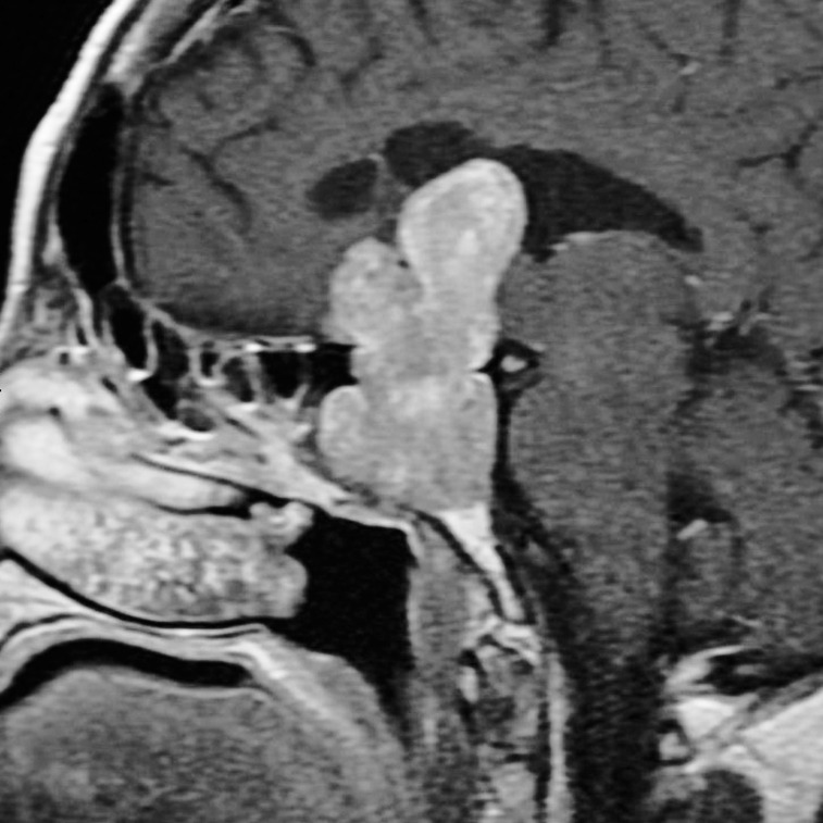
Vascularized flaps have greatly enhanced the capabilities for endoscopic skull base reconstruction. These flaps, including nasoseptal flaps and turbinate flaps, are vascularized via an axial blood supply that allows for increased flap survival. By comparison, intranasal free grafts have a random blood supply that limits their versatility when large grafts are needed. The nasoseptal flap is one of the most widely used vascularized flaps for skull base defect repairs (Figure 3).22 Its ease of harvest, large mucosal surface, favorable arc of rotation, and ability to cover sellar, suprasellar, clival, and anterior skull base defects make the nasoseptal flap a commonly used option for vascularized reconstruction. Doppler sonography can be used to assess the viability of a proposed flap site.28 Posterior and central sinonasal defects can be repaired with the inferior turbinate flap, particularly with a posterior pedicle.29 This flap uses the posterior lateral nasal artery, a branch of the sphenopalatine artery, for vascularization. Large anterior fossa defects can be repaired with a vascularized lateral nasal wall flap that involves the inferior turbinate and nasal floor mucosa with an anteriorly based pedicle.30 The middle turbinate flap is an additional option when the septum or inferior turbinate is not available as a donor site.31 Adverse effects of flap use include flap necrosis, flap displacement, and donor-site morbidity.
Figure 3.
Sagittal magnetic resonance imaging shows a pituitary macroadenoma prior to removal with an endoscopic transsphenoidal approach. The large postsurgical defect in the skull base required a vascularized nasoseptal flap as part of the reconstruction.
Tissue sealants can be used during reconstruction to add stability to a multilayered repair. Fibrin matrix–based and synthetic compounds are available. However, synthetic tissue sealants are rarely used as the primary material to close CSF leaks.32 Both types of material are effective but are relatively expensive.
MANAGEMENT OF CSF FISTULAS BY ANATOMIC LOCATION
The location of the skull base defect is a significant factor in predicting repair success.28 Leaks most commonly occur at the cribriform plate (35%), sphenoid sinus (26%), anterior ethmoid sinus (18%), frontal sinus (10%), posterior ethmoid sinus (9%), and inferior clivus (2%).28 While ethmoid and sphenoid leak sites may be managed with an endoscopic nasal approach, the management of frontal sinus leaks tends to require an open procedure.28
Frontal sinus repairs have been consistently found to have the highest failure rate (44%), with superior and lateral extension of the defect on the posterior table being the major limiting factor to successful repair.33 A narrow anterior-posterior diameter of the frontal recess and the inability to access a far-lateral defect are factors that may necessitate an open approach.33
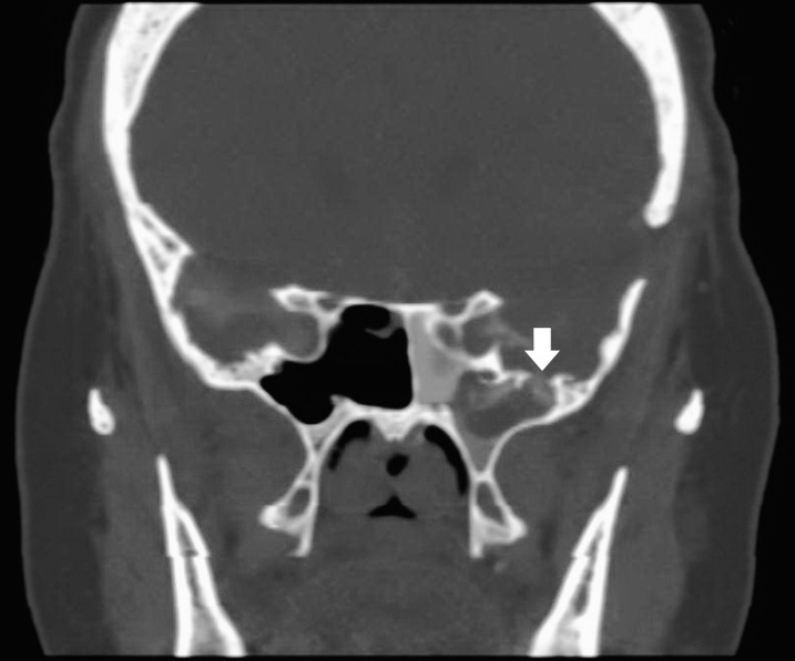
Defects in the lateral recess of the sphenoid sinus also pose a challenge (Figure 4). The endoscopic transpterygoid approach provides far-lateral access; the posterior face of the maxillary sinus is removed to access the pterygopalatine fossa, and the contents of the fossa are displaced to access the sphenoid sinus.34
Figure 4.
Coronal computed tomography shows a defect in the floor of the middle cranial fossa producing a cerebrospinal fluid leak into the pneumatized sphenoid wing (arrow). The left sphenoid sinus is filled with contrast fluid after the injection of intrathecal contrast.
Repair of cribriform and anterior ethmoid defects typically requires a mucosal graft overlaid with bioabsorbable materials and nonabsorbable packing to support the graft.35 Exposure of bony edges is followed by the removal of surrounding mucosa and application of reconstructive materials.
PREDICTORS OF SUCCESS IN ENDOSCOPIC SURGERY
Multiple factors influence the success of CSF leak repairs. Studies support the importance of distinguishing between low-flow and high-flow CSF leaks before treatment plans are developed.36
Large dural defects have more successful rates of CSF leak closure when a vascularized reconstructive approach is used compared to free graft reconstruction.37 Resection of pituitary macroadenomas and clival chordomas via an endoscopic approach also has higher leak closure rates compared to sublabial microscopic procedures.38-40 In comparison, craniopharyngiomas and meningiomas have lower rates of CSF leak closure success, as would be expected from their intradural location.40 Following intraarachnoidal dissection and endoscopic tumor resection, the nasoseptal flap has been noted to reduce case complications, particularly in the hands of an experienced surgeon.41 The presence of hydrocephalus or benign intracranial hypertension has the potential to significantly change patient outcomes. In an evaluation of patients who underwent CSF repair, all patients with recurrent leaks were found to have hydrocephalus.12
Patients with increased ICP typically benefit from intraoperative placement of a lumbar drain that can remove CSF, briefly decompressing the dura mater, elevating the bony defect, and allowing for more accurate placement of grafts. Some authors have recommended lumbar drains to control ICP 24-48 hours postoperatively.1 However, risks such as meningitis, pneumocephalus, and chronic headache, as well as delay in patient mobility and discharge, indicate the need for careful assessment on a case-by-case basis. Lumbar drains should be considered for use in cases of complex skull base defects with high-volume preoperative and intraoperative leaks.42
Postoperatively, maintaining graft integrity is crucial. Patients must be discouraged from engaging in any activity that places stress on the graft or increases ICP, including heavy lifting, intense exercise, and other strenuous activities. The type of graft used has not been found to affect patient outcomes, provided the CSF leak is controlled, and the choice of graft is based primarily on physician experience.43 Quality of life, however, has been shown to be better postoperatively with autologous grafts compared to nonautologous grafts.44,45
CONCLUSION
Repair of CSF rhinorrhea has evolved from requiring an open craniotomy approach to a minimally invasive endoscopic procedure. Trauma causes the majority of cases of CSF rhinorrhea, and an active surgical approach to treating CSF leaks may provide better long-term outcomes in select patients compared to more conservative management. Spontaneous CSF fistulas have lower closure success rates compared to traumatic fistulas and require more monitoring because of the possibility of recurrence and increased ICP. A combination of biochemical assay with radiologic studies is typically required to secure a diagnosis and guide management. Adjuncts such as intrathecally injected fluorescein can aid diagnosis and treatment plans. When conservative management fails, endoscopic repair of CSF leaks is an option in the modern surgical armamentarium. Craniotomy remains a possible approach for repairing leaks that are severe, recurrent, or not amenable to minimally invasive repair. The approach to repair a CSF fistula depends on the location of the dehiscence, the fistula size, and the flow volume. Several reconstructive materials and techniques are available to optimize successful CSF leak closure. The management of CSF rhinorrhea continues to evolve with the introduction of new repair techniques, materials, and studies of long-term outcomes.
ACKNOWLEDGMENTS
The authors have no financial or proprietary interest in the subject matter of this article.
This article meets the Accreditation Council for Graduate Medical Education and the American Board of Medical Specialties Maintenance of Certification competencies for Patient Care and Medical Knowledge.
REFERENCES
- 1. Wise SK, Schlosser RJ. Evaluation of spontaneous nasal cerebrospinal fluid leaks. Curr Opin Otolaryngol Head Neck Surg. 2007. February; 15 1: 28- 34. [DOI] [PubMed] [Google Scholar]
- 2. Rangel-Castilla L, Gopinath S, Robertson CS. Management of intracranial hypertension. Neurol Clin. 2008. May; 26 2: 521- 541, x doi: 10.1016/j.ncl.2008.02.003. [DOI] [PMC free article] [PubMed] [Google Scholar]
- 3. Zlab MK, Moore GF, Daly DT, Yonkers AJ. Cerebrospinal fluid rhinorrhea: a review of the literature. Ear Nose Throat J. 1992. July; 71 7: 314- 317. [PubMed] [Google Scholar]
- 4. Lee TJ, Huang CC, Chuang CC, Huang SF. Transnasal endoscopic repair of cerebrospinal fluid rhinorrhea and skull base defect: ten-year experience. Laryngoscope. 2004. August; 114 8: 1475- 1481. [DOI] [PubMed] [Google Scholar]
- 5. Eljamel MS. Fractures of the middle third of the face and cerebrospinal fluid rhinorrhoea. Br J Neurosurg. 1994; 8 3: 289- 293. [DOI] [PubMed] [Google Scholar]
- 6. Bernal-Sprekelsen M, Alobid I, Mullol J, Trobat F, Tomás-Barberán M. Closure of cerebrospinal fluid leaks prevents ascending bacterial meningitis. Rhinology. 2005. December; 43 4: 277- 281. [PubMed] [Google Scholar]
- 7. Currarino G, Maravilla KR, Salyer KE. Transsphenoidal canal (large craniopharyngeal canal) and its pathologic implications. AJNR Am J Neuroradiol. 1985. Jan-Feb; 6 1: 39- 43. [PMC free article] [PubMed] [Google Scholar]
- 8. Tomazic PV, Stammberger H. Spontaneous CSF-leaks and meningoencephaloceles in sphenoid sinus by persisting Sternberg's canal. Rhinology. 2009. December; 47 4: 369- 374. doi: 10.4193/Rhin08.236. [DOI] [PubMed] [Google Scholar]
- 9. Kaufman B, Nulsen FE, Weiss MH, Brodkey JS, White RJ, Sykora GF. Acquired spontaneous, nontraumatic normal-pressure cerebrospinal fluid fistulas originating from the middle fossa. Radiology. 1977. February; 122 2: 379- 387. [DOI] [PubMed] [Google Scholar]
- 10. Woodworth BA, Prince A, Chiu AG, et al. Spontaneous CSF leaks: a paradigm for definitive repair and management of intracranial hypertension. Otolaryngol Head Neck Surg. 2008. June; 138 6: 715- 720. doi: 10.1016/j.otohns.2008.02.010. [DOI] [PubMed] [Google Scholar]
- 11. Basu D, Haughey BH, Hartman JM. Determinants of success in endoscopic cerebrospinal fluid leak repair. Otolaryngol Head Neck Surg. 2006. November; 135 5: 769- 773. [DOI] [PubMed] [Google Scholar]
- 12. Ommaya AK. Spinal fluid fistulae. Clin Neurosurg. 1976; 23: 363- 392. [DOI] [PubMed] [Google Scholar]
- 13. Thurtell MJ, Wall M. Idiopathic intracranial hypertension (pseudotumor cerebri): recognition, treatment, and ongoing management. Curr Treat Options Neurol. 2013. February; 15 1: 1- 12. doi: 10.1007/s11940-012-0207-4. [DOI] [PMC free article] [PubMed] [Google Scholar]
- 14. Psaltis AJ, Schlosser RJ, Banks CA, Yawn J, Soler ZM. A systematic review of the endoscopic repair of cerebrospinal fluid leaks. Otolaryngol Head Neck Surg. 2012. August; 147 2: 196- 203. doi: 10.1177/0194599812451090. [DOI] [PubMed] [Google Scholar]
- 15. Zweig JL, Carrau RL, Celin SE, et al. Endoscopic repair of cerebrospinal fluid leaks to the sinonasal tract: predictors of success. Otolaryngol Head Neck Surg. 2000. September; 123 3: 195- 201. [DOI] [PubMed] [Google Scholar]
- 16. Chaaban MR, Illing E, Riley KO, Woodworth BA. Spontaneous cerebrospinal fluid leak repair: a five-year prospective evaluation. Laryngoscope. 2014. January; 124 1: 70- 75. doi: 10.1002/lary.24160. [DOI] [PubMed] [Google Scholar]
- 17. McCudden CR, Senior BA, Hainsworth S, et al. Evaluation of high resolution gel β(2)-transferrin for detection of cerebrospinal fluid leak. Clin Chem Lab Med. 2013. February; 51 2: 311- 315. [DOI] [PubMed] [Google Scholar]
- 18. Schlosser RJ, Bolger WE. Management of multiple spontaneous nasal meningoencephaloceles. Laryngoscope. 2002. June; 112 6: 980- 985. [DOI] [PubMed] [Google Scholar]
- 19. Martin TJ, Loehrl TA. Endoscopic CSF leak repair. Curr Opin Otolaryngol Head Neck Surg. 2007. February; 15 1: 35- 39. [DOI] [PubMed] [Google Scholar]
- 20. Krudy AG. MR myelography using heavily T2-weighted fast spin-echo pulse sequences with fat presaturation. AJR Am J Roentgenol. 1992. December; 159 6: 1315- 1320. [DOI] [PubMed] [Google Scholar]
- 21. Stone JA, Castillo M, Neelon B, Mukherji SK. Evaluation of CSF leaks: high-resolution CT compared with contrast-enhanced CT and radionuclide cisternography. AJNR Am J Neuroradiol. 1999. April; 20 4: 706- 712. [PMC free article] [PubMed] [Google Scholar]
- 22. Ecin G, Oner AY, Tokgoz N, Ucar M, Aykol S, Tali T. T2-weighted vs. intrathecal contrast-enhanced MR cisternography in the evaluation of CSF rhinorrhea. Acta Radiol. 2013. July; 54 6: 698- 701. doi: 10.1177/0284185113478008. [DOI] [PubMed] [Google Scholar]
- 23. Tabaee A, Placantonakis DG, Schwartz TH, Anand VK. Intrathecal fluorescein in endoscopic skull base surgery. Otolaryngol Head Neck Surg. 2007. August; 137 2: 316- 320. [DOI] [PubMed] [Google Scholar]
- 24. Seth R, Rajasekaran K, Benninger MS, Batra PS. The utility of intrathecal fluorescein in cerebrospinal fluid leak repair. Otolaryngol Head Neck Surg. 2010. November; 143 5: 626- 632. doi: 10.1016/j.otohns.2010.07.011. [DOI] [PubMed] [Google Scholar]
- 25. Meier JC, Bleier BS. Novel techniques and the future of skull base reconstruction. Adv Otorhinolaryngol. 2013; 74: 174- 183. doi: 10.1159/000342294. [DOI] [PubMed] [Google Scholar]
- 26. Wigand ME. Transnasal ethmoidectomy under endoscopical control. Rhinology. 1981. March; 19 1: 7- 15. [PubMed] [Google Scholar]
- 27. Cukurova I, Cetinkaya EA, Aslan IB, Ozkul D. Endonasal endoscopic repair of ethmoid roof cerebrospinal fluid fistula by suturing the dura. Acta Neurochir (Wien). 2008. September; 150 9: 897- 900; discussion 900 doi: 10.1007/s00701-008-0005-7. [DOI] [PubMed] [Google Scholar]
- 28. Pinheiro-Neto CD, Carrau RL, Prevedello DM, et al. Use of acoustic Doppler sonography to ascertain the feasibility of the pedicled nasoseptal flap after prior bilateral sphenoidotomy. Laryngoscope. 2010. September; 120 9: 1798- 1801. doi: 10.1002/lary.20996. [DOI] [PubMed] [Google Scholar]
- 29. Fortes FS, Carrau RL, Snyderman CH, et al. The posterior pedicle inferior turbinate flap: a new vascularized flap for skull base reconstruction. Laryngoscope. 2007. August; 117 8: 1329- 1332. [DOI] [PubMed] [Google Scholar]
- 30. Hadad G, Rivera-Serrano CM, Bassagaisteguy LH, et al. Anterior pedicle lateral nasal wall flap: a novel technique for the reconstruction of anterior skull base defects. Laryngoscope. 2011. August; 121 8: 1606- 1610. doi: 10.1002/lary.21889. [DOI] [PubMed] [Google Scholar]
- 31. Prevedello DM, Barges-Coll J, Fernandez-Miranda JC, et al. Middle turbinate flap for skull base reconstruction: cadaveric feasibility study. Laryngoscope. 2009. November; 119 11: 2094- 2098. doi: 10.1002/lary.20226. [DOI] [PubMed] [Google Scholar]
- 32. Kumar A, Maartens NF, Kaye AH. Reconstruction of the sellar floor using Bioglue following transsphenoidal procedures. J Clin Neurosci. 2003. January; 10 1: 92- 95. [DOI] [PubMed] [Google Scholar]
- 33. Purkey MT, Woodworth BA, Hahn S, Palmer JN, Chiu AG. Endoscopic repair of supraorbital ethmoid cerebrospinal fluid leaks. ORL J Otorhinolaryngol Relat Spec. 2009; 71 2: 93- 98. doi: 10.1159/000193219. [DOI] [PubMed] [Google Scholar]
- 34. Bolger WE. Endoscopic transpterygoid approach to the lateral sphenoid recess: surgical approach and clinical experience. Otolaryngol Head Neck Surg. 2005. July; 133 1: 20- 26. [DOI] [PubMed] [Google Scholar]
- 35. McMains KC, Gross CW, Kountakis SE. Endoscopic management of cerebrospinal fluid rhinorrhea. Laryngoscope. 2004. October; 114 10: 1833- 1837. [DOI] [PubMed] [Google Scholar]
- 36. McCoul ED, Anand VK, Singh A, Nyquist GG, Schaberg MR, Schwartz TH. Long-term effectiveness of a reconstructive protocol using the nasoseptal flap after endoscopic skull base surgery. World Neurosurg. 2014. January; 81 1: 136- 143. doi: 10.1016/j.wneu.2012.08.011. [DOI] [PubMed] [Google Scholar]
- 37. Harvey RJ, Parmar P, Sacks R, Zanation AM. Endoscopic skull base reconstruction of large dural defects: a systematic review of published evidence. Laryngoscope. 2012. February; 122 2: 452- 459. doi: 10.1002/lary.22475. [DOI] [PubMed] [Google Scholar]
- 38. DeKlotz TR, Chia SH, Lu W, Makambi KH, Aulisi E, Deeb Z. Meta-analysis of endoscopic versus sublabial pituitary surgery. Laryngoscope. 2012. March; 122 3: 511- 518. doi: 10.1002/lary.22479. [DOI] [PubMed] [Google Scholar]
- 39. Komotar RJ, Starke RM, Raper DM, Anand VK, Schwartz TH. Endoscopic endonasal versus open repair of anterior skull base CSF leak, meningocele, and encephalocele: a systematic review of outcomes. J Neurol Surg A Cent Eur Neurosurg. 2013. July; 74 4: 239- 250. doi: 10.1055/s-0032-1325636. [DOI] [PubMed] [Google Scholar]
- 40. Raper DMS, Komotar RJ, Starke RM, et al. Endoscopic versus open approaches to the skull base: a comprehensive literature review. Oper Tech Otolaryngol Head Neck Surg. 2011; 22: 302- 307. [Google Scholar]
- 41. Kassam AB, Thomas A, Carrau RL. Endoscopic reconstruction of the cranial base using a pedicled nasoseptal flap. Neurosurgery. 2008. July; 63 1 Suppl 1: ONS44-52; discussion ONS52-3 doi: 10.1227/01.neu.0000335010.53122.75. [DOI] [PubMed] [Google Scholar]
- 42. Casiano RR, Jassir D. Endoscopic cerebrospinal fluid rhinorrhea repair: is a lumbar drain necessary? Otolaryngol Head Neck Surg. 1999. December; 121 6: 745- 750. [DOI] [PubMed] [Google Scholar]
- 43. Hegazy HM, Carrau RL, Snyderman CH, Kassam A, Zweig J. Transnasal endoscopic repair of cerebrospinal fluid rhinorrhea: a meta-analysis. Laryngoscope. 2000. July; 110 7: 1166- 1172. [DOI] [PubMed] [Google Scholar]
- 44. McCoul ED, Anand VK, Bedrosian JC, Schwartz TH. Endoscopic skull base surgery and its impact on sinonasal-related quality of life. Int Forum Allergy Rhinol. 2012. Mar-Apr; 2 2: 174- 181. doi: 10.1002/alr.21008. [DOI] [PubMed] [Google Scholar]
- 45. McCoul ED, Anand VK, Schwartz TH. Improvements in site-specific quality of life 6 months after endoscopic anterior skull base surgery: a prospective study. J Neurosurg. 2012. September; 117 3: 498- 506. doi: 10.3171/2012.6.JNS111066. [DOI] [PubMed] [Google Scholar]


