

Abstract
We present the case of a 35 year-old male with pain and swelling in his right thigh. By CT and sonography, an abscess was localized to the deep, anteromedial, mid-thigh within the quadriceps muscle, along with a 1.3 cm loose body. The infected loose body was removed under ultrasound guidance without complications.
Abbreviations: CT, computed tomography
Introduction
Retained loose bodies sometimes remain undetected in the initial evaluation of soft tissue wounds in the emergency room setting, especially if the loose body is localized deep to the skin surface or does not present as a radio-opaque density on radiography. Loose bodies retained in wounds may be associated with significant problems including inflammation, pain, and chronic infection refractory to antibiotics. In order to minimize the potential difficulties associated with surgical excision, effective removal of small loose bodies from soft tissues requires adequate detection techniques. Ultrasound can be a safe and cost-effective tool in surgical removal of loose bodies in soft tissue. The use of ultrasound for localization and guided removal of a soft tissue loose body is discussed.
Case Report
A 35-year old man presented to the emergency department with a 6 day history of right thigh pain and swelling, which had worsened over the past 2 or 3 days. He reported a similar problem approximately 5 years prior while living in Brazil but received no treatment at the time. He admitted to fevers and chills but denied shortness of breath, chest pain or nausea and vomiting. Vital signs at the time of admission showed a temperature of 103.1°. Physical exam was unremarkable except for tenderness without erythema or crepitus associated with a 12 to 15 cm area of induration in the anterior aspect of the mid right thigh.
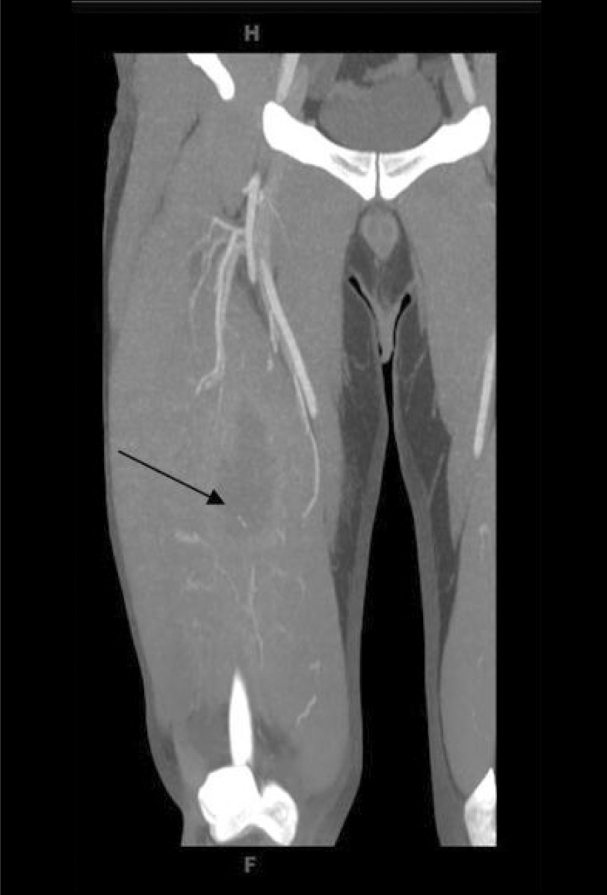
Deep venous thrombosis was suspected however; subsequent two dimensional, pulsed Doppler and color Doppler venous ultrasound of the right lower extremity showed no evidence of thrombus in the right femoral and popliteal veins. CT angiogram was then performed from the right hip through to the plantar arch during intravenous administration of contrast. No evidence of arterial stenosis was shown. Axial, coronal and sagittal images were obtained, and CT revealed a large 9.7 × 3.8 × 3.6 cm fluid collection in the deep anteromedial mid thigh within the quadriceps muscle with peripheral enhancement, likely representing abscess (Fig. 1). An elongated 1.3 cm hyperdensity was seen dependently within the abscess cavity, 5.6 cm deep to the skin surface, suspicious for foreign body. Soft tissue swelling was noted at this level, in addition to a small lateral effusion in the joint space of the right knee. Incision and drainage was performed and the patient was discharged home on oral antibiotics.
Figure 1.

Coronal CT image of the right thigh demonstrating a large 9.7 × 3.8 × 3.6 cm fluid collection in the deep, anteromedial, mid-thigh within the quadriceps muscle with peripheral enhancement.
Four days later upon evaluation in the surgical clinic for follow up, he was found to be febrile with a tense right thigh and mild to moderate edema. Local wound exploration done at the clinic was unsuccessful to fully delineate the extent of the fluid collection and concern for residual abscess prompted referral to the emergency department for further evaluation. In the ED the patient underwent multichannel axial imaging of the right lower extremity with intravenous contrast. Comparison with prior lower extremity CT angiogram showed evidence of large residual abscess associated with the right quadriceps with minimal interval change (Fig. 2). An elongated 1.3 cm hyperdensity was again demonstrated in the dependent aspect of the fluid collection, likely representing a foreign body. Soft tissue swelling was noted at this level with post drainage changes and a small amount of air in the soft tissues. The quadriceps muscle appeared expanded slightly more than on the previous study.
Figure 2.
Coronal CT of the right thigh depicting large residual abscess associated with the right quadriceps with minimal interval change.
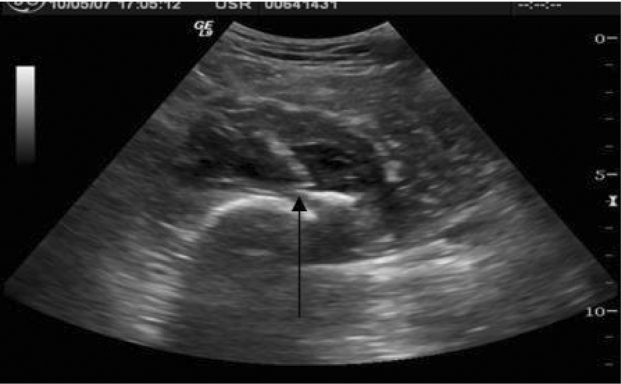
The patient was admitted and on the following day ultrasound guided drainage of the abscess with retrieval of the loose body was attempted. The patient was placed in the supine position on the examination table and scout scans were obtained. A linear, dense loose body measuring approximately 1 cm in length was seen within the fluid collection (Fig. 3).
Figure 3.

Axial Sonogram of the medial thigh shows a hyperechoic, loose body within the infero-medial aspect of a hypoechoic fluid collection.
The overlying skin was marked at an appropriate level. The skin was cleansed with antiseptic solution and local lidocaine was infiltrated down to the margin of the abscess cavity. A Cook 18 gauge Turner needle was used to enter the abscess cavity and confirmed the loose body to lie dependent within the inferior medial aspect of the abscess as the loose body was readily movable (Fig. 4A). Under real-time ultrasound, blunt dissection was performed to enter the cavity with a Kelly clamp through the initial needle puncture site. The loose body was then grasped with the clamp and removed along the previously created tract (Fig. 4B). Post procedure scans were obtained and showed no residual loose body (Fig. 5). Hyperechoic foci within the region of hypoechoic fluid are representative of air that had been introduced into the abscess cavity during procedural manipulation. The skin was then re-cleansed for ultrasound guided abscess drainage.
Figure 4A.
Sonographically guided loose body needle localization in the transverse plane.
Figure 4B.

Sonogram in transverse plane of the thigh showing guided removal of loose body.
Figure 5.
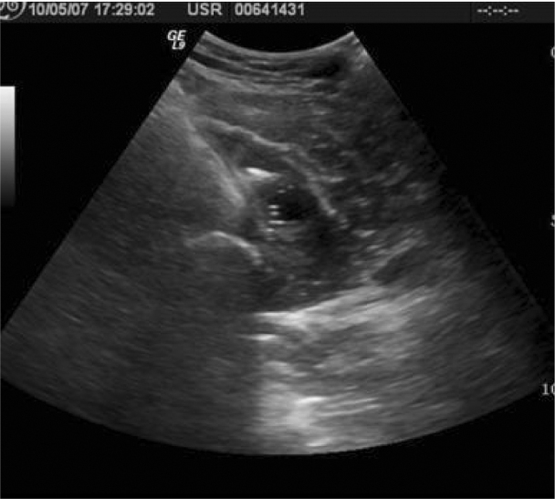
Post procedural transverse scan of the thigh shows hypoechoic fluid collection with absence of loose body.
To maintain sterility for fluid cultures, another Cook 18-gauge Turner needle was introduced into the fluid collection and aspiration of the fluid sample was obtained and sent for evaluation. Aspiration of approximately 55 cc of a purulent, thick fibrinous fluid was accomplished. A 10 Fr. Pigtail catheter was locked in place and connected to gravity drainage and a dressing was applied. Subsequent culture of the abscess fluid revealed Staphylococcus aureus. The patient was discharged on oral antibiotics and returned 10 days later for follow up. CT imaging revealed the abscess cavity to be markedly decreased in size compared to the prior study, measuring 1.4 × 1.0 × 1.0 cm (Fig. 5). The radio-opaque loose body seen previously within the fluid collection was no longer identified.
The loose body was identified upon removal as ossified tissue, possibly representing an avulsed bone fragment. Such a fragment could act as a nidus for refractory infection, similar to a foreign body. However, the origin of the loose body was never definitively identified and the item was not sent for laboratory analysis.
Discussion
The major challenge to loose body removal from soft tissues is precise location of the loose body to minimize dissection. Sonography can be an effective guide to removal of loose bodies, requiring smaller dissection and decreased time for their removal. If the site is old and the original track has closed, determining the loose body position by ultrasound can also result in a smaller incision with decreased trauma to the surrounding structures [1]. Sonography has been shown to have a positive impact in the care of patients who present with soft tissue loose bodies either in conjunction with or separate from radiographs. Although sonography has been demonstrated to be very effective in localization and removal of loose bodies, limitations still exist to its successful use, including operator experience, size and depth of the loose body, proximity to bony structures and the presence of air within the wound, which can often mimic the appearance of a loose body. Diligence must also be taken to avoid pitfalls in distinguishing loose bodies from other body tissue such as ossified cartilage, sesamoid bones, keratin plugs and scar tissue [1, 2]. In cases where deep location of the loose body precludes visualization and removal by ultrasound, fluoroscopy has long been shown to serve as a valuable adjunct. Because the exact position of an object buried in soft tissue is difficult to determine using two-dimensional imaging techniques, the major advantage of sonography is accurate three-dimensional preoperative localization of a loose body [3] and real time imaging. In contrast to conventional radiographic and fluoroscopic techniques, sonography can locate even loose bodies that are radiolucent and without radiation exposure [4].
Although a number of ultrasound procedures for superficial loose body removal have been described in the literature [5, 6, 7, 8, 9], we found few references describing successful removal of a loose body from the thigh [10] and no cases where removal was successful from deep soft tissue in the absence of the original track site.
Several factors allowed for the successful ultrasound-guided localization and removal of the loose body in our case. The loose body in our case measured 1.3 cm in length, making it easily distinguishable from the surrounding tissues. Also, the echogenic loose body was surrounded by a hypo-echoic fluid collection further contrasting the loose body and surrounding tissue/fluid. Presence of a hypoechoic rim around an echogenic loose body has been demonstrated to improve the sensitivity and specificity of ultrasound examination [11]. Furthermore, the calcified nature of the loose body itself allowed for easy visualization by ultrasound. The dependent location of the echogenic focus within a pocket of fluid allowed for demonstration of its mobility, which confirmed it as representing an intracavitary loose body.
Figure 6.
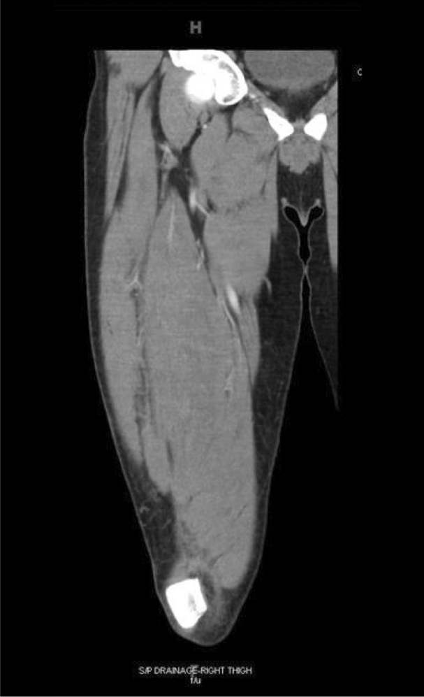
Post procedural coronal CT examination of the right thigh revealing significant decrease in size of right leg abscess cavity.
Conclusion
Retained loose bodies, overlooked at the initial physical examination, have a predisposition to develop into serious infections, which may be unresponsive to antibiotic therapy alone. We have demonstrated that ultrasound guided therapy provides a safe, cost-effective modality in localization and removal of retained loose bodies in deep soft tissue, even in the presence of soft tissue gas when the original track site is absent.
Footnotes
Published: July 8, 2008
References
- 1.Shiels WE, 2nd, Babcock DS, Wilson JL, Burch RA. Localization and guided removal of soft-tissue foreign bodies with sonography. AJR. 1990;155:1277–1281. doi: 10.2214/ajr.155.6.2122680. [PubMed] [DOI] [PubMed] [Google Scholar]
- 2.Gooding GA, Hardiman T, Sumers M, Stess R, Graf P, Grunfeld C. Sonography of the hand and foot in foreign body detection. J Ultrasound Med. 1987 Aug;6(8):441–447. doi: 10.7863/jum.1987.6.8.441. [PubMed] [DOI] [PubMed] [Google Scholar]
- 3.Fornage BD, Schernberg FL. Sonographic diagnosis of foreign bodies of the distal extremities. AJR Am J Roentgenol. 1986; Sep;147(3):567–569. doi: 10.2214/ajr.147.3.567. [PubMed] [DOI] [PubMed] [Google Scholar]
- 4.Blankstein A, Cohen I, Heiman Z, Salai M, Diamant L, Heim M, Chechick A. Ultrasonography as a diagnostic modality and therapeutic adjuvant in the management of soft tissue foreign bodies in the lower extremities. Isr Med Assoc J. 2001; Jun;3(6):411–413. [PubMed] [PubMed] [Google Scholar]
- 5.Crawford R, Matheson AB. Clinical value of ultrasonography in the detection and removal of radiolucent foreign bodies. Injury. 1989; Nov;20(6):341–343. doi: 10.1016/0020-1383(89)90008-9. [PubMed] [DOI] [PubMed] [Google Scholar]
- 6.Blankstein A, Cohen I, Heiman Z, Salai M, Heim M, Checkick A. Localization, detection and guided removal of soft tissue in the hands using sonography. Arch Orthop Trauma Surg. 2000;120(9):514–517. doi: 10.1007/s004020000173. [PubMed] [DOI] [PubMed] [Google Scholar]
- 7.Gibbs TS. The use of sonography in identification, localization, and removal of soft tissue foreign bodies. J Diag Med Sono. 2006;22(1):5–21. [Google Scholar]
- 8.Nelson AL, Sinow RM. Real-time ultrasonographically guided removal of nonpalpable and intramuscular Norplant capsules. Am J Obstet Gynecol. 1998;178:1185–1193. doi: 10.1016/s0002-9378(98)70321-7. [PubMed] [DOI] [PubMed] [Google Scholar]
- 9.McArthur T, Abell BA, Levsky ME. A Procedure for Soft Tissue Foreign Body Removal under Real-Time Ultrasound Guidance. Military Medicine. 2007;172(8):858–859. doi: 10.7205/milmed.172.8.858. [PubMed] [DOI] [PubMed] [Google Scholar]
- 10.Yiengpruksawan A, Mariadason J, Ganepola GA, Freeman HP. Localization and retrieval of bullets under ultrasound guidance. Arch Surg. 1987; Sep;122(9):1082–1084. doi: 10.1001/archsurg.1987.01400210120020. [PubMed] [DOI] [PubMed] [Google Scholar]
- 11.Boyse TD, Fessel DP, Jacobson JA, Lin J, van Holsbeeck MT, Hayes CW. US of soft-tissue foreign bodies and associated complications with surgical correlation. Radiographics. 2001; Sep-Oct;21(5):1251–1256. doi: 10.1148/radiographics.21.5.g01se271251. [PubMed] [DOI] [PubMed] [Google Scholar]