Abstract
Metastatic disease is the leading cause of cancer-related deaths and involves critical interactions between tumor cells and the microenvironment. Hypoxia is a potent microenvironmental factor promoting metastatic progression. Clinically, hypoxia and the expression of the hypoxia-inducible transcription factors HIF-1 and HIF-2 are associated with increased distant metastasis and poor survival in a variety of tumor types. Moreover, HIF signaling in malignant cells influences multiple steps within the metastatic cascade. Here we review research focused on elucidating the mechanisms by which the hypoxic tumor microenvironment promotes metastatic progression. These studies have identified potential biomarkers and therapeutic targets regulated by hypoxia that could be incorporated into strategies aimed at preventing and treating metastatic disease.
Tumor metastasis is a major challenge in the clinical management of cancer. Metastatic disease is responsible for more than 90% of all cancer-related deaths and is often associated with high patient mortality because it is difficult to treat surgically or with conventional chemotherapy and radiation therapy. Metastasis is a complex and dynamic process that selects for highly aggressive tumor cells that acquire the ability to disseminate from their tissue of origin, survive within foreign tissue microenvironments, and grow at distant sites. Recent studies have demonstrated that cellular and molecular constituents regulated by the microenvironment in both the primary tumor and distant tissue profoundly influence the propensity of tumors to metastasize.
Oxygen is a microenvironmental factor that controls developmental processes, as well as normal tissue homeostasis. At the cellular level, oxygen is required for oxidative metabolism, the generation of adenosine 5′-triphosphate (ATP), and cell survival. To maintain oxygen homeostasis, metazoan organisms use the hypoxic signaling pathway to facilitate oxygen delivery and cellular adaptation to oxygen deprivation (1). Hypoxia is emerging as a key microenvironmental factor in the regulation of metastasis. Here we review our current knowledge of the roles that hypoxia and hypoxic signaling play in metastasis and highlight recent advances within this field.
Hypoxic signaling
The hypoxia-inducible transcription factors HIF-1 and HIF-2 coordinate the adaptive cellular response to low oxygen tensions by activating gene expression programs controlling glucose uptake, metabolism, angiogenesis, erythropoiesis, cell proliferation, differentiation, and apoptosis (1). Both initiation and progression of tumorigenesis are promoted by HIF signaling, and tumors use multiple strategies to activate this pathway.
Hypoxia, or low oxygen tensions, is a hallmark feature of the tumor microenvironment. It has been estimated that 50 to 60% of solid tumors contain regions of hypoxia and/or anoxia that arise as a result of an imbalance between oxygen delivery and oxygen consumption (2). Within the tumor microenvironment, oxygen delivery is impaired because of abnormalities in the tumor vasculature, including distended capillaries characterized by leaky and sluggish blood flow (3). At the same time, oxygen consumption rates are high because of the demands of proliferating tumor cells and infiltrating immune cells. Clinically, hypoxia is associated with HIF activation, metastasis, and resistance to chemotherapy and radiotherapy, as well as poor patient survival, indicating that hypoxia may contribute to tumor progression and resistance to therapy (4–6).
“Clinically, [tumor] hypoxia is associated with HIF activation, metastasis, and resistance to chemotherapy and radiotherapy, as well as poor patient survival…”
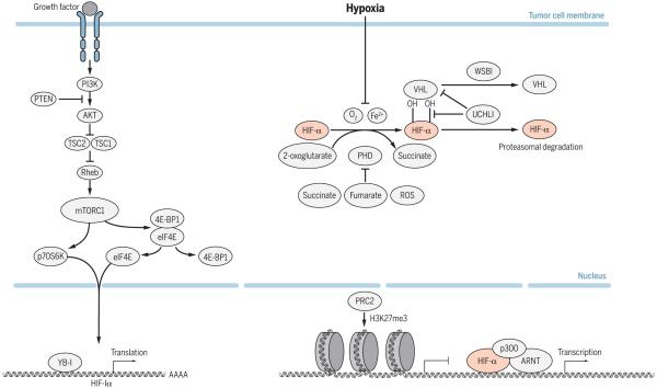
Hypoxia activates HIF signaling by promoting the protein stability of HIF-α subunits. Under normoxic conditions, prolyl hydroxylase enzymes (EGLN 1–3, also known as PHD 1–3) use oxygen as a substrate to hydroxylate key proline residues within the HIF-1 and HIF-2 α subunits (7–9). This hydroxylation event allows the von Hippel–Lindau tumor suppressor protein (VHL), the substrate recognition component of an E3 ubiquitin ligase complex, to bind to HIF-α and target it for proteasomal degradation (10–14). Under hypoxic conditions, PHD activity is inhibited, resulting in HIF-α stabilization and translocation to the nucleus. In the nucleus, HIF-α subunits dimerize with aryl hydrocarbon receptor nuclear translocator (ARNT) and bind to hypoxia-responsive elements in target genes to activate gene transcription by recruiting transcriptional coactivators including p300/CBP (15–17). Hundreds of genes are activated in response to HIF-1 and HIF-2; these factors allow cells to survive and adapt to low oxygen tensions, as well as confer changes in both and host that promote metastasis (Fig. 1).
Fig. 1. Mechanisms of HIF-1 and HIF-2 activation in tumor cells.
Hypoxia is a common mechanism of HIF activation in cancer. Under normoxic conditions, PHD enzymes (also called EGLN 1–3) utilize oxygen as a substrate to hydroxylate key proline residues located within the HIF-α subunit. This hydroxylation event mediates pVHL binding and subsequent ubiquitination and degradation by the 26S proteasome. Under conditions of hypoxia or loss of pVHL, HIF-α is stabilized and translocates to the nucleus, where it heterodimerizes with ARNT and binds to hypoxia response elements (HREs) within regulatory regions of target genes. The HIF heterodimer activates gene expression at these sites upon cofactor (p300/CBP) recruitment. PRC2-mediated histone methylation can inhibit the activation of HIF target genes involved in metastasis. HIF activity can also be induced in tumor cells through mechanisms that enhance HIF-α production (TORC1, YB-1) or prevent its degradation (UCHL1, WSB1).
In addition to activation by hypoxia, the PHD-VHL-HIF signaling pathway can be regulated by genetic events and other microenvironmental factors. The enzymatic activity of the EGLN 1–3 requires iron and 2-oxoglutarate to hydroxylate the HIF-α subunits. As a result, EGLN 1–3 activity and HIF-α stability are affected by iron availability and can be inhibited by Krebs cycle intermediates, including succinate and fumarate, which compete with 2-oxoglutarate (18–22). In tumor cells, mutations in the genes encoding succinate dehydrogenase (SDH) or fumarate hydratase (FH) cause the accumulation of cytosolic succinate or fumarate; this inhibits the activity of prolyl hydroxylases and leads to HIF stabilization under normoxic conditions [for a recent review, see (22)]. Additionally, PHD activity can be inhibited by reactive oxygen species (ROS) produced by the mitochondria; however, the mechanisms by which this occurs under hypoxia remain to be defined (Fig. 1) (21, 23).
As noted above, VHL regulates hypoxic signaling by controlling the ubiquitination and degradation of HIF-α subunits. Genetic mutations or epigenetic inactivation of VHL causes VHL disease, which is associated with constitutive activation of HIF-1 and HIF-2 and the development of highly vascularized tumors, including hemangioblastomas, pheochromocytomas, pancreatic islet cell tumors, and clear cell renal cell carcinoma (ccRCC). Renal cell carcinoma is a major cause of morbidity in VHL patients, owing to the ability of these tumors to metastasize to other tissue sites (24). In addition, most sporadic ccRCCs lose VHL function through genomic alterations (25). Although genetic inactivation of VHL is thought to be an early event in the pathogenesis of ccRCC, recent work has identified mechanisms by which tumors further potentiate HIF activity to promote metastatic progression. By analyzing metastatic subpopulations of VHL-deficient ccRCC cells, Vanharanta and colleagues discovered that epigenetic modifications within prometastatic genes allow for enhanced expression of metastasis-associated HIF target genes (26). For example, loss of polycomb repressive complex 2 (PRC2)–dependent histone H3 Lys-27 trimethylation activates HIF-mediated chemokine (C-X-C motif) receptor 4 (CXCR4) expression to promote invasion and metastasis (26). These findings open new areas of research aimed at understanding how epigenetic changes at prometastatic loci are altered during tumor progression. Recent studies have also indicated that VHL activity in tumors can be regulated by WSB1, an E3 ligase that targets pVHL for ubiquitination and proteasomal degradation (27). WSB1 expression in tumor cells leads to HIF stabilization under normoxic and hypoxic conditions and correlates with metastasis in cancer patients (Fig. 1) (27).
Finally, hypoxic signaling can be activated by factors that promote HIF-α production or prevent its degradation. Both HIF-α mRNA transcription and translation are regulated by TORC1 activity [reviewed in (22)]. Inactivating mutations in phosphatase and tensin homolog (PTEN) or the tuberosis sclerosis complex (TSC) complex, as well as activating mutations in phosphatidylinositol 3-kinase (PI3K) or AKT, lead to increased TORC1 activity that promotes HIF-α stabilization (22, 28–31). Additionally, activation of PI3K and AKT signaling through growth factor receptor signaling can promote HIF-α stabilization and activity under normoxic conditions (32, 33). HIF-1 mRNA translation can be enhanced under normoxic and hypoxic conditions through the binding of YB-1, an RNA and DNA binding protein; this leads to increased HIF-1 protein and prometastatic activity in sarcoma cells (34). HIF activity can also be promoted through the up-regulation of factors that prevent HIF-α degradation. Using an elegant screening approach to identify factors that would lead to HIF-1 transcriptional activity under normoxic conditions, Goto et al. recently identified the ubiquitin C-terminal hydrolase-L1 (UCHL1) as a HIF-1 deubiquitinating enzyme that promotes HIF-1 activity under normoxic and hypoxic conditions by preventing VHL-mediated degradation of HIF-1 (Fig. 1) (35). UCHL1 is associated with HIF-1 and distant metastasis in cancer patients, suggesting that UCHL1 may promote metastasis through HIF (35). In support of this concept, genetic inactivation of HIF-1 in UCHL1-overexpressing cells reversed the metastatic potential of these cells (35). In summary, these studies highlight the diverse mechanisms by which HIF signaling can be activated under hypoxic and normoxic conditions and may contribute to HIF activation during metastatic progression (Fig. 1).
Clinically, HIF-1 and HIF-2 are highly expressed in primary tumors and metastases. Immunohistochemical analysis of human primary tumor samples has revealed an association between high HIF-1 expression and metastasis in patients with gynecological, pancreatic, esophageal, lung, and prostate cancers (36–38). HIF-2 expression in primary tumors is also associated with distant metastasis in patients with small cell lung and breast cancers (39, 40). Moreover, increased HIF expression is often linked with increased patient mortality in a variety of human cancers (41). In experimental models, overexpression of HIF in tumor cells promotes metastasis (42, 43), whereas inactivation of HIF decreases the metastatic potential of tumor cells (43–47). Together, these clinical and experimental findings demonstrate an important role for HIF signaling in metastatic tumor progression. However, before this knowledge can be translated into safe and effective antimetastatic therapies, it is critical to understand how this pathway is used by cancer cells and stromal cells to support metastasis.
Mechanisms of HIF-mediated metastasis
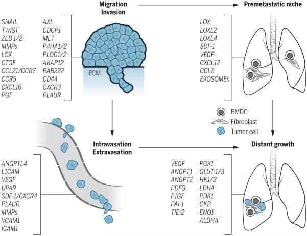
Hypoxia and activation of HIF signaling influence multiple steps within the metastatic cascade, including invasion and migration, intravasation and extravasation, and establishment of the premetastatic niche, as well as survival and growth at the distant site [Fig. 2; for recent reviews, see (48, 49)]. Here, we highlight recent studies that have revealed novel mechanisms by which HIF signaling promotes metastatic progression.
Fig. 2. HIF signaling regulates multiple steps within the metastatic cascade.
Highlighted in brackets are direct target genes of HIF that promote each step of metastasis (to the lung, in the example shown). ECM, extracellular matrix; BMDC, bone marrow–derived cell.
Hypoxic signaling and immune evasion
A critical step in metastatic tumor progression is the ability of tumor cells to evade immune attack. Tumor hypoxia is thought to promote the immunosuppressive phenotypes of both tumor cells and infiltrating immune cells. In preclinical models, respiratory hyperoxia (60% O2) can promote tumor regression, reduce metastatic disease, and prolong animal survival (50). In these studies, respiratory hyperoxia was associated with a decrease in intratumoral hypoxia and an immunopermissive tumor microenvironment characterized by abundant CD8+ T cell infiltration and decreased regulatory T cell infiltration (50). Understanding the mechanisms by which hypoxia promotes immunosuppression is an active area of investigation and may have important therapeutic implications in the treatment of metastatic disease.
Hypoxia promotes tumor resistance to immune attack through several HIF-dependent mechanisms. Normoxic and hypoxic stabilization of HIF signaling in tumor cells promotes resistance to cytotoxic T lymphocyte–mediated lysis (51–54). Hypoxia and HIF-mediated activation of autophagy in tumor cells also regulates natural killer (NK) cell–mediated antitumor responses; this occurs through the hypoxic degradation of NK-derived granzyme B in autophagosomes and the induction of inositol 1,4,5-trisphosphate receptor, type 1 (55, 56). Hypoxic signaling can promote resistance to T cell–mediated killing by increasing the expression of programmed death–ligand 1 (PD-L1) on tumor cells and myeloid-derived suppressor cells (MDSCs), and by enhancing CTLA-4 expression on CD8+ T cells, respectively [reviewed in (57)]. Additionally, hypoxic tumor cells evade innate immune recognition through the up-regulation of CD47, a cell surface molecule that interacts with signal regulatory protein alpha (SIRP α) on the surface of macrophages to block phagocytosis (58).
Hypoxia and HIF promote an immunosuppressive microenvironment by recruiting regulatory T cells (Tregs), MDSCs, and macrophages into the tumor microenvironment [reviewed in (57, 59)]. Hypoxic tumor cells recruit Tregs expressing CCR10 (CC chemokine receptor type 10) and NP-1 (neuropilin-1) into the tumor microenvironment through the secretion of CCL28, transforming growth factor–beta (TGF-β), and vascular endothelial growth factor [VEGF; reviewed in (57)]. Once in the tumor microenvironment, Tregs promote immune tolerance and angiogenesis, which supports metastatic tumor growth (60). Similarly, hypoxic tumor cells recruit myeloid cells into the tumor microenvironment through the secretion of chemokines and cytokines, including CCL5, C-X-C motif chemokine 12 (CXCL12 or SDF-1), VEGF, and endothelins [ET-1 and ET-2; reviewed in (57)]. Hypoxic signaling in MDSCs and macrophages directly contributes to tumor progression by promoting an immunosuppressive phenotype and stimulating angiogenesis [reviewed in (57, 61–63)]. Moreover, hypoxia inhibits the effector functions of tumor-infiltrating lymphocytes largely through the accumulation of extracellular adenosine. Adenosine signals through A2A receptors on T cells to increase cyclic adenosine 3′,5′-monophosphate, which inhibits T cell proliferation, expansion, and cytokine secretion [reviewed in (59)]. Collectively, these findings illustrate the diverse ways in which HIF signaling within tumor cells, MDSCs, and tumor-associated macrophages help establish an immunosuppressive tumor microenvironment.
HIF signaling in EMT, invasion, and migration
In the early stages of metastasis, tumor cells acquire invasive and migratory properties that allow them to exit the localized primary tumor mass and enter neighboring and distant host tissues. Tumor cells take advantage of many mechanisms to migrate and invade, including both individual and collective cell-migration strategies (64). In vitro tumor cell migration is associated with an epithelial-mesenchymal transition (EMT). EMT is characterized by changes in cell morphology, as well as cell-cell and cell-matrix adhesions. Cell-cell adhesions are mediated primarily by cadherin proteins expressed at intercellular junctions (65). Loss of E-cadherin allows cells to detach from their neighbors and begin their migratory route toward the circulatory or lymphatic system to seek out new terrain. Reduced expression of E-cadherin is often observed in metastatic tumors, and work with experimental models has demonstrated that loss of E-cadherin is sufficient to promote metastasis of malignant cells (66).
Hypoxia or overexpression of HIF is sufficient to induce EMT and invasion in multiple cell types (42, 67–71). There are both direct and indirect mechanisms by which HIF signaling promotes EMT. A direct role for HIF in the regulation of key EMT transcription factors such as ZEB1, Snail, and Twist has been demonstrated through the identification of functional hypoxia response elements (HREs) within the regulatory elements of these genes (Fig. 2) (42, 72, 73). The HIF pathway also indirectly promotes EMT through a number of cell signaling pathways, including Notch (74, 75), TGF-β (76), integrin-linked kinase (77), certain tyrosine kinase receptors (48), Wnt (73), and Hedgehog (78, 79). We recently identified the AXL receptor tyrosine kinase as a novel HIF target driving EMT, invasion, and metastasis in VHL-deficient and hypoxic cancer cells (80). A growing body of literature supports an essential role for AXL in promoting metastasis within many tumor types, including breast (81), ovarian (82), and lung (83). Moreover, AXL inhibition has been reported to sensitize drug-resistant mesenchymal tumor cells to anticancer agents, including antimitotic agents, epidermal growth factor receptor (EGFR) inhibitors, and PI3K inhibitors (83–86). Loss of AXL does not appear to have deleterious effects on embryonic development or normal adult tissue function in mice (87, 88). Moreover, biological inhibitors of AXL, such as soluble AXL decoy receptors, are specific and are not associated with normal tissue or hematologic toxicities in mice (82, 89). AXL thus appears to be a promising therapeutic target for the treatment of advanced cancer, and several inhibitors of AXL signaling are currently in preclinical and clinical development.
HIF signaling and the premetastatic niche
The late stages of metastasis are governed by the ability of disseminated tumor cells to colonize, survive, and grow within the distant tissue microenvironment. Over the past decade, experiments in mice have convincingly shown that formation of a tumor-promoting premetastatic niche in secondary organs is essential for the extravasation and growth of metastatic tumor cells at the distant site (90). The finding that the premetastatic niche is in part established by tumor-secreted factors has stimulated great interest in identifying the factors that govern tissue-specific metastasis for potential therapeutic targeting. The role of tumor microenvironmental factors such as hypoxia in the regulation of these secreted factors is likewise an area of intense investigation.
HIF signaling in the primary tumor contributes to the production of secreted factors involved in premetastatic niche formation (Fig. 2). In breast cancer cells, HIF signaling results in the increased expression and secretion of lysyl oxidase (LOX) and LOX-like proteins (LOXL2 and 4). These proteins modify the collagen matrix in the lung to recruit bone marrow–derived cells (BMDCs) that prime the lung for metastatic colonization. The BMDCs promote metastasis by producing chemokines that recruit tumor cells to the lung by mechanisms that include stimulation of tumor cell extravasation (44, 90–95). Recent studies have shown that LOX is also a key factor in the establishment of the premetastatic niche in bone. Proteomic analysis of the hypoxic secretome in bone tropic MDA-231 human breast cancer cells compared to MDA-231 parental cells identified LOX as one of the most highly up-regulated secreted proteins in bone-tropic cells (96). Additionally, LOX expression is clinically associated with estrogen receptor negative (ER−) breast cancer metastasis to bone. In preclinical models, LOX expression in MDA-231 bone-tropic cells within the primary tumor is both necessary and sufficient to induce osteolytic bone lesions and cortical bone loss in immunocompromised mice even before the arrival of tumor cells. LOX regulates osteoclastogenesis, and high expression of LOX in primary tumors was found to be associated with the formation of osteolytic bone lesions in mice (96). These findings suggest that LOX may be an important biomarker to identify ER− negative patients who are at risk for bone relapse. They also raise the possibility that LOX inhibition may prevent bone and lung relapse in breast cancer patients. Given that LOX also promotes tumor cell proliferation, invasion, and angiogenesis, LOX is an attractive therapeutic target for cancer therapy (97).
Another mechanism by which breast cancer cells condition the premetastatic niche is through tumor-lymphatic vessel cross-talk. Lymphatic vessels at the distant site support metastatic colonization in breast, melanoma, prostate, gastric, and colon cancer patients (98). Lymphatic endothelial cells (LECs) are a specialized endothelium that line lymphatic vessels and promote lymph metastases by actively recruiting tumor cells to the lymphatic vessels. LECs accomplish this by producing chemoattractants, including SDF-1 and CCL21, that bind to cognate receptors CXCR4 and CCR7, respectively, which are expressed by tumor cells (99). LECs within premetastatic organs are conditioned by tumor-derived factors to facilitate tumor cell recruitment, extravasation, and colonization (98). IL-6 secretion by the tumor activates signal transducer and activator of transcription 3 (STAT3) signaling in LECs localized within the lymph node and lung to promote (i) CCL5-mediated recruitment of CCR5-positive breast cancer cells into the lymphatic system and (ii) VEGF-mediated lung vascular permeability and lymph node angiogenesis (98). Interestingly, IL-6–induced VEGF expression in LECs is associated with the activation of HIF-1, indicating that tumor-derived factors may promote lymphatic metastasis at least in part through the activation of HIF signaling in LECs (98). Collectively, these studies demonstrate that hypoxia and HIF signaling mediates tumor-stromal cross-talk by activating the expression of circulating factors that can prime and direct metastatic growth.
An emerging area of interest is the role of exosomes in metastasis and whether hypoxia and HIF signaling might promote formation of the premetastatic niche by this mechanism. Exosomes are extracellular vesicles that regulate cell-cell communication by carrying and transferring molecules including proteins, lipids, microRNAs, and mRNAs (100). Exosomes have recently been implicated in the establishment of the premetastatic niche. They are present at elevated levels in the serum of patients with cancer and are associated with advanced disease. Moreover, tumor-derived exosomes preferentially localize to sites of metastasis, including liver, lung, and bone marrow, where they promote vascular permeability and increase BMDC recruitment (101–104). However, the factors within the primary tumor that regulate exosome content and release remain largely unknown. Recent studies have shown that exosomes mediate hypoxia-dependent stimulation of angiogenesis in glioblastoma multiforme (GBM) through the activation of paracrine signaling in endothelial cells and pericytes (105). Interestingly, exosomes isolated from the plasma of GBM patients contain increased amounts of hypoxia-regulated proteins— including MMP9, MMP8, platelet-derived growth factor (PDGF), PAI1, and insulin-like growth factor–binding protein 3 (IGFBP3)—compared to exosomes isolated from plasma of sex- and age-matched controls (105). This work suggests that hypoxia can influence exosome cargo content to promote angiogenesis. Additionally, these findings suggest that the hypoxic signature of exosomes may serve as a noninvasive biomarker to predict the aggressiveness of malignant tumors. In addition to regulating exosome cargo content, hypoxia also promotes microvesicle shedding. Hypoxic cells up-regulate the small guanosine triphosphatase RAB22A in a HIF-dependent manner to promote microvesicle shedding, invasion, and metastasis (106).
“…hypoxia within the liver microenvironment selects for disseminated tumor cells that have the ability to metabolically adapt to the hypoxic stress.”
HIF signaling and cellular growth and survival at the distant site
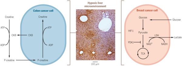
Successful metastatic colonization requires disseminated tumor cells to adapt to the microenvironment of the distant tissue, which can be very different from that of the tissue harboring the primary tumor. To illustrate this point, we discuss liver, a common metastatic site for colorectal cancer. The liver maintains overall energy balance by controlling carbohydrate and lipid metabolism. Within the liver, an oxygen gradient is established that provides boundaries for metabolic activities within the organ (Fig. 3). The partial pressure of oxygen in periportal blood is 60 to 65 mm Hg and in the perivenous blood it is 30 to 35 mm Hg (107). Because oxygen is an essential electron acceptor for oxidative metabolism, hepatocytes that perform glucose or fatty acid oxidation are located in the aerobic periportal zone, whereas oxygen-independent metabolic functions such as glucose uptake, glycolysis, and fatty acid synthesis are predominantly performed by perivenous hepatocytes (108). Given that colon cancer cells metastasize to the liver through the portal circulation, Loo and colleagues (109) hypothesized that these cells would experience acute hypoxia and competition for glycolytic substrates. Using a HIF-1 transcriptional luciferase reporter mouse, they determined that colon cancer cells experience hypoxia early after hepatic dissemination. They found that the disseminated cancer cells increase their release of the enzyme creatine kinase brain-type (CKB), which helps them survive within the hypoxic microenvironment of the liver (109). CKB controls the amount of rapidly mobilized high-energy phosphates by catalyzing the transfer of a high-energy phosphate group from ATP to the metabolite creatine, producing phosphocreatine (110). Under conditions of metabolic stress, such as hypoxia, tumor cells use phosphocreatine stores as a source of high-energy phosphate that can be transferred to adenosine 5′-diphosphate (ADP) to generate ATP (110). Depletion of CKB increased caspase-mediated cell death in hypoxic tumor cells within the liver, suggesting that hepatic hypoxia is a barrier for colon cancer cells early in metastatic dissemination and that cells overcome this metabolic stress by generating ATP from phosphocreatine reserves (Fig. 3) (109).
Fig. 3. Mechanisms of metabolic reprogramming in liver metastasis.
The liver microenvironment is hypoxic. Pimonidazole staining (brown) demonstrates areas of hypoxia in the liver (middle panel). Metastatic tumor cells enter hypoxic regions of the liver and must metabolically adapt to survive the metabolic stress associated with hypoxia. Disseminated breast cancer cells (right) metabolically adapt by increasing the expression of pyruvate dehydrogenase kinase 1 (PDK1) and promoting glycolytic reprogramming. Disseminated colon cancer cells (left) metabolically adapt by increasing the expression and secretion of creatine kinase brain-type (CKB). This enzyme controls the amount of rapidly mobilized high-energy phosphates by catalyzing the transfer of a high-energy phosphate group from ATP to the metabolite creatine, producing phosphocreatine. Under conditions of metabolic stress, such as hypoxia, tumor cells utilize phosphocreatine stores as a source of high-energy phosphate that can be transferred to ADP to generate ATP.
Consistent with the concept that hepatic hypoxia and the associated glycolytic phenotype in periportal regions of the liver provide a barrier for metastatic colonization, Dupuy and colleagues observed that breast metastases to the liver are highly dependent on glycolysis for survival in comparison to bone and lung metastases (111). The metabolic reprogramming of liver-metastatic breast cancer cells was largely dependent on the HIF-1 target gene pyruvate dehydrogenase kinase (PDK1). PDK1 represses mitochondrial function by antagonizing the function of pyruvate dehydrogenase (PDH), a rate-limiting enzyme for pyruvate conversion to acetyl–coenzyme A and entry into the tricarboxylic acid cycle (112). Notably, PDK1 expression was elevated in breast cancer liver metastases compared to primary tumor specimens, further supporting the notion that PDK1 expression is particularly important for the growth and survival of breast cancer cells within the liver microenvironment. These findings suggest that breast cancer cells adapt to the hypoxic liver microenvironment through the activation of HIF signaling and glycolytic reprogramming mediated by PDK1 (Fig. 3). Thus, these studies demonstrate that hypoxia within the liver microenvironment selects for disseminated tumor cells that have the ability to metabolically adapt to the hypoxic stress.
Finally, HIF promotes late stages of metastasis at the distant site by stimulating angiogenesis. Like primary tumors, metastases require angiogenesis to support their growth. VEGF-A is a proangiogenic factor produced by tumor cells that stimulates the recruitment and proliferation of endothelial cells, as well as promoting pericyte proliferation and migration (113). VEGF-A is a well-established HIF target, and its expression is induced by HIF signaling in both primary tumors and metastases (114).
Conclusions
Hypoxia and HIF-dependent signaling play an important role in metastatic tumor progression. Although hypoxia is an important factor that activates HIF signaling within tumors, recent studies have demonstrated that malignant cells use multiple strategies to enhance HIF signaling during progression by promoting HIF-α mRNA translation, protein stability, and downstream target gene expression.
The hypoxic tumor microenvironment influences both the early and late stages of metastasis. Hypoxic stress plays an important role in immune escape by promoting immune suppression and tumor resistance. Within the primary tumor, HIF-dependent gene expression controls EMT, invasion, migration, and angiogenesis to support the early stages of metastasis. Recent work has identified the receptor tyrosine kinase, AXL, as a critical mediator of HIF-dependent invasion and metastasis, as well as a potential therapeutic target for the prevention and treatment of metastatic disease. In addition, HIF signaling promotes the production of secreted factors such as LOX, LOX-like proteins, and exosomes to establish a prometastatic environment within the lung and bones of breast cancer patients. Hypoxia at the distant site also plays a substantial role in selecting for metastatic cells that can survive the metabolic stress associated with hypoxia. Metastatic breast cancer cells activate HIF signaling within the hypoxic environment of the liver to metabolically adapt to the glycolytic environment. Additionally, colon cancer cells adapt to the metabolic stress within the hypoxic liver by releasing CKB into the extracellular space to generate and import phosphocreatine as a source of ATP generation in this microenvironment.
In summary, recent studies have begun to highlight the diverse mechanisms by which hypoxia and HIF-dependent signaling promote metastatic tumor progression. As a result, a number of new biomarkers and therapeutic targets have been identified that could potentially be valuable for the detection and treatment of metastatic disease.
ACKNOWLEDGMENTS
This work was supported by NIH Grants CA-198291, CA-67166, and CA-197713, the Silicon Valley Foundation, the Sydney Frank Foundation, and the Kimmelman Fund (A.J.G.); and the Department of Defense Ovarian Cancer Research Academy OC140611 (E.B.R.). We apologize to those colleagues whose work we could not cite owing to space constraints.
REFERENCES
- 1.Semenza GL. Cell. 2012;148:399–408. doi: 10.1016/j.cell.2012.01.021. [DOI] [PMC free article] [PubMed] [Google Scholar]
- 2.Vaupel P, Mayer A. Cancer Metastasis Rev. 2007;26:225–239. doi: 10.1007/s10555-007-9055-1. [DOI] [PubMed] [Google Scholar]
- 3.Brown JM, Giaccia AJ. Cancer Res. 1998;58:1408–1416. [PubMed] [Google Scholar]
- 4.Rankin EB, Giaccia AJ. Cell Death Differ. 2008;15:678–685. doi: 10.1038/cdd.2008.21. [DOI] [PMC free article] [PubMed] [Google Scholar]
- 5.Schindl M, et al. Clin. Cancer Res. 2002;8:1831–1837. [PubMed] [Google Scholar]
- 6.Yamamoto Y, et al. Breast Cancer Res. Treat. 2008;110:465–475. doi: 10.1007/s10549-007-9742-1. [DOI] [PubMed] [Google Scholar]
- 7.Epstein AC, et al. Cell. 2001;107:43–54. doi: 10.1016/s0092-8674(01)00507-4. [DOI] [PubMed] [Google Scholar]
- 8.Bruick RK, McKnight SL. Science. 2001;294:1337–1340. doi: 10.1126/science.1066373. [DOI] [PubMed] [Google Scholar]
- 9.Ivan M, et al. Proc. Natl. Acad. Sci. U.S.A. 2002;99:13459–13464. doi: 10.1073/pnas.192342099. [DOI] [PMC free article] [PubMed] [Google Scholar]
- 10.Jaakkola P, et al. Science. 2001;292:468–472. doi: 10.1126/science.1059796. [DOI] [PubMed] [Google Scholar]
- 11.Ivan M, et al. Science. 2001;292:464–468. doi: 10.1126/science.1059817. [DOI] [PubMed] [Google Scholar]
- 12.Maynard MA, et al. J. Biol. Chem. 2003;278:11032–11040. doi: 10.1074/jbc.M208681200. [DOI] [PubMed] [Google Scholar]
- 13.Maxwell PH, et al. Nature. 1999;399:271–275. doi: 10.1038/20459. [DOI] [PubMed] [Google Scholar]
- 14.Tanimoto K, Makino Y, Pereira T, Poellinger L. EMBO J. 2000;19:4298–4309. doi: 10.1093/emboj/19.16.4298. [DOI] [PMC free article] [PubMed] [Google Scholar]
- 15.Schödel J, et al. Blood. 2011;117:e207–e217. doi: 10.1182/blood-2010-10-314427. [DOI] [PMC free article] [PubMed] [Google Scholar]
- 16.Wang GL, Jiang BH, Rue EA, Semenza GL. Proc. Natl. Acad. Sci. U.S.A. 1995;92:5510–5514. doi: 10.1073/pnas.92.12.5510. [DOI] [PMC free article] [PubMed] [Google Scholar]
- 17.Wang GL, Semenza GL. Proc. Natl. Acad. Sci. U.S.A. 1993;90:4304–4308. doi: 10.1073/pnas.90.9.4304. [DOI] [PMC free article] [PubMed] [Google Scholar]
- 18.Pan Y, et al. Mol. Cell. Biol. 2007;27:912–925. doi: 10.1128/MCB.01223-06. [DOI] [PMC free article] [PubMed] [Google Scholar]
- 19.Nandal A, et al. Cell Metab. 2011;14:647–657. doi: 10.1016/j.cmet.2011.08.015. [DOI] [PMC free article] [PubMed] [Google Scholar]
- 20.Jones DT, Trowbridge IS, Harris AL. Cancer Res. 2006;66:2749–2756. doi: 10.1158/0008-5472.CAN-05-3857. [DOI] [PubMed] [Google Scholar]
- 21.Majmundar AJ, Wong WJ, Simon MC. Mol. Cell. 2010;40:294–309. doi: 10.1016/j.molcel.2010.09.022. [DOI] [PMC free article] [PubMed] [Google Scholar]
- 22.Kaelin WG., Jr. Cold Spring Harb. Symp. Quant. Biol. 2011;76:335–345. doi: 10.1101/sqb.2011.76.010975. [DOI] [PMC free article] [PubMed] [Google Scholar]
- 23.Kaelin WG., Jr. Cell Metab. 2005;1:357–358. doi: 10.1016/j.cmet.2005.05.006. [DOI] [PubMed] [Google Scholar]
- 24.Lonser RR, et al. Lancet. 2003;361:2059–2067. doi: 10.1016/S0140-6736(03)13643-4. [DOI] [PubMed] [Google Scholar]
- 25.Gnarra JR, et al. Nat. Genet. 1994;7:85–90. doi: 10.1038/ng0594-85. [DOI] [PubMed] [Google Scholar]
- 26.Vanharanta S, et al. Nat. Med. 2013;19:50–56. doi: 10.1038/nm.3029. [DOI] [PMC free article] [PubMed] [Google Scholar]
- 27.Kim JJ, et al. Genes Dev. 2015;29:2244–2257. doi: 10.1101/gad.268128.115. [DOI] [PMC free article] [PubMed] [Google Scholar]
- 28.Hudson CC, et al. Mol. Cell. Biol. 2002;22:7004–7014. doi: 10.1128/MCB.22.20.7004-7014.2002. [DOI] [PMC free article] [PubMed] [Google Scholar]
- 29.Arsham AM, Howell JJ, Simon MC. J. Biol. Chem. 2003;278:29655–29660. doi: 10.1074/jbc.M212770200. [DOI] [PubMed] [Google Scholar]
- 30.Brugarolas JB, Vazquez F, Reddy A, Sellers WR, Kaelin WG., Jr. Cancer Cell. 2003;4:147–158. doi: 10.1016/s1535-6108(03)00187-9. [DOI] [PubMed] [Google Scholar]
- 31.Zundel W, et al. Genes Dev. 2000;14:391–396. [PMC free article] [PubMed] [Google Scholar]
- 32.Laughner E, Taghavi P, Chiles K, Mahon PC, Semenza GL. Mol. Cell. Biol. 2001;21:3995–4004. doi: 10.1128/MCB.21.12.3995-4004.2001. [DOI] [PMC free article] [PubMed] [Google Scholar]
- 33.Zhong H, et al. Cancer Res. 2000;60:1541–1545. [PubMed] [Google Scholar]
- 34.El-Naggar AM, et al. Cancer Cell. 2015;27:682–697. doi: 10.1016/j.ccell.2015.04.003. [DOI] [PubMed] [Google Scholar]
- 35.Goto Y, et al. Nat. Commun. 2015;6:6153. doi: 10.1038/ncomms7153. [DOI] [PMC free article] [PubMed] [Google Scholar]
- 36.Jin Y, et al. PLOS ONE. 2015;10:e0127229. doi: 10.1371/journal.pone.0127229. [DOI] [PMC free article] [PubMed] [Google Scholar]
- 37.Matsuo Y, et al. J. Hepatobiliary Pancreat. Sci. 2014;21:105–112. doi: 10.1002/jhbp.6. [DOI] [PubMed] [Google Scholar]
- 38.Ping W, Sun W, Zu Y, Chen W, Fu X. Tumour Biol. 2014;35:4401–4409. doi: 10.1007/s13277-013-1579-0. [DOI] [PubMed] [Google Scholar]
- 39.Luan Y, et al. Pathol. Res. Pract. 2013;209:184–189. doi: 10.1016/j.prp.2012.10.017. [DOI] [PubMed] [Google Scholar]
- 40.Wang HX, Qin C, Han FY, Wang XH, Li N. GMR. 2014;13:2817–2826. doi: 10.4238/2014.January.22.6. [DOI] [PubMed] [Google Scholar]
- 41.Semenza GL. Oncogene. 2010;29:625–634. doi: 10.1038/onc.2009.441. [DOI] [PMC free article] [PubMed] [Google Scholar]
- 42.Yang MH, et al. Nat. Cell Biol. 2008;10:295–305. doi: 10.1038/ncb1691. [DOI] [PubMed] [Google Scholar]
- 43.Hiraga T, Kizaka-Kondoh S, Hirota K, Hiraoka M, Yoneda T. Cancer Res. 2007;67:4157–4163. doi: 10.1158/0008-5472.CAN-06-2355. [DOI] [PubMed] [Google Scholar]
- 44.Wong CC, et al. Proc. Natl. Acad. Sci. U.S.A. 2011;108:16369–16374. doi: 10.1073/pnas.1113483108. [DOI] [PMC free article] [PubMed] [Google Scholar]
- 45.Zhang H, et al. Oncogene. 2012;31:1757–1770. doi: 10.1038/onc.2011.365. [DOI] [PMC free article] [PubMed] [Google Scholar] [Retracted]
- 46.Liao D, Corle C, Seagroves TN, Johnson RS. Cancer Res. 2007;67:563–572. doi: 10.1158/0008-5472.CAN-06-2701. [DOI] [PubMed] [Google Scholar]
- 47.Hanna SC, et al. J. Clin. Invest. 2013;123:2078–2093. doi: 10.1172/JCI66715. [DOI] [PMC free article] [PubMed] [Google Scholar]
- 48.De Bock K, Mazzone M, Carmeliet P. Nat. Rev. Clin. Oncol. 2011;8:393–404. doi: 10.1038/nrclinonc.2011.83. [DOI] [PubMed] [Google Scholar]
- 49.Semenza GL. Biochim. Biophys. Acta. 2016;1863:382–391. doi: 10.1016/j.bbamcr.2015.05.036. [DOI] [PMC free article] [PubMed] [Google Scholar]
- 50.Hatfield SM, et al. Sci. Transl. Med. 2015;7:277ra30. doi: 10.1126/scitranslmed.aaa1260. [DOI] [PMC free article] [PubMed] [Google Scholar]
- 51.Lee YH, et al. Clin. Cancer Res. 2015;21:1438–1446. doi: 10.1158/1078-0432.CCR-14-1979. [DOI] [PMC free article] [PubMed] [Google Scholar]
- 52.Noman MZ, et al. J. Immunol. 2009;182:3510–3521. doi: 10.4049/jimmunol.0800854. [DOI] [PubMed] [Google Scholar]
- 53.Barsoum IB, et al. Cancer Res. 2011;71:7433–7441. doi: 10.1158/0008-5472.CAN-11-2104. [DOI] [PubMed] [Google Scholar]
- 54.Noman MZ, et al. Cancer Res. 2011;71:5976–5986. doi: 10.1158/0008-5472.CAN-11-1094. [DOI] [PubMed] [Google Scholar]
- 55.Baginska J, et al. Proc. Natl. Acad. Sci. U.S.A. 2013;110:17450–17455. doi: 10.1073/pnas.1304790110. [DOI] [PMC free article] [PubMed] [Google Scholar]
- 56.Messai Y, et al. Cancer Res. 2014;74:6820–6832. doi: 10.1158/0008-5472.CAN-14-0303. [DOI] [PubMed] [Google Scholar]
- 57.Palazon A, Goldrath AW, Nizet V, Johnson RS. Immunity. 2014;41:518–528. doi: 10.1016/j.immuni.2014.09.008. [DOI] [PMC free article] [PubMed] [Google Scholar]
- 58.Zhang H, et al. Proc. Natl. Acad. Sci. U.S.A. 2015;112:E6215–E6223. doi: 10.1073/pnas.1520032112. [DOI] [PMC free article] [PubMed] [Google Scholar]
- 59.Kumar V, Gabrilovich DI. Immunology. 2014;143:512–519. doi: 10.1111/imm.12380. [DOI] [PMC free article] [PubMed] [Google Scholar]
- 60.Facciabene A, et al. Nature. 2011;475:226–230. doi: 10.1038/nature10169. [DOI] [PubMed] [Google Scholar]
- 61.Corzo CA, et al. J. Exp. Med. 2010;207:2439–2453. doi: 10.1084/jem.20100587. [DOI] [PMC free article] [PubMed] [Google Scholar]
- 62.Colegio OR, et al. Nature. 2014;513:559–563. doi: 10.1038/nature13490. [DOI] [PMC free article] [PubMed] [Google Scholar]
- 63.Chaturvedi P, Gilkes DM, Takano N, Semenza GL. Proc. Natl. Acad. Sci. U.S.A. 2014;111:E2120–E2129. doi: 10.1073/pnas.1406655111. [DOI] [PMC free article] [PubMed] [Google Scholar]
- 64.Friedl P, Wolf K. Nat. Rev. Cancer. 2003;3:362–374. doi: 10.1038/nrc1075. [DOI] [PubMed] [Google Scholar]
- 65.Cavallaro U, Christofori G. Nat. Rev. Cancer. 2004;4:118–132. doi: 10.1038/nrc1276. [DOI] [PubMed] [Google Scholar]
- 66.Derksen PW, et al. Cancer Cell. 2006;10:437–449. doi: 10.1016/j.ccr.2006.09.013. [DOI] [PubMed] [Google Scholar]
- 67.Higgins DF, et al. J. Clin. Invest. 2007;117:3810–3820. doi: 10.1172/JCI30487. [DOI] [PMC free article] [PubMed] [Google Scholar]
- 68.Kim WY, et al. J. Clin. Invest. 2009;119:2160–2170. doi: 10.1172/JCI38443. [DOI] [PMC free article] [PubMed] [Google Scholar]
- 69.Krishnamachary B, et al. Cancer Res. 2003;63:1138–1143. [PubMed] [Google Scholar]
- 70.Krishnamachary B, et al. Cancer Res. 2006;66:2725–2731. doi: 10.1158/0008-5472.CAN-05-3719. [DOI] [PubMed] [Google Scholar]
- 71.Liu Y, et al. Tumour Biol. 2014;35:8103–8114. doi: 10.1007/s13277-014-2056-0. [DOI] [PubMed] [Google Scholar]
- 72.Luo D, Wang J, Li J, Post M. Mol. Cancer Res. 2011;9:234–245. doi: 10.1158/1541-7786.MCR-10-0214. [DOI] [PubMed] [Google Scholar]
- 73.Zhang W, et al. PLOS ONE. 2015;10:e0129603. doi: 10.1371/journal.pone.0129603. [DOI] [PMC free article] [PubMed] [Google Scholar]
- 74.Chen J, Imanaka N, Chen J, Griffin JD. Br. J. Cancer. 2010;102:351–360. doi: 10.1038/sj.bjc.6605486. [DOI] [PMC free article] [PubMed] [Google Scholar]
- 75.Sahlgren C, Gustafsson MV, Jin S, Poellinger L, Lendahl U. Proc. Natl. Acad. Sci. U.S.A. 2008;105:6392–6397. doi: 10.1073/pnas.0802047105. [DOI] [PMC free article] [PubMed] [Google Scholar]
- 76.Copple BL. Liver Int. 2010;30:669–682. doi: 10.1111/j.1478-3231.2010.02205.x. [DOI] [PMC free article] [PubMed] [Google Scholar]
- 77.Chou CC, Chuang HC, Salunke SB, Kulp SK, Chen CS. Oncotarget. 2015;6:8271–8285. doi: 10.18632/oncotarget.3186. [DOI] [PMC free article] [PubMed] [Google Scholar]
- 78.Spivak-Kroizman TR, et al. Cancer Res. 2013;73:3235–3247. doi: 10.1158/0008-5472.CAN-11-1433. [DOI] [PMC free article] [PubMed] [Google Scholar]
- 79.Lei J, et al. Mol. Cancer. 2013;12:66. doi: 10.1186/1476-4598-12-66. [DOI] [PMC free article] [PubMed] [Google Scholar]
- 80.Rankin EB, et al. Proc. Natl. Acad. Sci. U.S.A. 2014;111:13373–13378. doi: 10.1073/pnas.1404848111. [DOI] [PMC free article] [PubMed] [Google Scholar]
- 81.Gjerdrum C, et al. Proc. Natl. Acad. Sci. U.S.A. 2010;107:1124–1129. doi: 10.1073/pnas.0909333107. [DOI] [PMC free article] [PubMed] [Google Scholar]
- 82.Rankin EB, et al. Cancer Res. 2010;70:7570–7579. doi: 10.1158/0008-5472.CAN-10-1267. [DOI] [PMC free article] [PubMed] [Google Scholar]
- 83.Byers LA, et al. Clin. Cancer Res. 2013;19:279–290. doi: 10.1158/1078-0432.CCR-12-1558. [DOI] [PMC free article] [PubMed] [Google Scholar]
- 84.Zhang Z, et al. Nat. Genet. 2012;44:852–860. doi: 10.1038/ng.2330. [DOI] [PMC free article] [PubMed] [Google Scholar]
- 85.Elkabets M, et al. Cancer Cell. 2015;27:533–546. doi: 10.1016/j.ccell.2015.03.010. [DOI] [PMC free article] [PubMed] [Google Scholar]
- 86.Wilson C, et al. Cancer Res. 2014;74:5878–5890. doi: 10.1158/0008-5472.CAN-14-1009. [DOI] [PubMed] [Google Scholar]
- 87.Lu Q, et al. Nature. 1999;398:723–728. doi: 10.1038/19554. [DOI] [PubMed] [Google Scholar]
- 88.Angelillo-Scherrer A, et al. Nat. Med. 2001;7:215–221. doi: 10.1038/84667. [DOI] [PubMed] [Google Scholar]
- 89.Kariolis MS, et al. Nat. Chem. Biol. 2014;10:977–983. doi: 10.1038/nchembio.1636. [DOI] [PMC free article] [PubMed] [Google Scholar]
- 90.Kaplan RN, et al. Nature. 2005;438:820–827. doi: 10.1038/nature04186. [DOI] [PMC free article] [PubMed] [Google Scholar]
- 91.Erler JT, et al. Nature. 2006;440:1222–1226. doi: 10.1038/nature04695. [DOI] [PubMed] [Google Scholar]
- 92.Erler JT, et al. Cancer Cell. 2009;15:35–44. doi: 10.1016/j.ccr.2008.11.012. [DOI] [PMC free article] [PubMed] [Google Scholar]
- 93.Yang L, et al. Cancer Cell. 2004;6:409–421. doi: 10.1016/j.ccr.2004.08.031. [DOI] [PubMed] [Google Scholar]
- 94.Lyden D, et al. Nat. Med. 2001;7:1194–1201. doi: 10.1038/nm1101-1194. [DOI] [PubMed] [Google Scholar]
- 95.Gao D, et al. Science. 2008;319:195–198. doi: 10.1126/science.1150224. [DOI] [PubMed] [Google Scholar]
- 96.Cox TR, et al. Nature. 2015;522:106–110. doi: 10.1038/nature14492. [DOI] [PMC free article] [PubMed] [Google Scholar] [Retracted]
- 97.Cox TR, Gartland A, Erler JT. Cancer Res. 2016;76:188–192. doi: 10.1158/0008-5472.CAN-15-2306. [DOI] [PubMed] [Google Scholar]
- 98.Lee E, et al. Nat. Commun. 2014;5:4715. doi: 10.1038/ncomms5715. [DOI] [PMC free article] [PubMed] [Google Scholar]
- 99.Albrecht I, Christofori G. Int. J. Dev. Biol. 2011;55:483–494. doi: 10.1387/ijdb.103226ia. [DOI] [PubMed] [Google Scholar]
- 100.Peinado H, Lavotshkin S, Lyden D. Semin. Cancer Biol. 2011;21:139–146. doi: 10.1016/j.semcancer.2011.01.002. [DOI] [PubMed] [Google Scholar]
- 101.Peinado H, et al. Nat. Med. 2012;18:883–891. doi: 10.1038/nm.2753. [DOI] [PMC free article] [PubMed] [Google Scholar]
- 102.Costa-Silva B, et al. Nat. Cell Biol. 2015;17:816–826. doi: 10.1038/ncb3169. [DOI] [PMC free article] [PubMed] [Google Scholar]
- 103.Hoshino A, et al. Nature. 2015;527:329–335. doi: 10.1038/nature15756. [DOI] [PMC free article] [PubMed] [Google Scholar]
- 104.Umezu T, et al. Blood. 2014;124:3748–3757. doi: 10.1182/blood-2014-05-576116. [DOI] [PMC free article] [PubMed] [Google Scholar]
- 105.Kucharzewska P, et al. Proc. Natl. Acad. Sci. U.S.A. 2013;110:7312–7317. doi: 10.1073/pnas.1220998110. [DOI] [PMC free article] [PubMed] [Google Scholar]
- 106.Wang T, et al. Proc. Natl. Acad. Sci. U.S.A. 2014;111:E3234–E3242. doi: 10.1073/pnas.1410041111. [DOI] [PMC free article] [PubMed] [Google Scholar]
- 107.Jungermann K, Kietzmann T. Hepatology. 2000;31:255–260. doi: 10.1002/hep.510310201. [DOI] [PubMed] [Google Scholar]
- 108.Jungermann K. Semin. Liver Dis. 1988;8:329–341. doi: 10.1055/s-2008-1040554. [DOI] [PubMed] [Google Scholar]
- 109.Loo JM, et al. Cell. 2015;160:393–406. doi: 10.1016/j.cell.2014.12.018. [DOI] [PMC free article] [PubMed] [Google Scholar]
- 110.Wyss M, Kaddurah-Daouk R. Physiol. Rev. 2000;80:1107–1213. doi: 10.1152/physrev.2000.80.3.1107. [DOI] [PubMed] [Google Scholar]
- 111.Dupuy F, et al. Cell Metab. 2015;22:577–589. doi: 10.1016/j.cmet.2015.08.007. [DOI] [PubMed] [Google Scholar]
- 112.Papandreou I, Cairns RA, Fontana L, Lim AL, Denko NC. Cell Metab. 2006;3:187–197. doi: 10.1016/j.cmet.2006.01.012. [DOI] [PubMed] [Google Scholar]
- 113.Joyce JA, Pollard JW. Nat. Rev. Cancer. 2009;9:239–252. doi: 10.1038/nrc2618. [DOI] [PMC free article] [PubMed] [Google Scholar]
- 114.Maxwell PH, et al. Proc. Natl. Acad. Sci. U.S.A. 1997;94:8104–8109. doi: 10.1073/pnas.94.15.8104. [DOI] [PMC free article] [PubMed] [Google Scholar]


