


Abstract
A 52-year-old man presented to the emergency department with a one-day history of epigastric pain. The patient reported a remote history of a “difficult” laparoscopic cholecystectomy that was converted to an open cholecystectomy in 1994. Further operative details were unavailable. Multiple radiologic studies were obtained, all demonstrating a saccular cystic structure in the gallbladder fossa containing calculi. A completion open cholecystectomy, or “recholecystectomy,” revealed a remnant gallbladder with cholecystitis and cholelithiasis. Multimodality imaging findings are reviewed.
Abbreviations: CT, computed tomography; MRCP, magnetic resonance cholangiopancreatography; ERCP, endoscopic retrograde cholangiopancreatography
Introduction
Incomplete gallbladder surgery, involving leaving a long cystic duct or gallbladder remnant, for example, can occur in both open and laparoscopic procedures (1). Incomplete surgery can lead to postcholecystectomy syndrome, which is a recurrence of symptoms similar to those that led to the initial cholecystectomy. The syndrome has been reported in 10–40% of postoperative patients and is seen more commonly in women (1). In this case, the patient suffered recurrent cholecystitis with cholelithiasis secondary to a retained gallbladder remnant, 14 years after a cholecystectomy.
Case report
A 52-year-old male presented to the emergency department with a one-day history of epigastric pain. The patient denied any associated nausea, vomiting, diarrhea, constipation, chest pain, or shortness of breath. He reported having a “difficult” cholecystectomy that was converted to an open procedure in 1994. Further details of the operation were not available. Additional surgical history included a prostatectomy for prostate cancer. Medical history included hypertension, cardiomyopathy, and hyperlipidemia.
Figure 1.
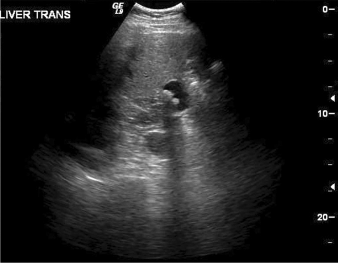
52-year-old man with recurrent cholecystitis and cholelithiasis. Transverse sonographic image at the level of the gallbladder fossa reveals a fluid-filled, saclike structure containing shadowing calculi, consistent with a remnant gallbladder.
Figure 2A-D.

52-year-old man with recurrent cholecystitis and cholelithiasis. Contrast-enhanced axial CT slices through the level of the gallbladder fossa demonstrate a saclike structure in continuity with the biliary system containing multiple calculi (arrows).
Physical exam revealed a mildly obese, soft abdomen with minimal tenderness, particularly in the right upper quadrant. The remainder of the physical exam was unremarkable. Laboratory results were significant for an elevated bilirubin of 3.9, ALT of 1057, AST of 395, and LDH of 332.
Abdominal ultrasound obtained on the day of admission revealed a fluid-filled saclike structure in the region of the gallbladder fossa resembling a gallbladder and containing shadowing calculi. Review of a recent radiograph before admission revealed right-upper-quadrant surgical clips, confirming the history of prior cholecystectomy. Additionally, there was mild biliary dilatation and an echogenic liver. The possibility of retained gallstones within a dilated cystic duct remnant was raised. Abdominal CT the same day demonstrated a saclike structure in the gallbladder fossa, containing two calculi and appearing to have a connection with the biliary tree. Mild common bile-duct dilatation was again noted. The following day, an MRCP demonstrated at least two small calculi in the distal common bile duct, with associated mild intrahepatic and extrahepatic biliary dilatation. There was a 2.8-cm saccular, fluid-filled structure contiguous with the cystic duct containing two calculi. The patient was diagnosed with cholelithiasis in a gallbladder remnant and choledocholithiasis as the cause of his postcholecystectomy syndrome.
Figure 3.
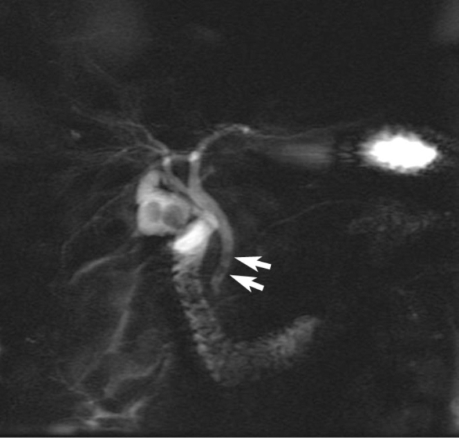
52-year-old man with recurrent cholecystitis and cholelithiasis. MRCP maximum-intensity projection image displays a fluid-filled sac in continuity with the cystic duct. Small filling defects are seen in the distal common bile duct, consistent with calculi (arrows).
The patient subsequently underwent an ERCP, sphincterotomy, and balloon sweep to clear the common bile duct stones.
A few days later, an open completion cholecystectomy and intraoperative cholangiogram was performed. Intraoperatively, a very small gallbladder remnant was noted. Palpation of the remnant gallbladder revealed two gallstones. The gallbladder was dissected to the level of the hilum, and the cystic duct was noted to be somewhat fused to the common bile duct. The cystic duct was dissected free to its base. An intraoperative cholangiogram revealed excellent flow and no filling defects. The cystic duct was then ligated, and the proximal cystic duct and gallbladder remnant were sent as specimens. A liver biopsy was also obtained.
Surgical pathology revealed a gallbladder remnant with chronic cholecystitis and cholelithiasis as well as a portion of the cystic duct. Liver biopsy revealed fatty change with no evidence of hepatitis, fibrosis, or malignancy.
Discussion
When gallbladder surgery is incomplete, long cystic duct remnants are more frequently seen in the laparoscopic approach, where the cystic duct is usually divided closer to the gallbladder to avoid iatrogenic common bile duct damage (2, 3). A common cause for leaving a gallbladder remnant is failure to properly identify the gallbladder-cystic junction, which can occur with incomplete mobilization of the cystic duct (3, 4). This complication is thought to be more common in the setting of acute cholecystitis (4). Incomplete surgery can lead to postcholecystectomy syndrome, described as a recurrence of symptoms similar to those that led to the initial cholecystectomy (1, 2, 4, 5). This usually presents as right-upper-quadrant pain and dyspepsia without jaundice (2).
Although it is extremely rare that incomplete surgery causes significant symptoms, one should always suspect a gallbladder remnant or long cystic duct in a patient with postcholecystectomy right-upper-quadrant symptoms (6). In one study of 500 cholecystectomized patients, 82% of patients with severe postoperative biliary distress had either a long cystic duct (>1cm) or a gallbladder remnant, where only 40% of asymptomatic or mildly symptomatic cholecystectomized patients had a long duct or remnant (7).
Other causes of postcholecystectomy syndrome include neuromas in the surgical bed, scar tissue around the cystic duct stump, biliary strictures, Sphincter of Oddi dyskinesia, and psychological conditions such as affective disorders (1, 2, 3, 4, 5, 8). Symptom onset can arise two days to 25 years after surgery (2).
Due to the numerous causes of postcholecystectomy syndrome, the etiology can be difficult to determine (2). Radiographic studies, such as ultrasound, CT, MRCP, and ERCP are helpful in evaluating the cause of postcholecystectomy syndrome. Abdominal ultrasound, often the initial test for evaluating right-upper-quadrant pain, can miss nearly half of biliary tract abnormalities compared to ERCP and MRCP. Both ERCP and MRCP have a sensitivity that has been reported anywhere between 85%–100% for choledocolithiasis; ERCP is more specific and should be used when intervention is clearly indicated (2). In a patient presenting with postcholecystectomy symptoms, with a gallbladder-like structure seen on radiographic studies, retained gallbladder remnant and/or retained cystic duct remnant should be the primary consideration.
Figure 4A-D.

52-year-old man with recurrent cholecystitis and cholelithiasis. T2 coronal images demonstrate a saclike structure contiguous with the cystic duct containing two large calculi (arrow). Multiple smaller calculi appear in the distal common bile duct (arrowhead). Mild extrahepatic and minimal intrahepatic biliary dilatation appears.
Figure 5.

52-year-old man with recurrent cholecystitis and cholelithiasis. Intraoperative cholangiogram shows no residual filling defects in the common bile duct.
Footnotes
Published: March 30, 2010
References
- 1.Demetriades H, Pramateftakis MG, Kanellos I, Angelopoulos S, Mantzoros I, Betsis D. Retained Gallbladder Remnant After Laparoscopic Cholecystectomy. Journal of Laparoendoscopic & Advanced Surgical Techniques. 2008 April 1;18(2):276–279. doi: 10.1089/lap.2006.0210. [PubMed] Doi:10.1089/lap.2006.0210. [DOI] [PubMed] [Google Scholar]
- 2.Lum YW, House MG, Hayanga AJ, Schweitzer M. Postcholecystectomy Syndrome in the Laparoscopic Era. Journal of Laparoendoscopic & Advanced Surgical Techniques. 2006;16(5):482–485. doi: 10.1089/lap.2006.16.482. [PubMed] [DOI] [PubMed] [Google Scholar]
- 3.Walsh RM, Chung RS, Grundfest-Broniatowski S. Incomplete excision of the gallbladder during laparoscopic cholecystectomy. Surg Endosc. 1995;9:67–70. doi: 10.1007/BF00187890. [PubMed] [DOI] [PubMed] [Google Scholar]
- 4.Walsh RM, Ponsky JL, Dumot J. Retained gallbladder/cystic duct remnant calculi as a cause of postcholecystectomy pain. Surg Endosc. 2002;16:981–984. doi: 10.1007/s00464-001-8236-1. [PubMed] Doi: 10.1007/s00464-001-8236-1. [DOI] [PubMed] [Google Scholar]
- 5.Tantia O, Jain M, Khanna S, Sen B. Post cholecystectomy syndrome: Role of cystic duct stump and re-intervention by laparoscopic surgery. Journal of Minimal Access Surgery. 2008;4(3):71–75. doi: 10.4103/0972-9941.43090. [PubMed] [DOI] [PMC free article] [PubMed] [Google Scholar]
- 6.Whitson BA, Wolpert SI. Cholelithiasis and Cholecystitis in a Retained Gallbladder Remnant after Cholecystectomy. Journal of the American College of Surgeons. 2007 December;205(6):814–815. doi: 10.1016/j.jamcollsurg.2007.03.032. [PubMed] Doi: 10.1016/j.jamcollsurg.2007.03.032. [DOI] [PubMed] [Google Scholar]
- 7.Bodvall B, Overgaar B. Cystic Duct Remnant After Cholecystectomy: Incidence Studied by Cholegraphy in 500 cases, and Significance in 103 Reoperations. Annals of Surgery. 1966 March;163(3):382–390. doi: 10.1097/00000658-196603000-00009. [PubMed] PMCID: PMC1476884. [DOI] [PMC free article] [PubMed] [Google Scholar]
- 8.Topazian M, Salem RR, Robert ME. Painful Cystic Duct Remnant iagnosed by Endoscopic Ultrasound. Am J Gastroenterol. 2005;100:491–495. doi: 10.1111/j.1572-0241.2005.41153.x. [PubMed] Doi: 10.11111/j.1572-0241.2005.41153.x. [DOI] [PubMed] [Google Scholar]