Abstract
Purpose
The purpose of this study is to compare the optical characteristics of the novel PanOptix presbyopia-correcting trifocal intraocular lens (IOL) and the multifocal ReSTOR +3.0 D IOL, through in vitro bench investigations.
Methods
The optical characteristics of AcrySof® IQ PanOptix™ (PanOptix) and AcrySof® IQ ReSTOR +3.0 D (ReSTOR +3.0 D) IOLs were evaluated by through-focus Badal images, simulated headlight images, and modulation transfer function (MTF) measurements which determine resolution, photic phenomena, and image quality. Through-focus Badal images of an Early Treatment of Diabetic Retinopathy Study chart were recorded at both photopic and mesopic pupil sizes. Simulated headlight images were taken on an MTF bench with a 50-μm pinhole target and a 5.0 mm pupil at the distance focus of the IOL. MTF curves were measured with a 3.0 mm pupil, and spatial frequencies equivalent to 20/40 and 20/20 visual acuities were recorded to illustrate the through-focus MTF curves. Far-, intermediate-, and near-focus MTF values were obtained.
Results
Bench Badal image testing and MTF measurements showed that PanOptix has a near focus at a distance of 42 cm and an additional intermediate focus at a distance of about 60 cm. The near focus for ReSTOR +3.0 D is at 45 cm. PanOptix and ReSTOR +3.0 D have comparable photopic distances and near MTF values. Additionally, PanOptix provided a substantial continuous range of vision from distance to intermediate and to near compared with ReSTOR +3.0 D. The halo propensity for PanOptix was slightly higher than that for ReSTOR +3.0 D.
Conclusion
Laboratory-based in vitro simulations showed that PanOptix trifocal IOL has comparable resolution and image quality performance in distance and near foci compared with ReSTOR +3.0 D IOL. PanOptix showed better resolution and image quality performance at the intermediate focus than ReSTOR +3.0 D IOL.
Keywords: multifocal, trifocal, modulation transfer function, Badal image, visual acuity, headlight images
Introduction
Cataract surgery techniques and technologies have improved the surgery from a procedure that could prevent blindness to a procedure that maximizes visual performance.1–3 The overwhelming majority of intraocular lens (IOL) implants after phacoemulsification surgery and cataract removal are monofocal IOLs, which have been specifically designed to improve the distance of optical resolution and have very few complications associated with the material or the technology.1–3 However, most of the patients will still require spectacles for near and intermediate tasks, including computer work. The ongoing need for spectacle use after surgery has, in turn, decreased the overall patient satisfaction and perceived quality of life,1–4 especially in younger patients who typically have more demands for spectacle-free vision for their lifestyle, reading, and computer work.5
The advent of multifocal IOLs introduced an effective solution for spectacle independence after cataract surgery and added to the surgical options for the correction of presbyopia.1,6–10 Compared with monofocal IOL capabilities, multifocal IOLs have been shown to increase the depth of vision, maintain distance vision, and improve near vision.1,3,6,7,11 The first-generation multifocal IOLs are apodized diffractive lenses that send energy to two focal points in small pupils and only to distance points in larger pupils, using the zero and first diffraction orders for distance and near foci, respectively. Because of their design, multifocal IOLs are generally ineffective in improving intermediate vision tasks.6,9,10 Also, these lenses have been associated with halos, reduced contrast sensitivity, and increased dysphotopsia, which can lead to ongoing patient dissatisfaction.1,7,12,13 Whether the potential visual complications from multifocal lenses outweigh the gain of additional spectacle independence depends on patient preference and adaptability.
Trifocality in IOL designs has been found to provide good near, intermediate, and distance visual performances and increase spectacle independence.14 AcrySof® IQ PanOptix™ Presbyopia Correcting IOLs (PanOptix; Alcon Research, Fort Worth, TX, USA) are trifocal IOLs that have been CE Mark-approved in Europe. The lens is made up of the same hydrophobic and ultraviolet- and blue light-filtering acrylate/methacrylate copolymer material used in the AcrySof family of lenses (Alcon Research). The lens design is intended to improve the intermediate vision tasks and increase patient satisfaction, with a third focal point at an optimal intermediate distance of 60 cm. PanOptix is a nonapodized diffractive trifocal IOL that distributes light energy to three focal points in both small and large pupil conditions. It uses zeroth, second, and third nonsequential diffraction orders for distance, intermediate, and near foci, respectively, and the energy at the first diffractive order is redistributed to optimize the performance at three other focal points. This novel diffractive structure produces high light utilization, transmitting 88% of light at the simulated 3.0 mm pupil size to the retina.15 The light is split into two with one half allocated to the distance focus and the other half split between the near and intermediate focus. PanOptix is also designed with an intermediary 4.5 mm diffractive zone, making its performance less dependent on pupil size.
Optical bench evaluation is a well-known method to determine the optical quality of IOLs.9,11,16,17 This study compared the results of various bench simulations of visual performance: through-focus modulation transfer function (MTF), through-focus Badal image testing, and headlight image testing for PanOptix and ReSTOR +3.0 D. ReSTOR +3.0 D was selected as the comparator lens because it is a multifocal IOL with established good distance and near visual acuity (VA).18 This study chose those particular bench tests on the basis of their specificity: through-focus Badal images to test optical resolution, headlight images to assess the photic phenomena associated with IOLs, and through-focus MTF curves to assess image quality by quantifying the contrast passing through a system at a given spatial frequency.5,6,9
Methods
Intraocular lenses
ReSTOR +3.0 D IOLs (model SN6AD1; Alcon Research) and PanOptix IOLs that are used in this study had a 21.0 D base power. ReSTOR +3.0 D is an apodized diffractive multifocal IOL, whereas PanOptix is a nonapodized diffractive trifocal IOL.
ReSTOR +3.0 D and PanOptix have aspherical designs and aspheric corrections for a corneal spherical aberration of −0.1 μm.19 Table 1 lists the optical specifications of ReSTOR +3.0 D and PanOptix.
Table 1.
Characteristics of ReSTOR +3.0 D and PanOptix IOLs
| PanOptix | ReSTOR +3.0 D | |
|---|---|---|
| Technology | Trifocal | Multifocal |
| Diffractive zone | 4.5 mm | 3.6 mm |
| Central zone | Diffractive | Diffractive |
| Optic type | Nonapodized | Apodized |
| Near add powers | +3.25 D | +3.00 D |
| Intermediate add powers | +2.17 D | None |
| Active orders | Zeroth, second, and third | Zeroth and first |
| Asphericity | −0.1 μm | −0.1 μm |
| Lens color | Yellow | Yellow |
ReSTOR +3.0 D has active diffraction orders of the zeroth and first magnitude, and PanOptix IOL has active diffraction orders of the zeroth, second, and third magnitude. The optical technology of PanOptix uses nonsequential diffractive orders to create near (42 cm), distance, and intermediate (60 cm) foci.20 In PanOptix, energy at the first diffractive order is redistributed to optimize the performance at the other focal points.20
Experimental design
Badal imaging
A custom model eye was assembled as a Badal optometer, as previously described by Carson et al.5 The model eye used for testing both the study lenses was modified with 0.1 μm spherical aberration according to the design of the lenses. A bench simulation of visual performance using through-focus Badal image testing of an Early Treatment of Diabetic Retinopathy Study chart from −1.0 D to +3.0 D at 0.25 D increments was performed, and images simulating viewing distances from infinity to 40 cm were evaluated. An additional image was also taken at the best near focus of each lens in order to account for the differences in near add power. Badal images taken with the letter chart target placed at the simulated depth of foci of infinity, 80, 60, 40 cm, and best near focus were depicted to demonstrate the difference between the two lens models.
The IOLs were positioned within a model eye containing deionized water and a convex plano model cornea lens with a matching spherical aberration, as described previously;5 the IOL was held on a paddle that contained a 3.0 mm pupil. The target was a chrome-on-glass, 25 mm diameter Early Treatment of Diabetic Retinopathy Study VA chart that depicted nine rows, with the smallest row corresponding to a VA of 20/12. VA difference of no more than two letters is considered equivalent at a given focus distance (less than one-half of the clinically relevant value of 0.1 logarithm of the minimum angle of resolution).
Simulated headlight imaging
Photic phenomena of the two IOLs in the form of halo propensity assessment were measured using the Optikos MTF System (Optikos Corp., Wakefield, MA, USA) with OpTest™ software (version 5.2.2; Optikos Corp.) in a pseudophakic eye model with a spherical aberration matching International Organization of Standardizations model eye as previously described by Carson et al.5 An illuminated pinhole with a 50 μm aperture was used to simulate a car headlight viewed at a distance of ~250 m.21 All the images were taken at the distance foci of the specific IOL model with 5.0 mm pupil on the IOL under white light conditions by illumination on one side using a Fiber-Lite® DC-950 Fiber Optic Illuminator (Dolan-Jenner Industries, Boxborough, MA, USA). Test conditions, including the light intensity and the position of the IOL relative to the aperture and cornea, were the same for both IOL models. The light intensity level was adjusted until the halo structure could be clearly seen on a charge-coupled device camera.
MTF measurements
Through-focus MTF is an established method to determine the amount of contrast passed through a system at a given spatial frequency.22 The IOLs were used following ISO 11979-2 requirements and test methods in order to assess the optical properties of the multifocal IOLs using a validated Optikos MTF system according to the conditions described by Carson et al.5 MTF measurement was performed on the IOLs with spherical aberration-matching corneas to yield the best optical performance. Testing was conducted with a 3.0 mm lens aperture that corresponds to average photopic pupil size. Slit targets illuminated by a light source with a 550 nm narrow-band filter were imaged at infinity. Each target image was obtained from the IOLs, was relayed to the charge-coupled device camera, and was analyzed. MTF curves were generated from the mean vertical- and horizontal-slit values. Through-focus MTF curves at two spatial frequencies of 50 and 100 line pairs per millimeter (lp/mm) were used to determine the best foci for distance, near distance, and intermediate distance. The two frequencies correspond to cycle widths of 4 and 2 minutes, respectively, conventionally equated to acuities of 20/40 and 20/20, respectively, at least for a square-wave grating.23
Results
Badal Images
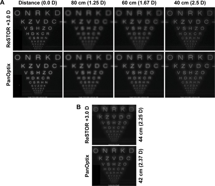
Through-focus Badal images captured at defocus distances of infinity (0.0 D), 80 cm (1.25 D), 60 cm (1.67 D), and 40 cm (2.50 D) are shown in Figure 1A. The trifocal IOL provided equivalent distance and near performance compared with the multifocal IOL, with a photopic pupil size of 3.0 mm. The intermediate visual performance was improved in the trifocal over the multifocal IOL, with approximately three lines of improvement at 60 and 80 cm defocus distances. The best PanOptix intermediate focus distance was 60 cm, whereas ReSTOR +3.0 D lacks an intermediate focus. Figure 1B shows the 3.0 mm pupil Badal images taken at the best near focus of ReSTOR +3.0 D and PanOptix, which were 44 cm (2.25 D) and 42 cm (2.37 D), respectively.
Figure 1.
Image quality of the ReSTOR +3.0 D and PanOptix IOLs.
Notes: Image quality of the ReSTOR +3.0 D and PanOptix IOLs at (A) focus distances of infinity (0.0 D), 80 cm (1.25 D), 60 cm (1.67 D), and 40 cm (2.5 D) with a 3.0 mm pupil; (B) best-near image for each IOL with a 3.0 mm pupil. The third line (with the text “R H S D V”) and the sixth line (with the text “C S R H N”) from the bottom are equivalent to visual acuities of 20/20 and 20/40, respectively.
Abbreviation: IOL, intraocular lens.
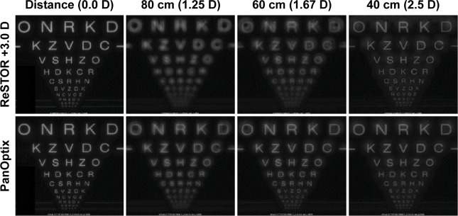
Results were similar for PanOptix in Badal images taken at infinity (0.0 D), 80 cm (1.25 D), 60 cm (1.67 D), and 40 cm (2.50 D) using the 4.5 mm pupil in Figure 2, whereas ReSTOR +3.0 D had less image quality at the 60 and 80 cm defocus positions.
Figure 2.
Image quality of the ReSTOR +3.0 D and PanOptix intraocular lenses at focus distances of infinity (0.0 D), 80 cm (1.25 D), 60 cm (1.67 D), and 40 cm (2.5 D) with a 4.5 mm pupil.
Note: The third line (with the text “R H S D V”) and the sixth line (with the text “C S R H N”) from the bottom are equivalent to visual acuities of 20/20 and 20/40, respectively.
The bench-simulated Badal images that were used to assess the resolution of ReSTOR +3.0 D and PanOptix IOLs were taken at defocus distances from 0.0 D to +3.5 D using the 3.0 mm pupil size. The following Video S1 shows the Badal images from ReSTOR +3.0 D (left) and PanOptix (right) over these defocus distances. The 20/40 line in PanOptix was resolvable in the 2.50 D (40 cm) to 1.25 D (80 cm) images.
Headlight images
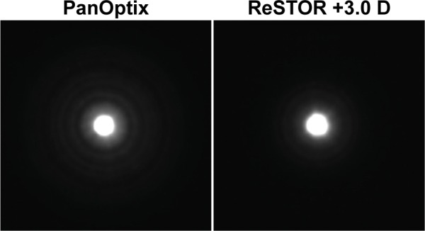
Representative simulated headlight images for the study lenses taken at the distance focus of the lenses using a 5.0 mm pupil are illustrated in Figure 3. The halos surrounding the headlight target diminished at a shorter distance from the central spot with the multifocal IOL compared with the trifocal IOL. Visually, halos were more distinct with PanOptix. The difference in halos can be explained by the difference in apodization between the lenses. In ReSTOR +3.0 D, apodization helps to direct most of the light energy (~85% by design) to distance focus in large pupil diameters. On the other hand, the nonapodized PanOptix design consistently splits the light energy to the three foci (distance, intermediate, and near) independent of the pupil diameter.
Figure 3.

Simulated headlight images through the PanOptix and ReSTOR +3.0 D intraocular lenses.
MTF measurements
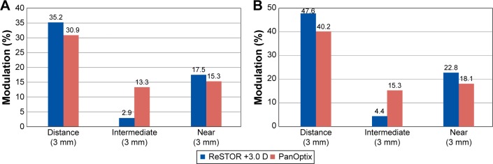
MTF measurements were taken at the designated foci of the two lens models with a 3.0 mm aperture and at spatial frequencies of 100 (Figure 4A) and 50 lp/mm (Figure 4B). Distance-focus and near-focus values were greater for ReSTOR +3.0 D than for PanOptix, but PanOptix had considerably higher intermediate MTF values.
Figure 4.
Modulation transfer function values of the ReSTOR +3.0 D and PanOptix intraocular lenses measured with a 3.0 mm pupil at (A) 100 and (B) 50 lp/mm (sample size, n=10).
Abbreviation: lp/mm, line pairs per millimeter.
At 100 and 50 lp/mm, the ReSTOR +3.0 D IOLs had distance MTF values of 35.2% and 47.6%, respectively, and the trifocal IOLs had distance MTF values of 30.9% and 40.2%, respectively. Near-focus MTF values at 100 and 50 lp/mm were 17.5% and 22.8%, respectively, for ReSTOR +3.0 D and 15.3% and 18.1%, respectively, for PanOptix. The intermediate-focus MTF values for PanOptix were higher than those for ReSTOR +3.0 D, as expected with the design of the trifocal IOL. Intermediate-focus MTF at 100 and 50 lp/mm were 2.9% and 4.4%, respectively, for ReSTOR +3.0 D and 13.3% and 15.3%, respectively, for PanOptix.
The through-focus MTF curves at spatial frequencies of 100 and 50 lp/mm using a 3.0 mm pupil are shown in Figure 5. The distance-focus MTF peak was greater for the multifocal IOL than the trifocal IOL. ReSTOR +3.0 D had a slightly greater near-focus MTF peak than PanOptix; the intermediate-focus peak was located at 60 cm, and ReSTOR +3.0 D did not possess an intermediate-focus peak.
Figure 5.
Through-focus modulation transfer function values of the ReSTOR +3.0 D and PanOptix intraocular lenses measured with a 3.0 mm pupil at (A) 100 and (B) 50 lp/mm.
Abbreviation: lp/mm, line pairs per millimeter.
Discussion
In this study, the new PanOptix trifocal IOL design was evaluated with standard bench measurements such as through-focus Badal images for resolution, through-focus MTF curves for image quality, and headlight images for photic phenomena (halo propensity), all compared with its multifocal counterpart, ReSTOR +3.0 D. The results showed that the PanOptix IOL had improved performance at an intermediate distance range of 60–80 cm and showed a greater than three lines of improvement in resolution at 60, 70, and 80 cm compared with the ReSTOR +3.0 D IOL. Laboratory testing also showed that distance and near resolution for the trifocal IOL is likely to be comparable to ReSTOR +3.0 D for photopic pupil (3.0 mm). The bench simulation that was used to measure image contrast showed equivalent distance and near performance for PanOptix and ReSTOR +3.0 D IOLs, but PanOptix fared much better in intermediate vision.
Monofocal IOLs have traditionally higher contrast sensitivity than their multifocal counterparts because the light from the out-of-focus image reduces the sharpness of the in-focus image in multifocal designs.23 However, monofocal lenses are not designed to provide spectacle-free vision in situations outside of distance vision, creating a challenge for patients who need good intermediate vision (eg, computer work) and near vision (eg, reading). Diffractive multifocal IOLs have compensated for this by decreasing near add powers,24 as in the case of the ReSTOR +3.0 D, which has a decreased near add power compared with its predecessor, the ReSTOR +4.0 D. However, in this study, PanOptix was shown to produce clearer images from 60 to 80 cm than even the ReSTOR +3.0 D, potentially overcoming the ongoing intermediate vision and contrast sensitivity issues associated with other bifocal lenses.5
The higher energy usage of PanOptix allowed the IOL to provide equivalent simulated distance vision to ReSTOR +3.0 D in this study. De Vries et al25 and Alfonso et al26 found that the reduced near add power of the ReSTOR +3.0 D, compared with the ReSTOR +4.0 D or other multifocal IOLs, produced better intermediate vision, but Gatinel and Houbrechts11 maintained that true intermediate VA can only be achieved by adding a third focal point. Additionally, the intermediate and near performances of PanOptix are independent of pupil size.
The through-focus MTF curves confirmed a distance and near focus for each lens, but the curve for PanOptix also had an intermediate focus at 60 cm. Other studies have found ReSTOR +3.0 D to have higher MTF values at near and distance focal points compared with other commercialized trifocal lenses.5 Of interest, the 60 cm intermediate focal point achieved by the PanOptix lens, as indicated by its MTF peak, was within the preferred viewing distance range, 45.7–61.0 cm, for computer terminals.27–29
In addition, ReSTOR +3.0 D demonstrated slightly lowered haloing effects compared with PanOptix. The slight differences in haloing effects between the two lenses can be accounted for by the central apodized diffractive zone of the multifocal IOL that ends at 3.6 mm diameter with a refractive outer zone.30 This allows the IOL to be more strongly distance-dominant with larger pupil sizes. In the nonapodized PanOptix lens, the addition of a third focus, by itself may increase halos. In large pupils, most of the light energy of ReSTOR +3.0 D (~85% by design) goes to distance focus, which may account for the variability between the two designs. It is not known whether this difference in halos between the study lenses is expected to be clinically significant, and future studies may be necessary to understand the clinical significance.
Other commercially available trifocal IOLs provide intermediate add powers at about 80 cm.17,31–34 However, this study showed that PanOptix provides an intermediate add power of about 60 cm in a unilateral bench test, which was intended to achieve the most suitable and comfortable intermediate distance for most patients.27–29
This study is limited by its laboratory nature; extrapolating the results into clinical practice may not be straightforward, and the findings cannot guarantee that the lenses will perform the same in vivo. Additionally, the study design was observational, limiting statistical analyses.
Conclusion
The design of PanOptix allows for the creation of three distinct foci. This novel, presbyopia-correcting lens is equivalent to ReSTOR +3.0 D in photopic near and distance performance but provides a substantial range of intermediate foci with an optimal intermediate focus at 60 cm. Although additional clinical studies are necessary, these bench analyses have shown that PanOptix may be a viable choice for patients who require optimal vision across all distances and minimal use of spectacle correction.
Acknowledgments
This study was funded by Alcon Research, Ltd. (Fort Worth, TX, USA). The authors thank Aldo Martinez, PhD, for his thorough review and contributions to manuscript preparation. Medical writing assistance was provided by Michelle Dalton and BelMed Professional Resources and was funded by Alcon.
Footnotes
Disclosure
All the authors are employees of Alcon Research, Ltd. The authors report no conflicts of interest in this work.
References
- 1.Calladine D, Evans JR, Shah S, Leyland M. Multifocal versus mono-focal intraocular lenses after cataract extraction. Cochrane Database Syst Rev. 2012;9:CD003169. doi: 10.1002/14651858.CD003169.pub3. [DOI] [PubMed] [Google Scholar]
- 2.Lundstrom M, Barry P, Henry Y, et al. Evidence-based guidelines for cataract surgery: guidelines based on data in the European Registry of Quality Outcomes for Cataract and Refractive Surgery database. J Cataract Refract Surg. 2012;38(6):1086–1093. doi: 10.1016/j.jcrs.2012.03.006. [DOI] [PubMed] [Google Scholar]
- 3.Tan N, Zheng D, Ye J. Comparison of visual performance after implantation of 3 types of intraocular lenses: accommodative, multifocal, and monofocal. Eur J Ophthalmol. 2014;24(5):693–698. doi: 10.5301/ejo.5000425. [DOI] [PubMed] [Google Scholar]
- 4.Luo BP, Brown GC, Luo SC, Brown MM. The quality of life associated with presbyopia. Am J Ophthalmol. 2008;145(4):618–622. doi: 10.1016/j.ajo.2007.12.011. [DOI] [PubMed] [Google Scholar]
- 5.Carson D, Hill WE, Hong X, Karakelle M. Optical bench performance of AcrySof(®) IQ ReSTOR(®), AT LISA(®) tri, and FineVision(®) intraocular lenses. Clin Ophthalmol. 2014;8:2105–2113. doi: 10.2147/OPTH.S66760. [DOI] [PMC free article] [PubMed] [Google Scholar]
- 6.Gatinel D, Pagnoulle C, Houbrechts Y, Gobin L. Design and qualification of a diffractive trifocal optical profile for intraocular lenses. J Cataract Refract Surg. 2011;37(11):2060–2067. doi: 10.1016/j.jcrs.2011.05.047. [DOI] [PubMed] [Google Scholar]
- 7.Javitt J, Brauweiler HP, Jacobi KW, et al. Cataract extraction with multifocal intraocular lens implantation: clinical, functional, and quality-of-life outcomes. Multicenter clinical trial in Germany and Austria. J Cataract Refract Surg. 2000;26(9):1356–1366. doi: 10.1016/s0886-3350(00)00636-2. [DOI] [PubMed] [Google Scholar]
- 8.Mesci C, Erbil H, Ozdoker L, et al. Visual acuity and contrast sensitivity function after accommodative and multifocal intraocular lens implantation. Eur J Ophthalmol. 2010;20(1):90–100. doi: 10.1177/112067211002000112. [DOI] [PubMed] [Google Scholar]
- 9.Montes-Mico R, Lopez-Gil N, Perez-Vives C, et al. In vitro optical performance of nonrotational symmetric and refractive-diffractive aspheric multifocal intraocular lenses: impact of tilt and decentration. J Cataract Refract Surg. 2012;38(9):1657–1663. doi: 10.1016/j.jcrs.2012.03.040. [DOI] [PubMed] [Google Scholar]
- 10.Ruiz-Alcocer J, Madrid-Costa D, Garcia-Lazaro S, et al. Optical performance of two new trifocal intraocular lenses: through-focus modulation transfer function and influence of pupil size. Clin Experiment Ophthalmol. 2014;42(3):271–276. doi: 10.1111/ceo.12181. [DOI] [PubMed] [Google Scholar]
- 11.Gatinel D, Houbrechts Y. Comparison of bifocal and trifocal diffractive and refractive intraocular lenses using an optical bench. J Cataract Refract Surg. 2013;39(7):1093–1099. doi: 10.1016/j.jcrs.2013.01.048. [DOI] [PubMed] [Google Scholar]
- 12.de Vries NE, Webers CA, Touwslager WR, et al. Dissatisfaction after implantation of multifocal intraocular lenses. J Cataract Refract Surg. 2011;37(5):859–865. doi: 10.1016/j.jcrs.2010.11.032. [DOI] [PubMed] [Google Scholar]
- 13.Lang A, Portney V. Interpreting multifocal intraocular lens modulation transfer functions. J Cataract Refract Surg. 1993;19(4):505–512. doi: 10.1016/s0886-3350(13)80615-3. [DOI] [PubMed] [Google Scholar]
- 14.Weeber HA, Meijer ST, Piers PA. Extending the range of vision using diffractive intraocular lens technology. J Cataract Refract Surg. 2015;41(12):2746–2754. doi: 10.1016/j.jcrs.2015.07.034. [DOI] [PubMed] [Google Scholar]
- 15.AcrySof [product information] Fort Worth, TX: Alcon Laboratories, Inc.; 2015. [Google Scholar]
- 16.Cochener B, Vryghem J, Rozot P, et al. Clinical outcomes with a trifocal intraocular lens: a multicenter study. J Refract Surg. 2014;30(11):762–768. doi: 10.3928/1081597X-20141021-08. [DOI] [PubMed] [Google Scholar]
- 17.Law EM, Aggarwal RK, Kasaby H. Clinical outcomes with a new trifocal intraocular lens. Eur J Ophthalmol. 2014;24(4):501–508. doi: 10.5301/ejo.5000407. [DOI] [PubMed] [Google Scholar]
- 18.Alfonso JF, Fernandez-Vega L, Blazquez JI, Montes-Mico R. Visual function comparison of 2 aspheric multifocal intraocular lenses. J Cataract Refract Surg. 2012;38(2):242–248. doi: 10.1016/j.jcrs.2011.08.034. [DOI] [PubMed] [Google Scholar]
- 19.Hong X, Zhang X. Optimizing distance image quality of an aspheric multifocal intraocular lens using a comprehensive statistical design approach. Opt Express. 2008;16(25):20920–20934. doi: 10.1364/oe.16.020920. [DOI] [PubMed] [Google Scholar]
- 20.He J, Carson D, Xu Z. Laboratory comparison of a novel presbyopia correcting IOL with two multifocal IOL models; European Society of Cataract and Refractive Surgeons; Barcelona, Spain. 2015. [Google Scholar]
- 21.Pieh S, Lackner B, Hanselmayer G, et al. Halo size under distance and near conditions in refractive multifocal intraocular lenses. Br J Ophthalmol. 2001;85(7):816–821. doi: 10.1136/bjo.85.7.816. [DOI] [PMC free article] [PubMed] [Google Scholar]
- 22.Madrid-Costa D, Ruiz-Alcocer J, Ferrer-Blasco T, et al. Optical quality differences between three multifocal intraocular lenses: bifocal low add, bifocal moderate add, and trifocal. J Refract Surg. 2013;29(11):749–754. doi: 10.3928/1081597X-20131021-04. [DOI] [PubMed] [Google Scholar]
- 23.Bennett AG, Rabbetts RB. Bennett & Rabbetts’ Clinical Visual Optics. 3rd ed. Oxford and Boston, MA: Butterworth-Heinemann; 1998. p. 48. [Google Scholar]
- 24.de Vries NE, Nuijts RM. Multifocal intraocular lenses in cataract surgery: literature review of benefits and side effects. J Cataract Refract Surg. 2013;39(2):268–278. doi: 10.1016/j.jcrs.2012.12.002. [DOI] [PubMed] [Google Scholar]
- 25.de Vries NE, Webers CA, Montes-Mico R, et al. Long-term follow-up of a multifocal apodized diffractive intraocular lens after cataract surgery. J Cataract Refract Surg. 2008;34(9):1476–1482. doi: 10.1016/j.jcrs.2008.05.030. [DOI] [PubMed] [Google Scholar]
- 26.Alfonso JF, Fernandez-Vega L, Puchades C, Montes-Mico R. Intermediate visual function with different multifocal intraocular lens models. J Cataract Refract Surg. 2010;36(5):733–739. doi: 10.1016/j.jcrs.2009.11.018. [DOI] [PubMed] [Google Scholar]
- 27.American Optometric Association . The Effects of Computer Use on Eye Health and Vision. St. Louis, MO: American Optometric Association; 1997. [Google Scholar]
- 28.Charness N, Dijkstra K, Jastrzembski T, et al. Monitor Viewing Distance for Younger and Older Workers: Proceedings of the Human Factors and Ergonomics Society 52nd Annual Meeting. 2008;52(19):1614–1617. [Google Scholar]
- 29.U.S. Department of Labor, Occupational Safety and Health Administration Working Safely with Video Display Terminals. [Accessed August 4, 2015]. (OSHA Publication 3092). revised 1997. Available from: https://www.osha.gov/Publications/osha3092.pdf.
- 30.Fisher BL. Presbyopia-correcting intraocular lenses in cataract surgery: literature review of the benefits and side effects. J Cataract Refract Surg. 2013;39(2):268–278. doi: 10.1016/j.jcrs.2012.12.002. [DOI] [PubMed] [Google Scholar]
- 31.Carballo-Alvarez J, Vazquez-Molini JM, Sanz-Fernandez JC, et al. Visual outcomes after bilateral trifocal diffractive intraocular lens implantation. BMC Ophthalmol. 2015;15:26. doi: 10.1186/s12886-015-0012-4. [DOI] [PMC free article] [PubMed] [Google Scholar]
- 32.Casas P, Cristobal JA, Angeles del Buey M. Objective and subjective visual performance of a diffractive trifocal implant. J Emmetropia. 2014;5:183–189. [Google Scholar]
- 33.Kretz FT, Breyer D, Diakonis VF, et al. Clinical outcomes after binocular implantation of a new trifocal diffractive intraocular lens. J Ophthalmol. 2015;2015:962891. doi: 10.1155/2015/962891. [DOI] [PMC free article] [PubMed] [Google Scholar]
- 34.Vryghem JC, Heireman S. Visual performance after the implantation of a new trifocal intraocular lens. Clin Ophthalmol. 2013;7:1957–1965. doi: 10.2147/OPTH.S44415. [DOI] [PMC free article] [PubMed] [Google Scholar]


