

Abstract
Background:
Bisphenol A (BPA) and nonylphenol (NP) have harmful effects on the endocrine system of humans and animals.
Aim:
We sought to investigate the effect of three doses of BPA and NP on the reproductive parameters of rats.
Materials and Methods:
Adult Wistar male rats weighing 150–200 g were used for a consecutive 35-day study. BPA and NP were given as gavage in three doses (5, 25, and 125 μg/kg). At the end of the study, the rats were anesthetized and 2 ml blood sample was obtained from the auxiliary venous plexus for the assessment of sex hormone levels. The testes were removed and kept in 10% formalin for the histomorphometric and histopathologic analyses.
Results:
BPA and NP significantly decreased the body weight of the animals compared to the controls (P < 0.05). The seminiferous tubule diameter and thickness of the seminiferous epithelium were significantly decreased in the groups receiving BPA and NP compared to the control (P < 0.05). The number of seminiferous tubules in every experimental group increased, except for the highest dose of NP (125 μg/kg), which showed a significant difference compared to the controls (P < 0.05). The number of spermatocytes and spermatogonia (54.97 ± 5.824, 35.78 ± 3.956, respectively) in the group receiving NP (125 μg/kg) was significantly decreased compared to the other groups. Serum levels of luteinizing hormone, Follicle stimulating hormone, and testosterone did not show any significant change compared to the controls.
Conclusion:
Based on the results, all three doses of BPA and NP significantly produced weight loss, as well as destruction of the testis tissue and impairment of the spermatogenesis.
Keywords: Bisphenol A, nonylphenol, reproductive, spermatocytes, spermatogenesis
Introduction
Nowadays, most of the chemicals that are used in our daily life cause the most damage to the environment. Some of these chemicals have severe toxic effects on humans and the environment. Plasticizers are among such chemicals. Plasticizers are substances that are used in disposable dishes, manufacture of polycarbonates, plastic industry, and dental materials to produce or increase plasticity and flexibility as well as to reduce fragility. In this regard, bisphenol A (BPA) and nonylphenol (NP) are the most commonly used organic plasticisers. Because of the semiestrogenic function of these substances, they are considered to be steroid and xenobiotic receptor-mediated transcription.[1] BPA and NP can have harmful effects on the endocrine system of humans and animals. BPA has a weak estrogenic activity and can interfere with the endocrine system, and can increase the possibility of testis and breast cancer.[2] Among the other harmful effects of these chemicals are deterioration in the quality of sperm parameters, anomalies in reproductive organs, decrease in sexual activities, and various fetal anomalies. NP is also an estrogenic-like substance that changes protein formation and induces hormonal impairments.[2,3,4]
Because of its ability to mimic the action of natural estrogen, NP is classified as an endocrine impairing agent as it interferes with the mammalian endocrine system.[5] This compound is mainly created from the decomposition of alkyl phenol ethoxylates, which are widely used in detergents (80%), latex dyes, as emulsifiers in pesticides, and in the textile industry.[6] In a study, a significant decrease in the body weight, in the weight of the reproductive organs, and in testosterone serum were observed in the rats that received 80 and 200 mg/kg of NP for 14 to 19 days.[7] It was shown that the administration of BPA at doses of 0.5 and 50 µg/kg to male rats (5 weeks’ old) for 4 to 8 weeks induced early puberty in the 6th or 7th week, as well as decrease in the level of free plasma testosterone.[4]
BPA is currently known to cause toxicity in the reproductive system.[8,9] It has been reported that in a study where male rats received 200–950 mg/kg of BPA for 44 days, there was a weight loss in the reproductive organs and damage to the seminiferous tubules.[10] Although a dose of 50 µg/kg/day BPA is believed to be standard daily tolerable dose for humans and a dose of 50 mg/kg is said to have no macroscopic harmful effects,[11] studies have shown that 3 mg/kg/day of BPA for 2 weeks causes a significant decrease in testosterone levels and increase in the luteinizing hormone (LH) level in the serum and testis of adult rats.[12]
Another investigation reported that the rats given BPA showed no changes in their reproductive organ functions.[13] Therefore, there appears to be controversy over the effect of BPA on reproductive system. Although studies have not revealed any link between exposure to BPA and reproductive hormones and sperm profiles in fertile men, a significant inverse relationship was reported between urinary concentration of BPA and free androgen level, even though it has no considerable effect on the reproductive function.[14]
Because of the controversial study reports and the heavy use of BPA and NP, the present study was designed to investigate the effect of three doses of BPA and NP on the reproductive parameters of adult rats, including the serum concentration of LH, follicle stimulating hormone (FSH), and testosterone.
Materials and Methods
BPA >99% pure (Daejung, Korea) NP >99% pure (Kento, Japan), Elisa Kit (Bioassay Technology Laboratory, China), and all other chemicals were purchased from local commercial sources.
Male Wistar rats (weighing 150–200 g) were obtained from the Babol University of Medical Sciences’ Animal room (Babol, Iran).
All rats were kept in plastic cages under controlled light and dark condition (12 h light: 12 h dark) at 22 ± 2°C and humidity of 50 ± 5%, with free access to chow and tap water ad libitum. The study was approved by the University Ethics Committee, and all the experiments were performed in accordance with the guidelines for the safe working of animals.
The rats were divided into a control group and three groups receiving three doses (5, 25, and 125 µg/kg) of BPA and three groups receiving three doses (5, 25, and 125 µg/kg) of NP. In each group, ten rats were used. All treatments continued for 35 days. BPA and NP were dissolved in olive oil as vehicle and were administered in rats by gavage. Control group was administered olive oil alone. On the 36th day, the rats were weighed and anesthetized with sodium thiopental. Tissue and blood samples were taken and kept at −80°C for hormone and histolopathological analyses.
Preparing the blood samples
Blood samples (1 ml) were obtained by cutting the auxilary artery for each rat and were transferred into 2-ml tubes. Then, the samples were separately centrifuged and the serums were kept at −20°C for further analysis.
Luteinizing hormone, follicle stimulating hormone, and testosterone assay
The frozen serum samples were allowed to thaw at laboratory temperature and vortexed and then were centrifuged at 1000 rpm for 5 min. The optical density of the samples was measured at wavelengths of 450 and 630 nm using ELISA reader (Rayto microplate RT-2100C). Then, the concentration of LH, FSH, and testosterone was determined.
Preparing testis tissue
The right testis of each rat was removed and separately weighed. The testis tissues were fixed in 10% formalin and three tissue slices were randomly prepared of each testis. Then, the selected slices were processed using a processing device (dewatering, shining, and staining) and were moulded in paraffin; 5-micron samples were prepared from the slices and stained using the Hematoxylin and eosin (H and E) staining method.
Histomorphometry
Figures were obtained from the smears using an Olympus optical microscope equipped with a Canon camera at a magnification of 4×, 10×, and 40× at four random points and a Motic application was used for extracting the data.
Measurement of seminiferous tubules diameter
Figures with a magnification of 10× were used to measure the diameter of the seminiferous tubules. For each animal, 10 seminiferous tubules were randomly chosen from various areas, and for each tubule, two diameters (long and short) perpendicular to each other were measured. The data were recorded and then analyzed using statistical tests.
Thickness of epithelium of seminiferous tubules
The thickness of the epithelium of the seminiferous tubules was measured in both the groups. The heights of the epithelium for 100 tubules in four directions and four angles were measured.
Surface area of the seminiferous tubules cross-sections
In order to measure the surface area of the seminiferous tubules cross-sections, the areas were individually calculated using a Motic software.
Cross-sectional area of epithelium of seminiferous tubules
The area of lumen of the seminiferous tubules was calculated using Motic software. Then, by subtracting this number from the area of the seminiferous tubules, the area of the epithelium of the seminiferous tubules was determined. In order to reduce the margin of error, the same tubules were used for each calculation.
Number of seminiferous tubules
In order to count the number of seminiferous tubules, figures with a magnification of 10× from various points of the smear were used. The counting started from 40 points for each animal. For an accurate recording, a square with 6.5 × 6.5 cm was drawn on a frame, using + symbols. All tubules in the middle and on the top right of the square were counted. Then, the number of seminiferous tubules was divided by the square area (42.25 cm2) to obtain the number of seminiferous tubules in 1 cm2 [Figure 1].
Figure 1.

Method of calculating the diameter, thickness of the epithelium and surface area of the epithelium in the seminiferous tubules using Motic software
Number of spermatocytes and spermatogonia in 1 cm2
In order to count the number of spermatocytes and spermatogonia in 1 cm2, the figures with a magnification of 40× from various points of the smear were used. Four figures were used for the cellular count of each rat. First, the area of the seminiferous tubule was calculated using a Motic software. Then, the number of spermatogonia, which looked round with dark circles in the outer layer of the epithelium, was determined. In the next step, the number of spermatocytes was recorded. Finally, the number of spermatocytes and spermatogonia was divided by the area of the seminiferous tubule to obtain the number of spermatocytes and spermatogonia in 1 cm2.
Statistical study
All the analyses were performed using the Statistical Package for the Social Sciences software version 16. The data were statistically analysed using one-way analysis of variance followed by post-hoc Tukey test. The significance of differences between data was assumed at P < 0.05.
Results
NP receiving rats showed a behaviour compared to BPA receiving groups. Statistical analysis showed a significant decrease in the weight of the rats after treatment in both BPA and NP compared to the control group (P < 0.05). Furthermore, no significant difference was seen between the BPA and the NP groups. Table 1 shows the mean weight of the rats in the control, BPA, and NP receiving groups at the baseline and the last day of the experiment.
Table 1.
Mean (±SD) of the weight changes at the baseline and on the last day of the study in control, Bisphenol A, and nonylphenol receiving groups

With progressing age, the weight increased, as seen in the control group, however, the body weight in rats receiving BPA and NP showed a considerable decrease [Table 1]. This decline in body weight was greater in the NP group compared to the BPA receiving group [Figure 2].
Figure 2.

Mean difference in body weight at baseline and after treatment of rats receiving the Bisphenol A and nonylphenol receiving groups compared to control (n = 10)
Level of luteinizing hormone, follicle stimulating hormone, and testosterone
The results of the ELISA analysis is shown in Table 2. The control group showed the highest amount of testosterone, whereas the group that had been given 5 µg/kg of the NP showed the least amount of testosterone.
Table 2.
Concentration of LH, FSH and testosterone in BPA, NP, and control groups

The results showed that serum concentration of testosterone in the BPA and NP groups in three doses significantly decreased compared to the control group (P < 0.05) [Table 2]. A significant difference was noted in the group which received 25 mg/kg of NP compared to other groups, except for the group which received 125 mg/kg of NP. There were no significant changes in the levels of LH or FSH in the BPA and NP groups compared to the control group [Table 2].
Morphometric findings of testis
The mean of the long and short diameters of the seminiferous tubules for all study controls were calculated. The highest diameter was seen in the control group and the least belonged to the rats received 25 µg/kg of NP. The mean of the four heights drawn for the epithelium of the seminiferous tubules was calculated for all the groups. The highest epithelium thickness belonged to the control group and the lowest belonged to the rats which received 25 µg/kg of the NP [Table 3]. The diameter of the seminiferous tubules and the thickness of the epithelium in the experimental group showed a significant decrease compared to those of the control group (P < 0.001). A significant difference was noted in the diameter of the seminiferous tubules and the thickness of the epithelium between groups receiving 5 and 25 µg/kg of the BPA and the group receiving 25 µg/kg of the NP (P < 0.0001). This difference was also observed between rats receiving 5 and 25 µg/kg of the NP and rats receiving 125 µg/kg of the NP (P < 0.05). However, no significant difference was noted within the groups receiving various doses of BPA.
Table 3.
Mean (±SD) of the diameter of seminiferous tubules, the thickness of the epithelium, surface are of the cross-sections of the seminiferous tubules and the epithelium of the seminiferous tubules, number of seminiferous tubules, count of spermatocytes and spermatogonia of control, BPA, and NP

The surface of area of cross-section of the epithelium of the seminiferous tubules was calculated, and is shown as the mean ± SD in Table 3. The highest surface area of the epithelium of the seminiferous tubules belongs to the control group and the lowest belongs to the group which received 25 µg/kg of NP. The surface area of the seminiferous tubules and epithelium of the seminiferous tubules in the experimental groups showed a significant decrease compared to the control (P < 0.05). There was a significant difference in the results of the groups which were administered 5 and 25 µg/kg of BPA and 5 and 25 µg/kg of NP (P < 0.05). The group received which 125 µg/kg of BPA showed a significant difference in the surface area of the seminiferous tubules and epithelium of the seminiferous tubules compared to all three doses of the NP receiving groups (P < 0.05).
The mean ± SD of seminiferous tubules in a surface unit (42.25 cm2) is presented in Table 3, with the highest belonging to the 25 µg/kg dose of the NP group and the lowest belonging to the 125 µg/kg dose of the NP group. Statistical analyses have shown that there was a significant difference in the number of seminiferous tubules in the 6.5 × 6.5 cm2 frame compared to the control group (P < 0.05) except for the 125 µg/kg dose of the BPA group. The groups receiving 5 and 25 µg/kg of BPA showed a significant difference from all the groups (P < 0.05) except among each other. A comparison of the experimental groups revealed that the group receiving 125 µg/kg of the BPA showed a significant difference from all the other groups (P < 0.05) except for the control group. There was a significant difference in the number of seminiferous tubules in the 6.5 × 6.5 cm2 frame in the groups receiving all three doses of the NP (P < 0.05).
The number of spermatocytes and spermatogonia in the seminiferous tubules was counted. The number per surface unit was calculated by dividing the total number by the area of the seminiferous tubules. Table 3 shows the mean ± SD of the number of spermatocytes and spermatogonia. The control showed the highest number, whereas the lowest number belonged to the group receiving 125 µg/kg of NP. Statistical analyses showed that the number of spermatocytes and spermatogonia in the group receiving 125 µg/kg of the NP showed a significant decrease compared to the control and other experimental groups (P < 0.001).
Histopathological changes
As can be seen in the microscopic photos of rat testes, in both the BPA (125 µg/kg) and the NP (125 µg/kg) groups, a higher extension of interstitial tissue was observed [Figure 3]. The result shows higher extension level for the NP group (125 µg/kg). In addition, a rupture in germinal tissue of the seminiferous tubules could be seen in this group, indicating a loss of integration of the structure in the germinal cells and the existence of gaps between them. This condition was less obvious in the germinal cells in the group treated with the 125 µg/kg of BPA.
Figure 3.
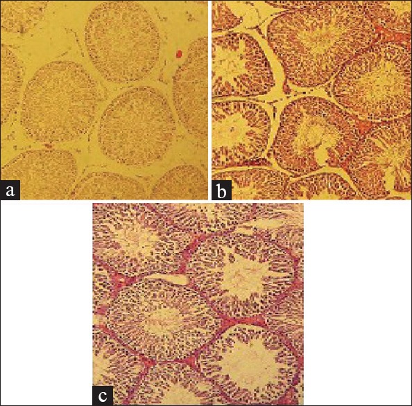
Effects on histopathology of testis with dose of 125 μg/kg Bisphenol A and Nonylphenol, (a) Control, (b) Nonylphenol and (c) Bisphenol A with 10× magnification
Discussion
Based on the present study, body weight of animals receiving BPA and NP was significantly decreased compared to the control group. Xiao et al. reported that long-term administration of NP in high dose can decrease the body weight of the rats compared to the control.[15] Mickae Couderc et al. also reported that pregnant female rats receiving NP showed a significant weight loss in comparison to the control group. This weight loss occurred during the first few days.[16] Although, in another study, no changes in body weight of the rats under treatment by NP were reported for a long duration.[17] The body weight of the experimental animals naturally increased with progressing age. According to the present study, this weight gain can be observed in control group. However, considerable weight loss in other experimental groups may confirm the toxic effects of long-term administration of BPA and NP.
The serum concentration of testosterone decreased in the experimental groups treated with BPA and NP. This decline might have been due to the blocking of the P450 cytochrome 17.[15]
The decrease in the level of testosterone could also have caused the weight loss by decreasing muscles and bone mass.[18] Toxic stresses can cause a decrease in the testosterone level by lowering the antioxidant capacities.[19] No noticeable change was observed in the levels of the LH and FSH hormones. The decrease in the level of testosterone was most probably due to a deterioration in the number and activity of the Leydig and Sertoli cells. However, because the pituitary was functioning properly, the levels of the FSH and LH hormones remained unchanged. Most probably, if the exposure to these chemicals had been prolonged, the levels of the FSH and LH hormones in the blood would have changed.
Mendiola et al. reported that exposure to environmental doses of BPA could increase the level of testosterone in adult male rats.[14] However, it has been reported that when adult rats were exposed to of the BPA (2 mg/kg) for 14 days, the levels of testosterone and FSH decreased while the level of LH increased.[20]
Along with the previous report, it was noted that in the group receiving 250 mg/kg of NP, the level of testosterone decreased but the LH and FSH increased. BPA and NP have an inhibitory effect on the P450 cytochrome, which is an important enzyme in the synthesis of testosterone in Leydig cells.[15] It has been suggested that 8-week-old male rats, which were treated with 250 mg/kg of the NP for 12 days, showed an increase in the levels of testosterone and FSH, however, the level of LH remained unchanged.[21] In another study, it was shown that high doses of BPA caused a significant decline in the level of testosterone.[8]
The BPA and NP doses used in this study were proper for evaluating the effects of low doses on reproductive function. BPA and NP caused a significant decrease in the diameter of seminiferous tubules, thickness of epithelium, and surface area of the cross-section of the epithelium of seminiferous tubules. It was also shown that number of spermatocytes and spermatogonia in NP (125 µg/kg) was significantly reduced compared to the other BPA and NP groups as well as the control group.
It has been reported that number of spermatocytes, spermatogonia, and spermatids and the concentration of testosterone may be reduced when male rats were exposed to environmental BPA. Furthermore, any decrease in the level of testosterone may decrease the number of sperms.[20] Treatment with NP increases the apoptosis Sertoli and germinal cells, reducing the production of sperms.[15]
Very high doses of the BPA in male rats (500 and 5000 mg/kg for 8 weeks) showed a significant decline in the number of sperms, however, there was no change in sperm's motility, diameter of seminiferous tubules, and the number of spermatocytes, spermatogonia, and Sertoli cells.[8]
Similar results were previously reported as well[8,22,23] that are consistent with the results of the present study.
Conclusion
It is concluded that BPA and NP can decrease the body weight of the rats and this may be due to the decrease in the level of testosterone. Furthermore, destructive effects of BPA and NP caused ruptures in the germinal tissues of seminiferous tubules and decreased the number of epithelium cells in the seminiferous tubules. Any decrease in the count of spermatocytes and spermatogonia, diameter and thickness of the epithelium, surface area of the seminiferous tubules and the epithelium, and testosterone level can confirm that spermatogenesis is decreased. These adverse effects of BPA and NP on spermatogenesis may suggest possible infertility effects of the compounds and the need to pay more attention to handling, usage, and exposure to estrogen-like plasticizers in the living environment.
Financial support and sponsorship
Nil.
Conflicts of interest
There are no conflicts of interest.
Acknowledgement
The authors wish to thank Dr. Soheil Ebrahimpour for his advisory cooperation. This investigation has been financially supported by the Research Affairs division of the Babol University of Medical Sciences.
References
- 1.Takeshita A, Koibuchi N, Oka J, Taguchi M, Shishiba Y, Ozawa Y. Bisphenol-A, an environmental estrogen, activates the human orphan nuclear receptor, steroid and xenobiotic receptor-mediated transcription. Eur J Endocrinol. 2001;145:513–7. doi: 10.1530/eje.0.1450513. [DOI] [PubMed] [Google Scholar]
- 2.Cao XL1, Corriveau J. Determination of bisphenol A in water by isotope dilution headspace solid-phase microextraction and gas chromatography/mass spectrometry without derivatization. J AOAC Int. 2008;91:622–9. [PubMed] [Google Scholar]
- 3.Mol HGJ, Sunarto S, Steijger OM. Determination of endocrine disruptors in water after derivatization with N-methyl-N-(tert.-butyldimethyltrifluoroacetamide) using gas chromatography with mass spectrometric detection. J Chromatogr A. 2000;83:742–7. doi: 10.1016/s0021-9673(00)00124-2. [DOI] [PubMed] [Google Scholar]
- 4.Benachour N, Aris A. Toxic effects of low doses of Bisphenol-A on human placental cells. Toxicol Appl Pharmacol. 2009;241:322–8. doi: 10.1016/j.taap.2009.09.005. [DOI] [PubMed] [Google Scholar]
- 5.Soares A, Guieysse B, Jefferson B, Cartmell E, Lester JN. Nonylphenol in the environment: A critical review on occurrence, fate, toxicity and treatment in wastewaters. Environ Int. 2008;34:1033–49. doi: 10.1016/j.envint.2008.01.004. [DOI] [PubMed] [Google Scholar]
- 6.Martínez-Zapata M, Aristizábal C, Peñuela G. Photodegradation of the endocrine-disrupting chemicals 4n-nonylphenol and triclosan by simulated solar UV irradiation in aqueous solutions with Fe (III) and in the absence/presence of humic acids. J Photochem Photobiol. 2013;251:41–9. [Google Scholar]
- 7.Jie X, Yang W, Jie Y, Hashim JH, Liu XY, Fan QY, et al. Toxic effect of gestational exposure to nonylphenol on F1 male rats. Birth Defects Res B Dev Reprod Toxicol. 2010;89:418–28. doi: 10.1002/bdrb.20268. [DOI] [PubMed] [Google Scholar]
- 8.Qiu LL, Wang X, Zhang XH, Zhang Z, Gu J, Liu L, et al. Decreased androgen receptor expression may contribute to spermatogenesis failure in rats exposed to low concentration of bisphenol A. Toxicol Letters. 2013;219:116–24. doi: 10.1016/j.toxlet.2013.03.011. [DOI] [PubMed] [Google Scholar]
- 9.Moriyama K, Tagami T, Akamizu T, Usui T, Saijo M, Kanamoto N, et al. Thyroid hormone action is disrupted by bisphenol A as an antagonist. J Clin Endocrinol Metab. 2002;87:5185–90. doi: 10.1210/jc.2002-020209. [DOI] [PubMed] [Google Scholar]
- 10.Takahashi O, Oishi S. Testicular toxicity of dietary 2,2-bis (4-hydroxyphenyl) propane (bisphenol A) in F344 rats. Arch Toxicol. 2001;75:42–51. doi: 10.1007/s002040000204. [DOI] [PubMed] [Google Scholar]
- 11.Doerge DR, Twaddle NC, Vanlandingham M, Brown RP, Fisher JW. Distribution of bisphenol A into tissues of adult, neonatal, and fetal Sprague-Dawley rats. Toxicol Appl Pharmacol. 2011;255:261–70. doi: 10.1016/j.taap.2011.07.009. [DOI] [PubMed] [Google Scholar]
- 12.Sun H, Xu LC, Chen JF, Song L, Wang XR. Effect of bisphenol A, tetrachlorobisphenol A and pentachlorophenol on the transcriptional activities of androgen receptor-mediated reporter gene. Food Chemical Toxicol. 2006;44:1916–21. doi: 10.1016/j.fct.2006.06.013. [DOI] [PubMed] [Google Scholar]
- 13.Sofie C, Marta A, Julie B, Anne MV, Gitte AP, Ulla H. Low-dose effects of bisphenol A on early sexual development in male and female rats. Reproduction. 2014;147:477–87. doi: 10.1530/REP-13-0377. [DOI] [PubMed] [Google Scholar]
- 14.Mendiola J, Jorgensen N, Andersson AM, Calafat AM, Ye X, Redmon JB, et al. Are environmental levels of bisphenol a associated with reproductive function in fertile men? Environ Health Perspect. 2010;118:1286–91. doi: 10.1289/ehp.1002037. [DOI] [PMC free article] [PubMed] [Google Scholar]
- 15.Xiao DH, Zhi GT, Yi G, Su NS, Xu YW, Li NK, et al. The toxic effects of nonylphenol on the reproductive system of male rats. Reprod Toxicol. 2004;19:215–21. doi: 10.1016/j.reprotox.2004.06.014. [DOI] [PubMed] [Google Scholar]
- 16.Couderc M, Gandar A, Kamari A, Allain Y, Zalouk-Vergnoux A, Herrenknecht C, et al. Neurodevelopmental and behavioral effects of nonylphenol exposure during gestational and breastfeeding period on F1 rats. Neurotoxicology. 2014;44:237–49. doi: 10.1016/j.neuro.2014.07.002. [DOI] [PubMed] [Google Scholar]
- 17.Chitra KC, Latchoumycandane C, Mathur PP. Induction of oxidative stress by bisphenol A in the epididymal sperm of rats. Toxicology. 2003;185:119–27. doi: 10.1016/s0300-483x(02)00597-8. [DOI] [PubMed] [Google Scholar]
- 18.Isidori AM, Giannetta E, Greco EA, Gianfrilli D, Bonifacio V, Isidori A, et al. Effects of testosterone on body composition, bone metabolism and serum lipid profile in middle-aged men: A meta-analysis. Clin Endocrinol. 2005;63:280–93. doi: 10.1111/j.1365-2265.2005.02339.x. [DOI] [PubMed] [Google Scholar]
- 19.Cox PJ. Cyclophosphamide cystitisidentification of acrolein as the causative agent. Biochem Pharmacol. 1979;28:2045–9. doi: 10.1016/0006-2952(79)90222-3. [DOI] [PubMed] [Google Scholar]
- 20.Jin P, Wang X, Chang F, Bai Y, Li Y, Zhou R, et al. Low dose bisphenol A impairs spermatogenesis by suppressing reproductive hormone production and promoting germ cell apoptosis in adult rats. J Biomed Res. 2013;27:135–44. doi: 10.7555/JBR.27.20120076. [DOI] [PMC free article] [PubMed] [Google Scholar]
- 21.Nagao T, Wada K, Marumo H, Yoshimura S, Ono H. Reproductive effects of nonylphenol in rats after gavage administration: A two-generation study. Reprod Toxicol. 2001;15:293–315. doi: 10.1016/s0890-6238(01)00123-x. [DOI] [PubMed] [Google Scholar]
- 22.Herath CB, Jin W, Watanabe G, Arai K, Suzuki AK, Taya K. Adverse effects of environmental toxicants, octylphenol and bisphenol A, on male reproductive functions in pubertal rats. Endocrine. 2004;25:163–72. doi: 10.1385/ENDO:25:2:163. [DOI] [PubMed] [Google Scholar]
- 23.Kazemi S, Mousavi SN, Aghapour F, Rezaee B, Sadeghi F, Moghadamnia AA. Induction effect of bisphenol A (BPA) on gene expression involving hepatic oxidative stress in rat. Oxidative Medicine and Cellular Longevity. 2016 doi: 10.1155/2016/6298515. [DOI] [PMC free article] [PubMed] [Google Scholar]